

Site-specific O-glucosylation of the epidermal growth factor-like (EGF) repeats of notch: efficiency of glycosylation is affected by proper folding and amino acid sequence of individual EGF repeats
- PMID: 22872643
- PMCID: PMC3464504
- DOI: 10.1074/jbc.M112.401315
Site-specific O-glucosylation of the epidermal growth factor-like (EGF) repeats of notch: efficiency of glycosylation is affected by proper folding and amino acid sequence of individual EGF repeats
Abstract
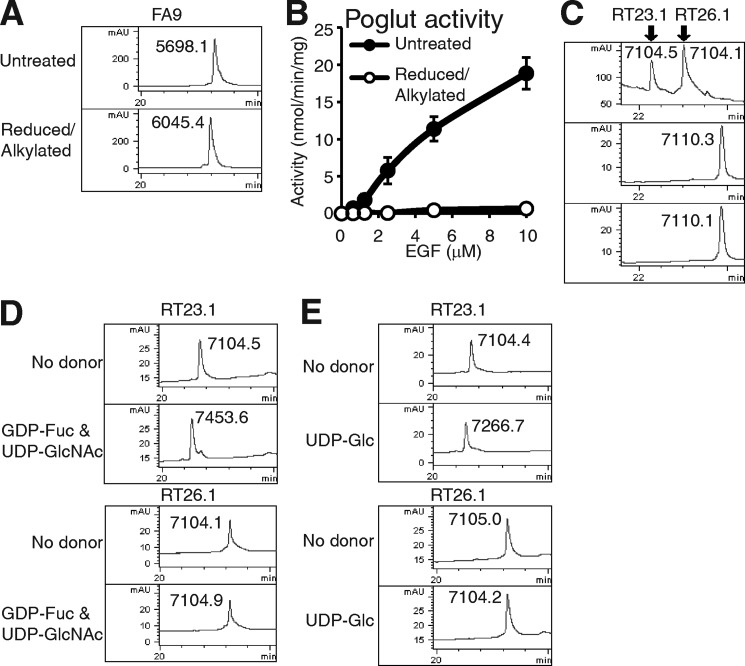
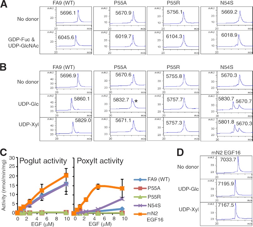
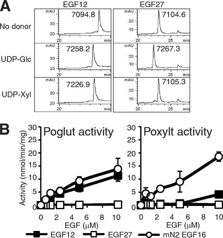
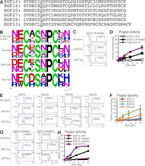
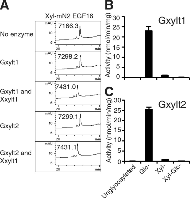
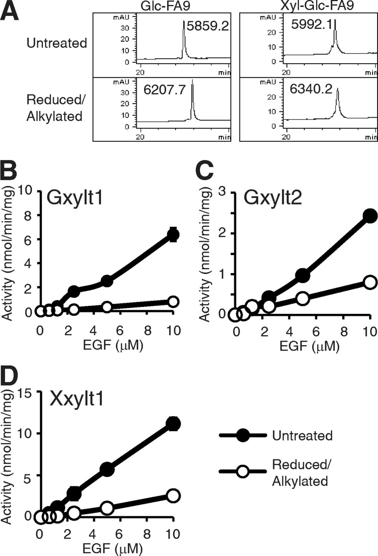
O-Glucosylation of epidermal growth factor-like (EGF) repeats in the extracellular domain of Notch is essential for Notch function. O-Glucose can be elongated by xylose to the trisaccharide, Xylα1-3Xylα1-3Glcβ1-O-Ser, whose synthesis is catalyzed by the consecutive action of three glycosyltransferases. A UDP-glucose:protein O-glucosyltransferase (Poglut/Rumi) transfers O-glucose to serine within the O-glucose consensus. Subsequently, either of two UDP-xylose:glucoside xylosyltransferases (Gxylt1 or Gxylt2) transfers xylose to O-glucose. Finally, a UDP-xylose:xyloside xylosyltransferase (Xxylt1) transfers xylose to Xylα1-3Glcβ1-O-EGF. Our prior site-mapping studies demonstrated that O-glucose consensus sites are modified at high but variable stoichiometries in mouse Notch1 and identified a novel glycosylation site with alanine in place of proline, suggesting a revised, broader consensus sequence (CXSX(P/A)C). Here we examined the molecular basis for this site specificity. A panel of EGF repeats from human coagulation factor 9 (FA9), mouse Notch1, and Notch2 were bacterially expressed and purified by reverse phase HPLC for use in in vitro enzyme assays. We demonstrate that proper folding of EGF repeats is essential for glycosylation by Poglut/Rumi, that alanine can substitute for proline in the context of coagulation factor 9 EGF repeat for O-glucose transfer, confirming the new consensus sequence, and that positively charged residues within the O-glucose consensus sequence reduce efficiency of glycosylation by Poglut/Rumi. Moreover, proper folding of EGF repeats is also important for the activities of Gxylt1, Gxylt2, and Xxylt1. These results indicate that protein folding and amino acid sequences of individual EGF repeats fundamentally affect both attachment and elongation of O-glucose glycans.
Figures
Similar articles
-
Molecular cloning of a xylosyltransferase that transfers the second xylose to O-glucosylated epidermal growth factor repeats of notch.J Biol Chem. 2012 Jan 20;287(4):2739-48. doi: 10.1074/jbc.M111.302406. Epub 2011 Nov 23. J Biol Chem. 2012. PMID: 22117070 Free PMC article.
-
Xylosyl Extension of O-Glucose Glycans on the Extracellular Domain of NOTCH1 and NOTCH2 Regulates Notch Cell Surface Trafficking.Cells. 2020 May 14;9(5):1220. doi: 10.3390/cells9051220. Cells. 2020. PMID: 32423029 Free PMC article.
-
Two novel protein O-glucosyltransferases that modify sites distinct from POGLUT1 and affect Notch trafficking and signaling.Proc Natl Acad Sci U S A. 2018 Sep 4;115(36):E8395-E8402. doi: 10.1073/pnas.1804005115. Epub 2018 Aug 20. Proc Natl Acad Sci U S A. 2018. PMID: 30127001 Free PMC article.
-
The multiple roles of epidermal growth factor repeat O-glycans in animal development.Glycobiology. 2015 Oct;25(10):1027-42. doi: 10.1093/glycob/cwv052. Epub 2015 Jul 14. Glycobiology. 2015. PMID: 26175457 Free PMC article. Review.
-
Protein O-glucosylation: another essential role of glucose in biology.Curr Opin Struct Biol. 2019 Jun;56:64-71. doi: 10.1016/j.sbi.2018.12.001. Epub 2019 Jan 18. Curr Opin Struct Biol. 2019. PMID: 30665188 Review.
Cited by
-
Synthesis and biological roles of O-glycans in insects.Glycoconj J. 2020 Feb;37(1):47-56. doi: 10.1007/s10719-019-09867-1. Epub 2019 Apr 1. Glycoconj J. 2020. PMID: 30937676 Review.
-
Marfan syndrome variation of the POGLUT2 and POGLUT3 consensus sequence can produce aberrant fibrillin-1 O-glucosylation.J Biol Chem. 2025 May;301(5):108411. doi: 10.1016/j.jbc.2025.108411. Epub 2025 Mar 17. J Biol Chem. 2025. PMID: 40107616 Free PMC article.
-
Analysis of endogenous NOTCH1 from POFUT1 S162L patient fibroblasts reveals the importance of the O-fucose modification on EGF12 in human development.Glycobiology. 2024 Jun 22;34(8):cwae047. doi: 10.1093/glycob/cwae047. Glycobiology. 2024. PMID: 38976017 Free PMC article.
-
Current Views on the Roles of O-Glycosylation in Controlling Notch-Ligand Interactions.Biomolecules. 2021 Feb 18;11(2):309. doi: 10.3390/biom11020309. Biomolecules. 2021. PMID: 33670724 Free PMC article. Review.
-
A Chemoenzymatic Method Based on Easily Accessible Enzymes for Profiling Protein O-GlcNAcylation.Anal Chem. 2020 Jul 21;92(14):9807-9814. doi: 10.1021/acs.analchem.0c01284. Epub 2020 Jul 7. Anal Chem. 2020. PMID: 32574038 Free PMC article.
References
-
- Fortini M. E. (2009) Notch signaling. The core pathway and its posttranslational regulation. Dev. Cell 16, 633–647 - PubMed
-
- Moloney D. J., Panin V. M., Johnston S. H., Chen J., Shao L., Wilson R., Wang Y., Stanley P., Irvine K. D., Haltiwanger R. S., Vogt T. F. (2000) Fringe is a glycosyltransferase that modifies Notch. Nature 406, 369–375 - PubMed
Publication types
MeSH terms
Substances
Grants and funding
LinkOut - more resources
Full Text Sources
Miscellaneous
