. 2012 Nov;26(11):4628-36.
doi: 10.1096/fj.12-215111. Epub 2012 Aug 7.

Sphingopeptides: dihydrosphingosine-based fusion inhibitors against wild-type and enfuvirtide-resistant HIV-1

Affiliations

Sphingopeptides: dihydrosphingosine-based fusion inhibitors against wild-type and enfuvirtide-resistant HIV-1

Avraham Ashkenazi et al. FASEB J. 2012 Nov.

Abstract

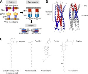
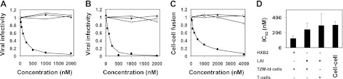
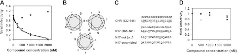
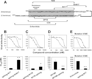
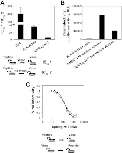
Understanding the structural organization of lipids in the cell and viral membranes is essential for elucidating mechanisms of viral fusion that lead to entry of enveloped viruses into their host cells. The HIV lipidome shows a remarkable enrichment in dihydrosphingomyelin, an unusual sphingolipid formed by a dihydrosphingosine backbone. Here we investigated the ability of dihydrosphingosine to incorporate into the site of membrane fusion mediated by the HIV envelope (Env) protein. Dihydrosphingosine as well as cholesterol, fatty acid, and tocopherol was conjugated to highly conserved, short HIV-1 Env-derived peptides with no antiviral activity otherwise. We showed that dihydrosphingosine exclusively endowed nanomolar antiviral activity to the peptides (IC(50) as low as 120 nM) in HIV-1 infection on TZM-bl cells and on Jurkat T cells, as well as in the cell-cell fusion assay. These sphingopeptides were active against enfuvirtide-resistant and wild-type CXCR4 and CCR5 tropic HIV strains. The anti-HIV activity was determined by both the peptides and their dihydrosphingosine conjugate. Moreover, their mode of action involved accumulation in the cells and viruses and binding to membranes enriched in sphingomyelin and cholesterol. The data suggest that sphingopeptides are recruited to the HIV membrane fusion site and provide a general concept in developing inhibitors of sphingolipid-mediated biological systems.


Figures

Figure 1.
Figure 1.
Model for HIV membrane fusion and the structure of lipids investigated in this study. A) HIV gp41 transmembrane protein has at least 3 major conformations during membrane fusion: the native nonfusogenic conformation, the prehairpin conformation, and the hairpin conformation. B) Side view of the recently determined hairpin structure of gp41 (Protein Data Bank identification number 2X7R; ref. 56). The trimeric inner coiled-coil of the N helices is shown in red; the 3 packing C helices are in blue. C, carboxyl terminus; N, amino terminus. Investigated short domains within the central core of the protein are indicated: the 17-mer conserved pocket sequence, termed N17; and the 19-mer N-helix binding sequence, termed DP19. C) Chemical structures of the hydrophobic conjugates investigated: the cellular moieties consisted of dihydrosphingosine (sphinganine), palmitic acid, and cholesterol; the noncellular moiety consisted of tocopherol.
Figure 2.
Figure 2.
Sphinganine exclusively endows antiviral activity to a short conserved gp41 N peptide in different HIV entry assays. Influence of sphinganine-N17 (■), palmitic acid-N17 (△), cholesterol-N17 (▲), and tocopherol-N17 (□) on HIV entry. A) TZM-bl cells that were infected with the wild-type fully infectious HXB2 strain. B) Jurkat T cells that were infected with a pseudotyped LAI strain. C) TZM-bl target cells that were fused to HIV Env-expressing effector cells. A representative viral infectivity curve is presented. D) IC50 of sphinganine-N17 in different viral entry systems. Results represent means ± sd (n=3).
Figure 3.
Figure 3.
Anti-HIV activity of sphingopeptides is determined by both the peptides and their lipid moiety. A) Viruses were constructed to have the same HIV core with 3 different Envs: LAI (CXCR4 tropic), AD8 (CCR5 tropic), and VSV-G. The 3 constructed viruses were allowed to infect TZM-bl cells in the presence of increasing concentrations of sphinganine-N17. Representative viral infection inhibitory curves are presented (n=2) for LAI (▲), AD8 (●), and VSV-G (■). Corresponding IC50 values of LAI and AD8 were 312 ± 83 and 403 ± 70 nM. B) Helical wheel representation demonstrates the interaction between the NHR regions of HIV gp41 in the core structure of the hairpin conformation, as observed in the crystal structures. Intermolecular association between the N helices occurs between positions a and d of the helical wheel. C) Mutational analysis of N17 interactions. In the N17mut(a,d) mutant (39), the residues at positions a and d of N17 were replaced by residues at positions f and c, respectively, of the CHR (residues are underscored). Residue numbers correspond to the HXB2 gp160 variant. In the scrambled N17 mutant, the residues of N17 were randomized. D) Sphingopeptides with mutated peptides, unable to self-interact or scrambled, lose their antiviral activity. TZM-bl cells were infected with fully infectious HXB2 HIV-1 in the presence of increasing concentrations of N17mut(a,d) (●), sphinganine-N17mut(a,d) (○), scrambled N17 (■), and sphinganine-scrambled N17 (□).
Figure 4.
Figure 4.
Sphinganine potentiates the activity of N and C peptides in wild-type and enfuvirtide-resistant viruses. A) Designation and location of the peptides within the gp41 core. The complex between N36 from the N helix and C34 from the C helix resembles the central core and is highlighted. The short domains N17 and DP19 are located within the central core, whereas the ShN17 extends from the core. Residue numbers correspond to the HXB2 gp160 variant. B–D) Viral infection inhibitory curves of the compounds and their corresponding IC50 values. TZM-bl cells were infected with fully infectious HXB2 HIV-1 in the presence of increased concentrations of the indicated compounds. B) Sphinganine (▲), sphinganine-N17 (sphing-N17; ♦), and sphinganine-ShN17 (sphing-ShN17; □). C) N-terminal conjugated N17K (sphing-N17K; ▲) or C-terminal conjugated N17K (N17K-sphing; △). D) DP19K alone (●) and DP19K-sphinganine (DP19K-sphing; ○). E) Cells were infected with fully infectious enfuvirtide-resistant LAI strain with an escape mutation in the gp41 core (V38E). Viral infection inhibitory curve of the drug enfuvirtide (●) and sphing-N17 (○) as well as their corresponding IC50 values are presented. IC50 values are presented as means ± sd, n ≥ 3. *P < 0.05, **P < 0.02.
Figure 5.
Figure 5.
Sphingopeptides strongly bind and accumulate in the cell membrane and in the viral membrane. A) Sphingopeptides are strongly anchored to the membrane and sustain a potent inhibitory effect on washing. C34, enfuvirtide, and sphing-N17 were preincubated with TZM-bl cells, followed by washing to remove unbound peptides and the addition of fully infectious viruses to start the infection. IC50 of the peptide with or without washing the cells (IC50 1 or 2, respectively) was calculated. Results are presented as the mean ± sd ratio of IC50 1/2 (n≥2), which reflects the fold increase in IC50 after cell washing. B) Infectivity of purified viruses that were pretreated with sphing-N17 or DMSO. Infectivity of the purified viruses was measured by luciferase activity in TZM-bl cells and is expressed in relative light units. C) Antiviral activity of sphing-N17 when it is preincubated with TZM-bl cells before the addition of fully infectious virus or when it is added to a mixture of the virus with cells.

Similar articles

Cited by

References

    1. White J. M. (1990) Viral and cellular membrane fusion proteins. Annu. Rev. Physiol. 52, 675–697 - PubMed
    1. Chernomordik L. V., Kozlov M. M. (2008) Mechanics of membrane fusion. Nat. Struct. Mol. Biol. 15, 675–683 - PMC - PubMed
    1. Nguyen D. H., Hildreth J. E. (2000) Evidence for budding of human immunodeficiency virus type 1 selectively from glycolipid-enriched membrane lipid rafts. J. Virol. 74, 3264–3272 - PMC - PubMed
    1. Ono A., Freed E. O. (2001) Plasma membrane rafts play a critical role in HIV-1 assembly and release. Proc. Natl. Acad. Sci. U. S. A. 98, 13925–13930 - PMC - PubMed
    1. Viard M., Parolini I., Sargiacomo M., Fecchi K., Ramoni C., Ablan S., Ruscetti F. W., Wang J. M., Blumenthal R. (2002) Role of cholesterol in human immunodeficiency virus type 1 envelope protein-mediated fusion with host cells. J. Virol. 76, 11584–11595 - PMC - PubMed

Publication types

MeSH terms

LinkOut - more resources