. 2012 Oct;206(8):1185-93.
doi: 10.1093/infdis/jis495. Epub 2012 Aug 7.

Staphylococcus aureus leukotoxin GH promotes inflammation

Affiliations

Staphylococcus aureus leukotoxin GH promotes inflammation

Natalia Malachowa et al. J Infect Dis. 2012 Oct.

Abstract

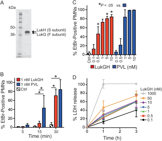
Background: Staphylococcus aureus produces numerous molecules that facilitate survival in the host. We recently identified a novel S. aureus leukotoxin (leukotoxin GH [LukGH]) using proteomics, but its role in virulence remains unclear. Here we investigated the role of LukGH in vivo.

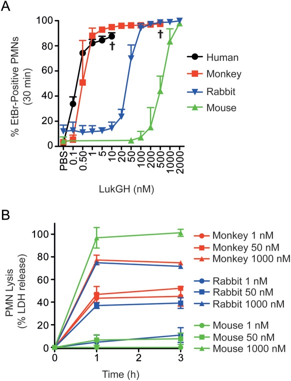
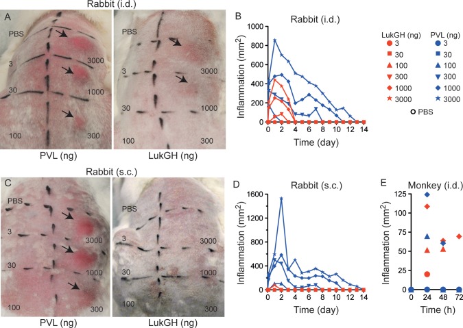
Methods: We tested cytotoxicity of LukGH toward polymorphonuclear leukocytes (PMNs) from mice, rabbits, monkeys, and humans. LukGH was administered to mice, rabbits, and a cynomolgus monkey by subcutaneous or intradermal injection to assess cytotoxicity or host response in vivo. The effects of LukGH in vivo were compared with those of Panton-Valentine leukocidin (PVL), a well-characterized S. aureus leukotoxin. The contribution of LukGH to S. aureus infection was tested using mouse and rabbit infection models.

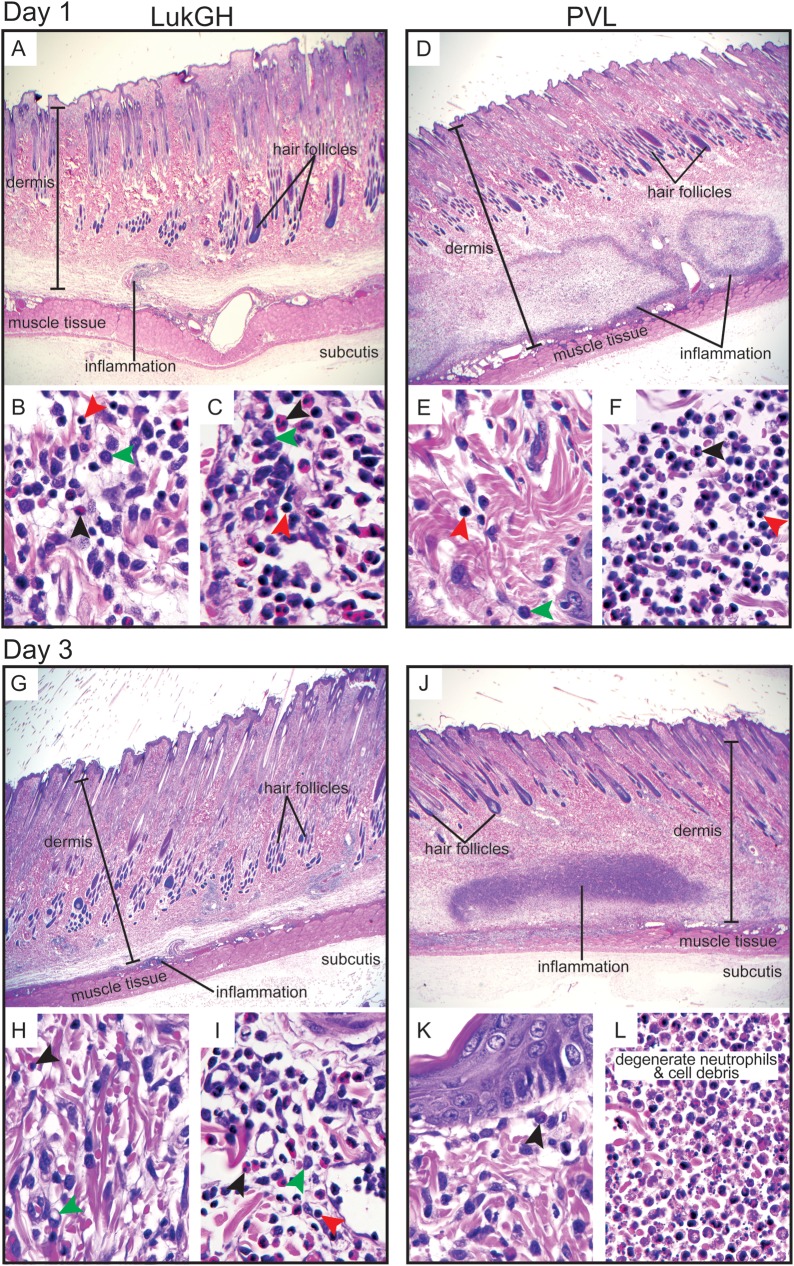
Results: Susceptibility of PMNs to LukGH was similar between humans and cynomolgus monkeys, and was greater than that of rabbits, which in turn was greater than that of mice. LukGH or PVL caused skin inflammation in rabbits and a monkey, but deletion of neither lukGH nor lukGH and lukS/F-PV reduced severity of USA300 infections in rabbits or mice. Rather, some disease parameters (eg, rabbit abscess size) were increased following infection with a lukGH and lukS/F-PV deletion strain.

Conclusions: Our findings indicate that S. aureus leukotoxins enhance the host inflammatory response and influence the outcome of infection.


Figures

Figure 1.
Figure 1.
Leukotoxin GH (LukGH)–mediated pore formation and polymorphonuclear leukocyte (PMN) cytolysis. A, Purified LukGH was resolved by 12% sodium dodecyl sulfate polyacrylamide gel electrophoresis, and the gel was stained with GelCode Blue. B, Permeability of human PMNs (plasma membrane pore formation) following exposure to 1 nM LukGH or Panton-Valentine leukocidin (PVL). *P < .0001 using a 2-way analysis of variance (ANOVA) and Bonferroni posttest. Ctrl, control assays containing assay media alone. For reference, 1 nM PVL is 0.072 μg/mL PVL (0.037 μg of LukF-PV plus 0.035 μg of LukS-PV per milliliter) and 1 nM LukGH is 0.079 μg/mL. C, Concentration-dependent pore formation in human PMNs following incubation with the indicated concentrations of LukGH or PVL for 30 minutes. Results are the mean (± SD) of 8 (B) or 10 (C) PMN donors. *P < .05 using a 1-way ANOVA and Tukey posttest. D, LukGH-mediated lysis of human PMNs. Results are the mean (± SD) of 4 PMN donors. Abbreviations: EtBr, ethidium bromide; LDH, lactate dehydrogenase.
Figure 2.
Figure 2.
Neutrophil sensitivity to leukotoxin GH (LukGH) is species-specific. A, LukGH-mediated pore formation. Polymorphonuclear leukocytes (PMNs) from humans, monkeys, rabbits, or mice were incubated for 30 minutes with 0–2000 nM LukGH. Daggers indicate pore formation could not be measured because of cell lysis and debris. B, LukGH-mediated PMN lysis. Results are the mean (± SD) of 3 experiments for A and B. Abbreviations: EtBr, ethidium bromide; LDH, lactate dehydrogenase; PBS, phosphate-buffered saline.
Figure 3.
Figure 3.
Leukotoxin GH (LukGH) causes skin inflammation. Inflammatory response in the skin of rabbits following intradermal (i.d.) (A and B) and subcutaneous (s.c.) (C and D) injection of purified LukGH or Panton-Valentine leukocidin (PVL). Images in A and C were taken 24 hours after injection. B and D, quantitation of skin inflammation for 14 days. Results are the mean of 2 rabbits. Note that hair was removed prior to administration of leukotoxins. E, Inflammatory response in the skin of a cynomolgus macaque following intradermal injection of LukGH or PVL as indicated. Results are presented as the area of inflammation from 1 monkey. Abbreviation: PBS, phosphate-buffered saline.
Figure 4.
Figure 4.
Histopathology of rabbit skin inflammation elicited by purified leukotoxin GH (LukGH) or Panton-Valentine leukocidin (PVL). Representative histopathological sections of the inflammatory lesions caused by intradermal injection of 1000 ng of purified LukGH (AC and GI) or PVL (DF and JL) are shown. AF, Day 1; GL, day 3. In A, D, G, and J, original magnification is ×20. B, E, H, and K show an upper layer of dermis; original magnification is ×1000. C, F, I, and L show cells of the inflammatory lesion; original magnification is ×1000. Black arrowhead, polymorphonuclear leukocyte; red arrowhead, lymphocyte; green arrowhead, mononuclear phagocyte.
Figure 5.
Figure 5.
Contribution of leukotoxin GH (LukGH) to virulence in animal models of USA300 infection. A, Rabbit abscesses were measured following subcutaneous inoculation with the indicated strains. Results are the mean of 6–10 abscesses (as indicated) from 3–5 rabbits (2 abscesses per animal). *P < .05 using a 1-way analysis of variance (ANOVA) and Dunnett posttest. B, A separate set of experiments (2 rabbits per strain and 4 abscesses in total) was used to determine bacterial colony-forming units (CFUs) in abscesses on day 6 after infection. C, Area of rabbit abscesses used for enumeration of CFUs in B. Each symbol represents a data point obtained from a single abscess, and the line indicates the mean. There were no significant differences in CFUs recovered from the mutant strains compared with the wild-type (wt) strain as determined using a 1-way ANOVA and Dunnett posttest. D, Mouse abscess model. E, Mouse bacteremia model. Each animal received 5 × 107 CFUs of Staphylococcus aureus in 0.1 mL of sterile saline by tail vein inoculation. Results in D and E are the mean of 15 animals per S. aureus strain. *P = .03 using a log-rank test for trend.

Similar articles

Cited by

References

    1. DeLeo FR, Diep BA, Otto M. Host defense and pathogenesis in Staphylococcus aureus infections. Infect Dis Clin North Am. 2009;23:17–34. - PMC - PubMed
    1. Prevost G, Cribier B, Couppie P, et al. Panton-Valentine leucocidin and gamma-hemolysin from Staphylococcus aureus ATCC 49775 are encoded by distinct genetic loci and have different biological activities. Infect Immun. 1995;63:4121–9. - PMC - PubMed
    1. Ventura CL, Malachowa N, Hammer CH, et al. Identification of a novel Staphylococcus aureus two-component leukotoxin using cell surface proteomics. PloS One. 2010;5:e11634. - PMC - PubMed
    1. Menestrina G, Dalla Serra M, Comai M, et al. Ion channels and bacterial infection: the case of β-barrel pore-forming protein toxins of Staphylococcus aureus. FEBS Lett. 2003;552:54–60. - PubMed
    1. Jayasinghe L, Bayley H. The leukocidin pore: evidence for an octamer with four LukF subunits and four LukS subunits alternating around a central axis. Protein Sci. 2005;14:2550–61. - PMC - PubMed

Publication types