

Clinical Implications of Micrometastasis Detection in Internal Mammary Nodes of Breast Cancer Patients
- PMID: 22872795
- PMCID: PMC3409387
- DOI: 10.1159/000339686
Clinical Implications of Micrometastasis Detection in Internal Mammary Nodes of Breast Cancer Patients
Abstract
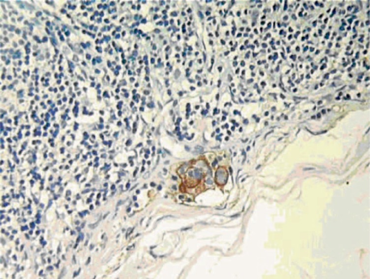
BACKGROUND: The aim of this study was to assess the efficiency of step-serial sectioning (SSS) combined with hematoxylin and eosin (H&E) and immunohistochemical (IHC) staining in detecting micrometastasis of internal mammary lymph nodes (IMLNs). PATIENTS AND METHODS: 135 IMLNs from 88 breast cancer patients were re-examined by SSS, combined with either H&E or IHC staining of the biomarkers cytokeratin-19 and epithelial membrane antigen. RESULTS: Of the 135 IMLNs, 6 nodes from 5 cases displayed 1 or more micrometastases. Histological grade and lymphovascular invasion status were significantly correlated with micrometastasis in the IMLNs (p = 0.018 and 0.001, respectively). Of the 6 nodes positive for micrometastasis, 1 node was detected by both H&E and IHC staining. The remaining 5 nodes from 4 cases showed evident tumor cells only by IHC staining. Finally 8 of the 83 patients (9.64%) without IMLN metastasis showed distant metastasis, while 2 of the 5 patients (40%) with IMLN metastasis showed distant metastasis within 28 months of operation. CONCLUSION: SSS combined with H&E and IHC staining is more efficient in detecting micrometastasis than classic routine single-slice H&E only.
Hintergrund: Ziel dieser Studie war die Bestimmung der Effizienz des Step-Serial Sectioning (SSS) kombiniert mit Hämatoxylin-Eosin (H&E)-Färbung bzw. immunhistochemischer (IHC) Färbung zur Detektion von Mikrometastasen in den Mammaria-interna-Lymphknoten.
Patientinnen und Methoden: 135 Mammaria-interna-Lymphknoten von 88 Mammakarzinompatientinnen wurden mittels SSS kombiniert mit H&E- bzw. IHC-Färbung erneut auf die Biomarker Cytokeratin-19 und epitheliales Membranantigen untersucht.
Ergebnisse: Von den 135 Mammaria-interna-Lymphknoten zeigten 6 Knoten von 5 Fällen eine oder mehrere Mikrometastasen. Der histologische Grad und lymphovasku-läre Invasionsstatus waren signifikant mit dem Vorliegen von Mikrometastasen in den Mammaria-interna-Lymphknoten korreliert (p = 0,018 bzw. 0,001). Einer der 6 positiven Lymphknoten konnte sowohl mit H&E- als auch mit IHC-Färbung dargestellt werden. Für die verbleibenden 5 Knoten von 4 Fällen konnte nur die IHC-Färbung Tumorzellen nachweisen. Insgesamt zeigten 8 der 83 Patientinnen (9,64%) ohne Metastasen in den Mammaria-interna-Lymphknoten Fernmetastasen, während 2 der 5 Patientinnen (40%) mit Metastasen in den Mammaria-interna-Lymphknoten innerhalb von 28 Monaten nach Operation Fernmetastasen aufwiesen.
Schlussfolgerung: SSS kombiniert mit H&E- bzw. IHC-Färbung ist beim Aufzeigen von Mikrometastasen effektiver als die routinemäßig eingesetzte, klassische Single-Slice-H&E-Färbung.
Figures


Similar articles
-
The efficiency of micrometastasis by sentinel node navigation surgery using indocyanine green and infrared ray laparoscopy system for gastric cancer.Gastric Cancer. 2012 Jul;15(3):287-91. doi: 10.1007/s10120-011-0105-6. Epub 2011 Oct 27. Gastric Cancer. 2012. PMID: 22041868 Clinical Trial.
-
Detection of sentinel and non-sentinel lymph node micrometastases by complete serial sectioning and immunohistochemical analysis for gastric cancer.J Exp Clin Cancer Res. 2008 May 30;27(1):7. doi: 10.1186/1756-9966-27-7. J Exp Clin Cancer Res. 2008. PMID: 18577253 Free PMC article.
-
Is lymph-node micrometastasis in gallbladder cancer a significant prognostic factor?Hepatogastroenterology. 2012 Jan-Feb;59(113):31-5. doi: 10.5754/hge10010. Hepatogastroenterology. 2012. PMID: 22251520
-
Clinical outcome of patients with lymph node-negative breast carcinoma who have sentinel lymph node micrometastases detected by immunohistochemistry.Cancer. 2005 Apr 15;103(8):1581-6. doi: 10.1002/cncr.20934. Cancer. 2005. PMID: 15747375 Review.
-
Breast cancer micrometastasis and axillary sentinel lymph nodes frozen section. Our experience and review of literature.Int J Surg. 2014;12 Suppl 1:S12-5. doi: 10.1016/j.ijsu.2014.05.044. Epub 2014 May 22. Int J Surg. 2014. PMID: 24859398 Review.
Cited by
-
Proteomics of Sentinel Lymph Nodes in Early Breast Cancer for Identification of Thymidylate Synthase as a Potential Biomarker to Flag Metastasis: A Preliminary Study.Cancer Manag Res. 2020 Jun 23;12:4841-4854. doi: 10.2147/CMAR.S255684. eCollection 2020. Cancer Manag Res. 2020. PMID: 32606973 Free PMC article.
-
Detection of cervical lymph node micrometastasis and isolated tumor cells in oral squamous cell carcinoma using immunohistochemistry and serial sectioning.J Oral Maxillofac Pathol. 2016 Sep-Dec;20(3):436-444. doi: 10.4103/0973-029X.190946. J Oral Maxillofac Pathol. 2016. PMID: 27721609 Free PMC article.
References
-
- Anim JT, John B, et al. Relationship between the expression of various markers and prognostic factors in breast cancer. Acta Histochem. 2005;107:87–93. - PubMed
-
- Kasami M, Uematsu T, et al. Comparison of estrogen receptor, progesterone receptor and Her-2 status in breast cancer pre- and post-neoadjuvant chemotherapy. Breast. 2008;17:523–527. - PubMed
-
- Jemal A, Siegel R, et al. Cancer statistics, 2007. CA Cancer J Clin. 2007;57:43–66. - PubMed
-
- Gooiker GA, van Gijn W, et al. A systematic review and meta-analysis of the volume-outcome relationship in the surgical treatment of breast cancer. Are breast cancer patients better of with a high volume provider? Eur J Surg Oncol. 2010;36((suppl 1)):S27–35. - PubMed
-
- Chen JJ, Wu J. Management strategy of early-stage breast cancer patients with a positive sentinel lymph node: with or without axillary lymph node dissection. Crit Rev Oncol Hematol. 2011;79:293–301. - PubMed
LinkOut - more resources
Full Text Sources
