

Current concepts in the mandibular condyle fracture management part I: overview of condylar fracture
- PMID: 22872830
- PMCID: PMC3408272
- DOI: 10.5999/aps.2012.39.4.291
Current concepts in the mandibular condyle fracture management part I: overview of condylar fracture
Abstract
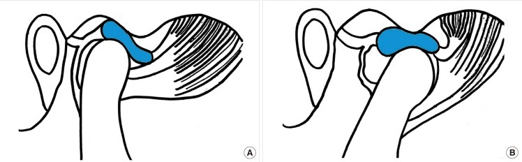
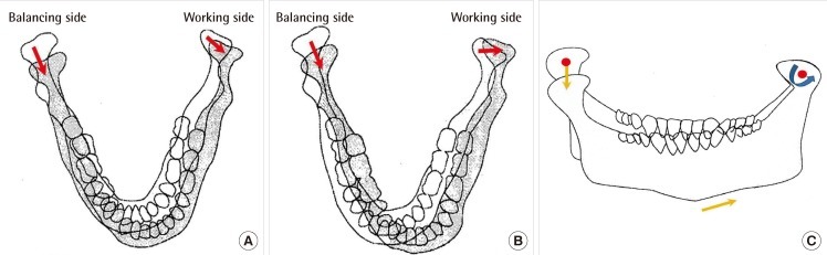
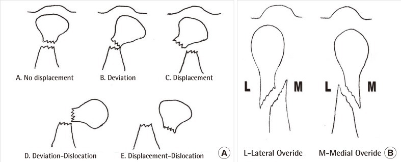
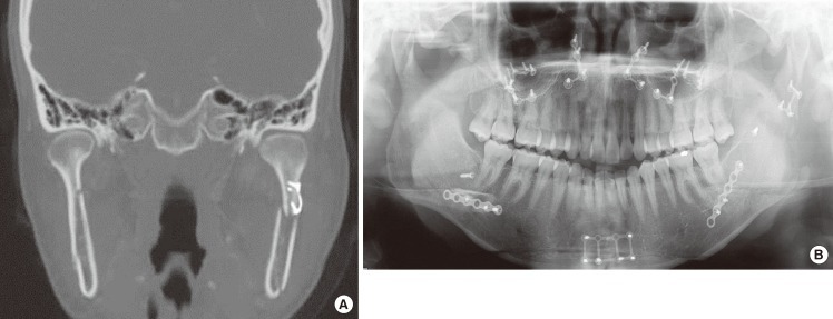
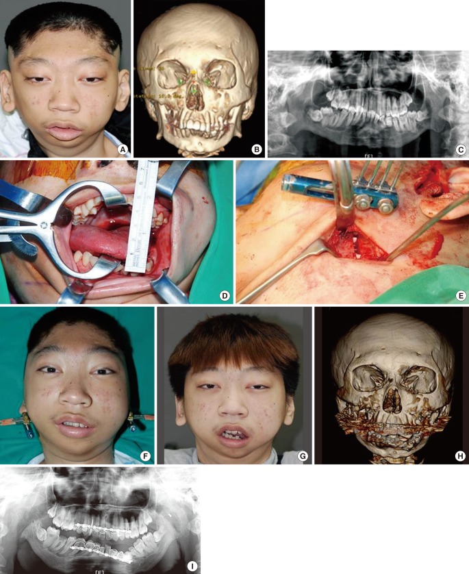
The incidence of condylar fractures is high, but the management of fractures of the mandibular condyle continues to be controversial. Historically, maxillomandibular fixation, external fixation, and surgical splints with internal fixation systems were the techniques commonly used in the treatment of the fractured mandible. Condylar fractures can be extracapsular or intracapsular, undisplaced, deviated, displaced, or dislocated. Treatment depends on the age of the patient, the co-existence of other mandibular or maxillary fractures, whether the condylar fracture is unilateral or bilateral, the level and displacement of the fracture, the state of dentition and dental occlusion, and the surgeonnds on the age of the patient, the co-existence of othefrom which it is difficult to recover aesthetically and functionally;an appropriate treatment is required to reconstruct the shape and achieve the function ofthe uninjured status. To do this, accurate diagnosis, appropriate reduction and rigid fixation, and complication prevention are required. In particular, as mandibular condyle fracture may cause long-term complications such as malocclusion, particularly open bite, reduced posterior facial height, and facial asymmetry in addition to chronic pain and mobility limitation, great caution should be taken. Accordingly, the authors review a general overview of condyle fracture.
Keywords: Mandibular condyle; Mandibular fractures; Temporomandibular joint.
Conflict of interest statement
No potential conflict of interest relevant to this article was reported.
Figures
Similar articles
-
Fractures of the mandibular condyle: a review of 466 cases. Literature review, reflections on treatment and proposals.J Craniomaxillofac Surg. 2006 Oct;34(7):421-32. doi: 10.1016/j.jcms.2006.07.854. Epub 2006 Oct 19. J Craniomaxillofac Surg. 2006. PMID: 17055280 Review.
-
Open reduction and rigid internal fixation of mandibular condylar fractures by an intraoral approach: a long-term follow-up study of 15 patients.J Oral Maxillofac Surg. 2006 Dec;64(12):1771-9. doi: 10.1016/j.joms.2005.12.069. J Oral Maxillofac Surg. 2006. PMID: 17113444
-
Dislocated pediatric condyle fractures - should conservative treatment always be the rule?J Craniomaxillofac Surg. 2020 Oct;48(10):933-941. doi: 10.1016/j.jcms.2020.08.001. Epub 2020 Aug 14. J Craniomaxillofac Surg. 2020. PMID: 32919835
-
Endoscope-assisted transoral reduction and internal fixation versus closed treatment of mandibular condylar process fractures--a prospective double-center study.J Oral Maxillofac Surg. 2012 Feb;70(2):384-95. doi: 10.1016/j.joms.2011.02.035. Epub 2011 Jun 12. J Oral Maxillofac Surg. 2012. PMID: 21664746
-
Contrast analysis of open reduction and internal fixation and non-surgical treatment of condylar fracture: a meta-analysis.J Craniofac Surg. 2014 Nov;25(6):2077-80. doi: 10.1097/SCS.0000000000001010. J Craniofac Surg. 2014. PMID: 25304143 Review.
Cited by
-
Comparison of intermaxillary fixation techniques for mandibular fractures with focus on patient experience.Arch Craniofac Surg. 2022 Feb;23(1):23-28. doi: 10.7181/acfs.2021.00549. Epub 2022 Feb 20. Arch Craniofac Surg. 2022. PMID: 35255592 Free PMC article.
-
Retro-Auricular Approach to the Fractures of the Mandibular Condyle: A Systematic Review.J Clin Med. 2021 Jan 11;10(2):230. doi: 10.3390/jcm10020230. J Clin Med. 2021. PMID: 33440626 Free PMC article. Review.
-
The application of the Risdon approach for mandibular condyle fractures.BMC Surg. 2013 Jul 6;13:25. doi: 10.1186/1471-2482-13-25. BMC Surg. 2013. PMID: 23829537 Free PMC article.
-
Long-term Stability after Reduction of Mandible Fracture by Keyhole Plate: Evaluation at the Time of Plate Removal.Maxillofac Plast Reconstr Surg. 2020 Mar 16;42(1):6. doi: 10.1186/s40902-020-00251-w. eCollection 2020 Dec. Maxillofac Plast Reconstr Surg. 2020. PMID: 32206665 Free PMC article.
-
A paediatric case of bilateral mandibular condyle fracture presenting with bloody otorrhoea following trauma.BMJ Case Rep. 2017 Apr 22;2017:bcr2016218995. doi: 10.1136/bcr-2016-218995. BMJ Case Rep. 2017. PMID: 28433980 Free PMC article.
References
-
- Turvey TA. Midfacial fractures: a retrospective analysis of 593 cases. J Oral Surg. 1977;35:887–891. - PubMed
-
- Fridrich KL, Pena-Velasco G, Olson RA. Changing trends with mandibular fractures: a review of 1,067 cases. J Oral Maxillofac Surg. 1992;50:586–589. - PubMed
-
- Celenza FV, Nasedkin JN. Occlusion: the state of the art. Chicago: Quintessence Pub. Co.; 1978.
-
- Mathog RH, Toma V, Clayman L, et al. Nonunion of the mandible: an analysis of contributing factors. J Oral Maxillofac Surg. 2000;58:746–752. - PubMed
-
- Mathog RH. Nonunion of the mandible. Otolaryngol Clin North Am. 1983;16:533–547. - PubMed
LinkOut - more resources
Full Text Sources