

doi: 10.5999/aps.2012.39.4.435.
Epub 2012 Jul 13.
Bilateral circular xanthelasma palpebrarum
Affiliations
- PMID: 22872853
- PMCID: PMC3408295
- DOI: 10.5999/aps.2012.39.4.435
Item in Clipboard
Bilateral circular xanthelasma palpebrarum
Arch Plast Surg.
2012 Jul.
No abstract available
Conflict of interest statement
No potential conflict of interest relevant to this article was reported.
Figures
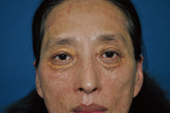
Preoperative view. A 53-year-old female presented bilateral circular yellowish skin lesions around the upper and lower eyelids.
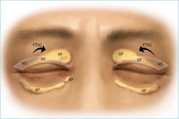
Schematic picture of the operation. The lesion of the lower eyelids were excised marginally and directly closed. Blepharoplasty with excision of xanthelasma palpebrarum was performed on the upper eyelids. The raw surface of the medial side was covered with the normal skin excised from the lateral side through the blepharoplasty incision. XP, xanthelasma palpebrarum; NS, normal skin; FTSG, full-thickness skin graft.

Postoperative 2-month view. The scars, which were made from marginal excision and direct closure, are shown in the lower eyelids. The sites of the full-thickness skin graft in the upper eyelids are shown with a good matching skin color and texture. There is no sign or symptom of ectropion.
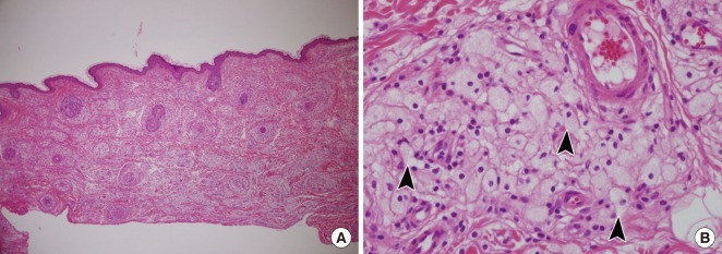
Histopathologic findings of the left upper eyelid lesion. (A) Pale areas containing foamy cells are dispersed throughout the dermis (H&E, ×40). (B) Xanthoma cells. These foamy histiocytes are polygonal or rounded with a distinct cell membrane. Their nuclei were small and eccentric and their cytoplasms are stuffed with lipid vacuoles (black arrowheads) (H&E, ×400).
References
-
- Jee MS, Chang SE, Choi JH, et al. Clinicopathologic study of 37 cases of xanthelasma palpebrarum; clinical significance of xanthelasma palpebrarum in hyperlipidemiae and cardiovascular diseases. Korean J Dermatol. 2003;41:333–337.
-
- Depot MJ, Jakobiec FA, Dodick JM, et al. Bilateral and extensive xanthelasma palpebrarum in a young man. Ophthalmology. 1984;91:522–527. - PubMed
-
- Yoon SY, Cho JH, Bae EY, et al. A case of bilateral extensive xanthelasma. Korean J Dermatol. 2005;43:675–677.
-
- Rohrich RJ, Janis JE, Pownell PH. Xanthelasma palpebrarum: a review and current management principles. Plast Reconstr Surg. 2002;110:1310–1314. - PubMed
LinkOut - more resources
Full Text Sources