. 2012 Nov;11(11):1263-73.
doi: 10.1074/mcp.M112.017194. Epub 2012 Aug 7.

Five friends of methylated chromatin target of protein-arginine-methyltransferase[prmt]-1 (chtop), a complex linking arginine methylation to desumoylation

Affiliations

Five friends of methylated chromatin target of protein-arginine-methyltransferase[prmt]-1 (chtop), a complex linking arginine methylation to desumoylation

Pavlos Fanis et al. Mol Cell Proteomics. 2012 Nov.

Abstract

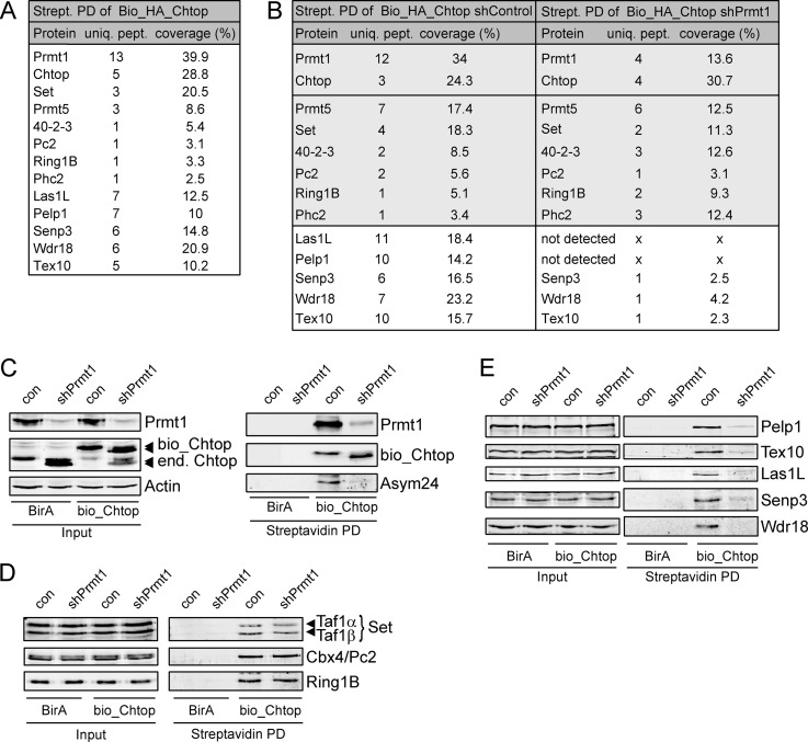
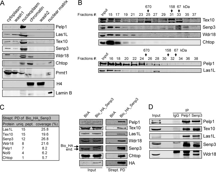
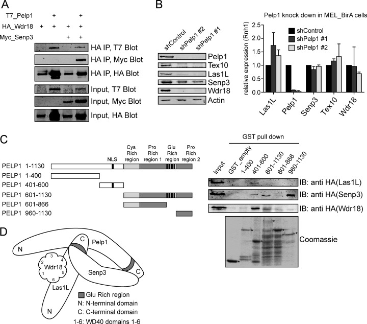
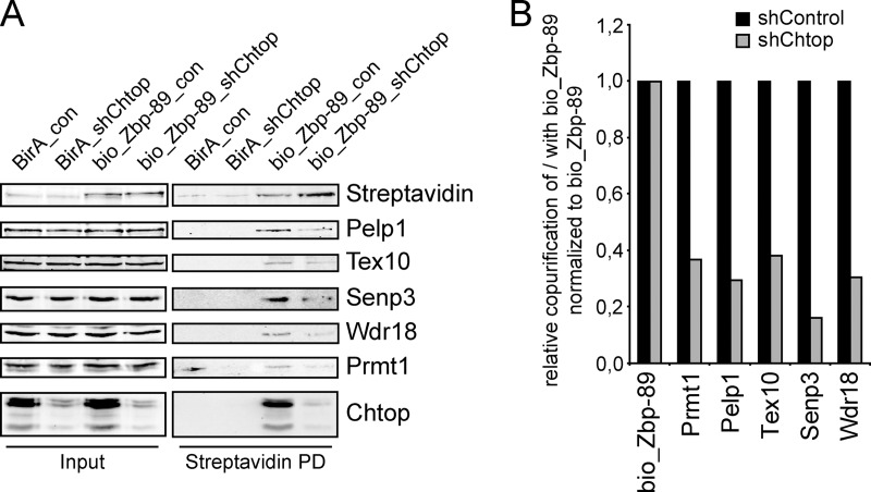
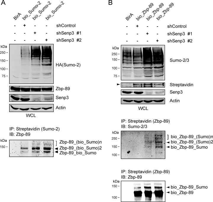
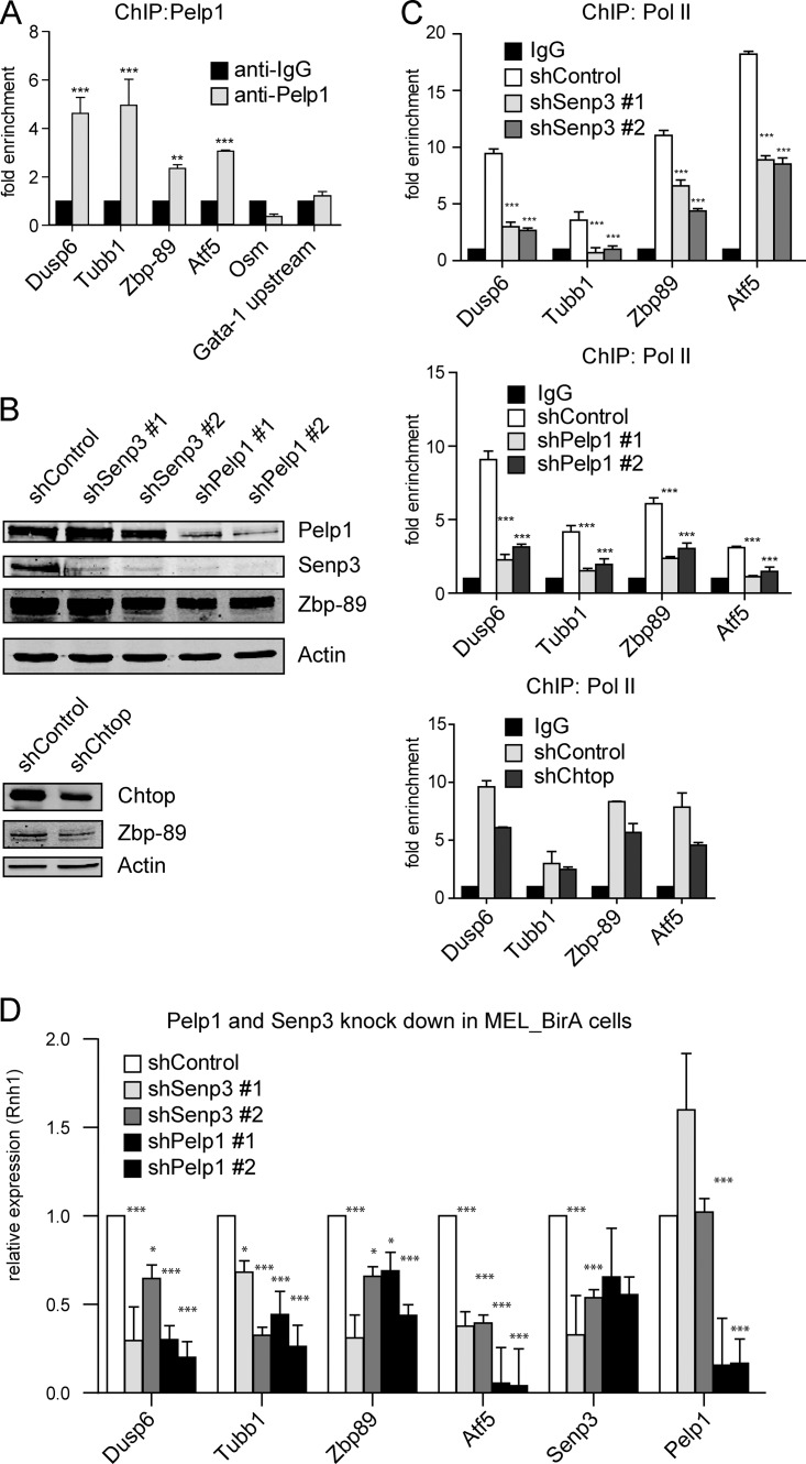
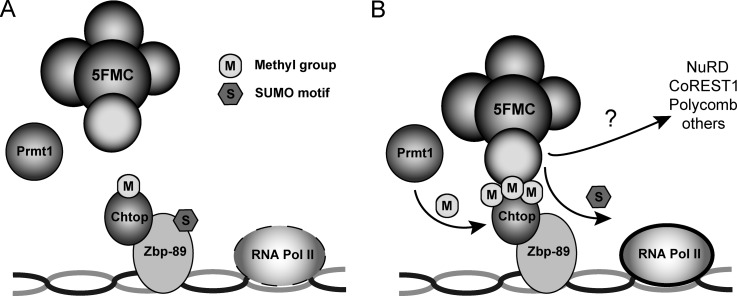
Chromatin target of Prmt1 (Chtop) is a vertebrate-specific chromatin-bound protein that plays an important role in transcriptional regulation. As its mechanism of action remains unclear, we identified Chtop-interacting proteins using a biotinylation-proteomics approach. Here we describe the identification and initial characterization of Five Friends of Methylated Chtop (5FMC). 5FMC is a nuclear complex that can only be recruited by Chtop when the latter is arginine-methylated by Prmt1. It consists of the co-activator Pelp1, the Sumo-specific protease Senp3, Wdr18, Tex10, and Las1L. Pelp1 functions as the core of 5FMC, as the other components become unstable in the absence of Pelp1. We show that recruitment of 5FMC to Zbp-89, a zinc-finger transcription factor, affects its sumoylation status and transactivation potential. Collectively, our data provide a mechanistic link between arginine methylation and (de)sumoylation in the control of transcriptional activity.


Figures

Fig. 1.
Fig. 1.
Identification of Chtop-interacting proteins. A, List of Chtop interacting proteins indentified by mass spectrometry (MS). Protein complexes from nuclear lysates of MEL_BirA cells ectopically expressing Bio_HA_Chtop were recovered by MS. Proteins, unique peptides (uniq. pept.) and percent coverage (coverage %) are indicated. B, List of Chtop interacting proteins in MEL_BirA cells treated with control lentivirus (shControl) and lentivirus expressing shRNA against Prmt1 (shPrmt1) identified by MS. Interactions with major differences between shControl and shPrmt1 treated cells are shown in a white box. C, Chtop is hypomethylated in the absence of Prmt1. Whole cell lysates (Input) and streptavidin pull downs (Streptavidin PD) were analyzed for Prmt1, Chtop and asymmetric dimethyl arginine (Asym24) residues. Actin staining serves as a loading control. Ectopically expressed (bio_Chtop) and endogenous Chtop (end. Chtop) are indicated. D, Chtop methylation-independent interactions. Whole cell lysates (Input) and streptavidin pull-downs (Streptavidin PD) were analyzed with the antibodies indicated. E, Chtop methylation-dependent interactions. Whole-cell lysates (Input) and streptavidin pull-downs (Streptavidin PD) were analyzed with the antibodies indicated.
Fig. 2.
Fig. 2.
5FMC is a nuclear complex. A, Chtop methylation dependent interaction proteins are localized mainly in the nucleoplasm. MEL_BirA cells were biochemically fractionated as described under “Experimental Procedures”. Cytoplasmic, nucleoplasmic, chromatin and nuclear matrix were tested using Pelp1, Las1L, Tex10, Senp3, Wdr18 antibodies against endogenous proteins. Chtop, Prmt1, H4, and Lamin B served as controls for individual fractions. B, MEL_BirA cell nuclear extracts were analyzed by sized-exclusion chromatography on a Superose 6 column. Proteins eluted from the indicated fractions were blotted with the indicated antibodies. Molecular mass markers are indicated at the top. C, Senp3 interactions in MEL cells. Whole cell lysates (Input) and streptavidin pull downs (Strept. PD) from MEL_BirA (BirA) and MEL_BirA cells expressing biotinylated Senp3 (Bio_HA_Senp3) were analyzed by MS (table) and Western blotting. Immunoblot probed with the indicated antibodies. Arrows indicate endogenous (end.) and biotinylated (Bio_HA) Senp3. D, 5FMC is a nuclear complex. Endogenous association between the 5FMC components. MEL_BirA cell nuclear lysates were analyzed by immunoprecipitation (IP) and Western blotting with the antibodies indicated.
Fig. 3.
Fig. 3.
Pelp1 is the core component of the 5FMC complex. A, Pelp1 is required for interaction between Senp3 and Wdr18. 293T cells were transiently transfected with expression vectors encoding T7_Pelp1, HA_Wdr18 and Myc_Senp3. Cell lysates were analyzed by IP and Western blotting with the antibodies indicated. B, Pelp1 is needed for the stability of the 5FMC complex. MEL_BirA cells were treated with the indicated shRNAs. Nuclear lysates were analyzed by Western blotting with the indicated antibodies. Actin staining serves as a loading control. Total RNA was extracted from MEL_BirA cells transduced with the indicated shRNA and analyzed by RT-QPCR for Las1L, Pelp1, Senp3, Tex10, and Wdr18. Error bars: S.D. of triplicate experiment. C, Mapping the interaction regions of Pelp1. Schematic representation of Pelp1 deletion constructs. GST fused Pelp1 domains and GST alone (GST_empty) were immobilized onto glutathione beads (lower panel stained with Coomassie) and used to pull down nuclear cell lysates from 293T cells expressing HA_Las1L, HA_Senp3 and HA_Wdr18. Asterisks indicate GST fusion proteins. D, Schematic representation of interaction between Pelp1, Las1L, Senp3, and Wdr18 protein domains.
Fig. 4.
Fig. 4.
Chtop recruits 5FMC to Zbp-89. A, Zbp-89 is associated with Chtop and 5FMC complex. Nuclear cell lysates (Input) of MEL_BirA cells (BirA) and MEL_BirA cells expressing biotinylated Zbp-89 (bio_Zbp-89) treated with lentivirus expressing shRNA against Chtop (shChtop) and control lentivirus (shControl) analyzed by streptavidin pull down (Streptavidin PD) and Western blotting with the antibodies indicated. B, Quantification of protein levels using the Odyssey Infrared Imaging System.
Fig. 5.
Fig. 5.
Senp3 regulates Zbp-89 sumoylation. A–B, Senp3 plays a role in Zbp-89 sumoylation. (A) MEL_BirA (BirA) and MEL_BirA cells expressing bio_HA_Sumo2 (bio_Sumo2) were treated with lentiviruses expressing two different shRNAs against Senp3 (shSenp3 #1, shSenp3 #2) and control lentivirus (shControl). Whole cell lysates (WCL) analyzed by Western blotting with the indicated antibodies. Actin staining serves as a loading control. Nuclear extracts were pull down using magnetic streptavidin beads and analyzed by Western blotting with anti-Zbp-89 antibody. B, MEL_BirA (BirA) and MEL_BirA cells expressing biotinylated Zbp-89 (bio_Zbp-89) were treated with lentiviruses expressing two different shRNAs against Senp3 (shSenp3 #1, shSenp3 #2) and control lentivirus (shControl). Whole cell lysates (WCL) analyzed by Western blotting with the antibodies indicated. Actin staining serves as a loading control. Nuclear extracts were pulled down using magnetic streptavidin beads and analyzed by Western blotting with anti-Zbp-89 and anti Sumo-2/3 antibodies.
Fig. 6.
Fig. 6.
5FMC is involved in the regulation of Zbp-89 target genes. A, Pelp1 is recruited at the promoter regions of Dusp6, Zbp-89, Atf5 and the coding region of Tubb1. MEL_BirA cells that ectopically expressed bio_Pelp1 analyzed by ChIP using Pelp1 antibody for the indicated gene promoter or coding regions. The promoter region of the Osm gene and the region upstream of Gata-1 promoter (Gata-1 upstream) were used as negative controls. *** indicates p < 0.001, ** indicates p < 0.01. Error bars: S.D. of triplicate experiment. B, Knockdown of Pelp1, Senp3 and Chtop in MEL_BirA cells. MEL_BirA cells were treated with the indicated shRNAs. Cell lysates were analyzed by Western blotting with the indicated antibodies. Actin staining serves as a loading control. C, Pelp1, Senp3 and Chtop knockdowns reduced RNA polymerase II (Pol II) occupancy at the promoter regions of Dusp6, Zbp-89, Atf5 and the coding region of Tubb1. MEL_BirA cells were treated as in (B). ChIP analysis at the indicated regions was performed using Poll II antibody. Error bars: S.D. of triplicate experiment. D, Regulation of Zbp-89 target genes by Senp3 and Pelp1. Total RNA was extracted from MEL_BirA cells treated as in (B) and analyzed by RT-QPCR for Dusp6, Tubb1, Zbp-89, Atf5, Senp3 and Pelp1. *** indicates p < 0.001, ** indicates p < 0.01, * indicates p < 0.05. Error bars: S.D. of triplicate experiment.
Fig. 7.
Fig. 7.
5FMC complex recruitment to Zbp-89 stimulates transcription of target genes. A–B, Model illustrating 5FMC complex function in Zbp-89 dependent gene expression. Methylated Chtop recruits the 5FMC complex to transcription factor Zbp-89. Upon 5FMC binding, Senp3 protease desumoylates Zbp-89 and possibly additional components of repressor complexes, resulting in the stimulation of transcription of target genes.

Similar articles

Cited by

References

    1. Verger A., Perdomo J., Crossley M. (2003) Modification with SUMO. A role in transcriptional regulation. EMBO Rep. 4, 137–142 - PMC - PubMed
    1. Bedford M. T., Clarke S. G. (2009) Protein arginine methylation in mammals: who, what, and why. Mol. Cell. 33, 1–13 - PMC - PubMed
    1. Pawlak M. R., Scherer C. A., Chen J., Roshon M. J., Ruley H. E. (2000) Arginine N-methyltransferase 1 is required for early postimplantation mouse development, but cells deficient in the enzyme are viable. Mol. Cell. Biol. 20, 4859–4869 - PMC - PubMed
    1. O'Brien K. B., Alberich-Jordà M., Yadav N., Kocher O., Diruscio A., Ebralidze A., Levantini E., Sng N. J., Bhasin M., Caron T., Kim D., Steidl U., Huang G., Halmos B., Rodig S. J., Bedford M. T., Tenen D. G., Kobayashi S. (2010) CARM1 is required for proper control of proliferation and differentiation of pulmonary epithelial cells. Development 137, 2147–2156 - PMC - PubMed
    1. Zhao X., Jankovic V., Gural A., Huang G., Pardanani A., Menendez S., Zhang J., Dunne R., Xiao A., Erdjument-Bromage H., Allis C. D., Tempst P., Nimer S. D. (2008) Methylation of RUNX1 by PRMT1 abrogates SIN3A binding and potentiates its transcriptional activity. Genes Dev. 22, 640–653 - PMC - PubMed

Publication types