. 2012 Aug 21;109(34):13561-6.
doi: 10.1073/pnas.1121572109. Epub 2012 Aug 7.

Keap1 degradation by autophagy for the maintenance of redox homeostasis

Affiliations

Keap1 degradation by autophagy for the maintenance of redox homeostasis

Keiko Taguchi et al. Proc Natl Acad Sci U S A. .

Abstract

The Kelch-like ECH-associated protein 1 (Keap1)-NF-E2-related factor 2 (Nrf2) system is essential for cytoprotection against oxidative and electrophilic insults. Under unstressed conditions, Keap1 serves as an adaptor for ubiquitin E3 ligase and promotes proteasomal degradation of Nrf2, but Nrf2 is stabilized when Keap1 is inactivated under oxidative/electrophilic stress conditions. Autophagy-deficient mice show aberrant accumulation of p62, a multifunctional scaffold protein, and develop severe liver damage. The p62 accumulation disrupts the Keap1-Nrf2 association and provokes Nrf2 stabilization and accumulation. However, individual contributions of p62 and Nrf2 to the autophagy-deficiency-driven liver pathogenesis have not been clarified. To examine whether Nrf2 caused the liver injury independent of p62, we crossed liver-specific Atg7::Keap1-Alb double-mutant mice into p62- and Nrf2-null backgrounds. Although Atg7::Keap1-Alb::p62(-/-) triple-mutant mice displayed defective autophagy accompanied by the robust accumulation of Nrf2 and severe liver injury, Atg7::Keap1-Alb::Nrf2(-/-) triple-mutant mice did not show any signs of such hepatocellular damage. Importantly, in this study we noticed that Keap1 accumulated in the Atg7- or p62-deficient mouse livers and the Keap1 level did not change by a proteasome inhibitor, indicating that the Keap1 protein is constitutively degraded through the autophagy pathway. This finding is in clear contrast to the Nrf2 degradation through the proteasome pathway. We also found that treatment of cells with tert-butylhydroquinone accelerated the Keap1 degradation. These results thus indicate that Nrf2 accumulation is the dominant cause to provoke the liver damage in the autophagy-deficient mice. The autophagy pathway maintains the integrity of the Keap1-Nrf2 system for the normal liver function by governing the Keap1 turnover.


Conflict of interest statement

The authors declare no conflict of interest.

Figures

Fig. 1.
Fig. 1.
Nrf2 is a definitive factor of liver injury in impaired autophagy. The phenotypes of Atg7::Keap1-Alb::p62−/− and Atg7::Keap1-Alb::Nrf2−/− mice were compared. Survival rate (A), liver/body weight ratio (B), blood markers for liver injury, ALT (C), and AST (D) are shown. Microscopic observations of liver stained with H&E (E). Control, Atg7flox/flox; A/K, Atg7flox/flox::Keap1flox/flox-Alb; A/K/P, Atg7flox/flox::Keap1flox/flox-Alb::p62−/−; A/K/N, Atg7flox/flox::Keap1flox/flox-Alb::Nrf2−/−. Data are the means ± SD. *P < 0.05, **P < 0.01 compared with each control.
Fig. 2.
Fig. 2.
Accumulation of p62 in Atg7::Keap1-Alb::Nrf2−/− liver. Immunoblot analysis of whole-cell extracts (Whole Cell) and nuclear extracts (Nucleus) of liver (A) and relative mRNA expression measured by quantitative real-time PCR (B) in livers from various genotypes of mice. αTubulin and Lamin B were used as loading controls. Lanes 1 Atg7flox/flox (Control), 2 Atg7flox/flox-Alb, 3 Atg7flox/flox::Keap1flox/flox-Alb, 4 Atg7flox/flox::Keap1flox/flox-Alb::p62−/−, and 5 Atg7flox/flox::Keap1flox/flox-Alb::Nrf2−/−. Data are the means ± SD. *P < 0.05, **P < 0.01 compared with control.
Fig. 3.
Fig. 3.
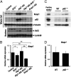
Increase of the Keap1 protein level in Atg7-Alb and p62−/− mice. Protein and mRNA expression levels of Keap1, Nrf2, and p62 in Nrf2−/−, Atg7-Alb, Atg7-Alb::Nrf2−/−, and Keap1-Alb mice (A and B) and p62−/− mice (C and D). Immunoblot analyses were carried out using whole-cell extracts (Whole Cell) and nuclear extracts (Nucleus) of liver (A and C). αTubulin and Lamin B were used as loading controls. Atg7-Alb was used as a positive control in C. Data are the means ± SD of three mice. *P < 0.05, **P < 0.01 compared with each control (B).
Fig. 4.
Fig. 4.
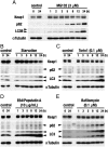
Effects of a proteasome inhibitor, starvation and autophagy inhibitors on the protein levels of Keap1 and p62. Immunoblot analyses were carried out using whole-cell extracts of HepG2 cells. Keap1, p62, and LC3 proteins were detected with specific antibodies. αTubulin was detected as a loading control. A proteasome inhibitor, MG132 (3 μM) (A), nutritional starvation in HBSS (B), and three kinds of autophagy inhibitors, Torin1 (0.1 μM) (C), E64/PepstatinA (10 μg/mL) (D), and Bafilomycin (0.1 μM) (E) were applied. These experiments were repeated multiple times and found to be reproducible. Representative results are shown.
Fig. 5.
Fig. 5.
Half-life of Keap1 protein in the presence or absence of electrophilic stimuli. Immunoblot analyses were carried out using whole-cell extracts of HepG2 cells treated with various electrophiles in the presence of CHX (A). Band intensities of Keap1 were quantified, normalized to those of αTubulin, and plotted (B). Representative results are shown. The half-life of Keap1 under each condition was calculated based on the data in A. Data are the means ± SD of three independent experiments. The half-life of Keap1 was determined in the presence of DEM (100 μM), 1,2-NQ (50 μM), and tBHQ (100 μM) with or without CHX (0.1 mg/mL).
Fig. 6.
Fig. 6.
Impaired autophagy accumulates Keap1 as well as p62 and leads to Nrf2-dependent liver injury. Autophagy is one of proteolytic systems. When the isolation membrane emerges, LC3 is attached to the membrane and binds p62, which in turn links to ubiquitinated substrate proteins. Autophagosome fuses with lysosome to form autolysosome, resulting in the protein degradation. Autophagy impaired by Atg7 deletion accumulates Keap1 as well as p62. Constitutive accumulation of Nrf2 is the dominant cause of the liver injury in impaired autophagy independent of p62.

Similar articles

Cited by

References

    1. Itoh K, et al. Keap1 represses nuclear activation of antioxidant responsive elements by Nrf2 through binding to the amino-terminal Neh2 domain. Genes Dev. 1999;13:76–86. - PMC - PubMed
    1. Itoh K, et al. Keap1 regulates both cytoplasmic-nuclear shuttling and degradation of Nrf2 in response to electrophiles. Genes Cells. 2003;8:379–391. - PubMed
    1. Kobayashi A, Ohta T, Yamamoto M. Unique function of the Nrf2-Keap1 pathway in the inducible expression of antioxidant and detoxifying enzymes. Methods Enzymol. 2004;378:273–286. - PubMed
    1. Kobayashi M, Yamamoto M. Molecular mechanisms activating the Nrf2-Keap1 pathway of antioxidant gene regulation. Antioxid Redox Signal. 2005;7:385–394. - PubMed
    1. Motohashi H, Yamamoto M. Nrf2-Keap1 defines a physiologically important stress response mechanism. Trends Mol Med. 2004;10:549–557. - PubMed

Publication types

MeSH terms

Substances