Podosomes in migrating microglia: components and matrix degradation
- PMID: 22873355
- PMCID: PMC3423073
- DOI: 10.1186/1742-2094-9-190
Podosomes in migrating microglia: components and matrix degradation
Abstract
Background: To perform their functions during development and after central nervous system injury, the brain's immune cells (microglia) must migrate through dense neuropil and extracellular matrix (ECM), but it is not known how they degrade the ECM. In several cancer cell lines and peripheral cells, small multi-molecular complexes (invadopodia in cancer cells, podosomes in nontumor cells) can both adhere to and dissolve the ECM. Podosomes are tiny multi-molecular structures (0.4 to 1 μm) with a core, rich in F-actin and its regulatory molecules, surrounded by a ring containing adhesion and structural proteins.
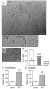
Methods: Using rat microglia, we performed several functional assays: live cell imaging for chemokinesis, degradation of the ECM component, fibronectin, and chemotactic invasion through Matrigel™, a basement membrane type of ECM. Fluorescent markers were used with high-resolution microscopy to identify podosomes and their components.
Results: The fan-shaped lamella at the leading edge of migrating microglia contained a large F-actin-rich superstructure composed of many tiny (<1 μm) punctae that were adjacent to the substrate, as expected for cell-matrix contact points. This superstructure (which we call a podonut) was restricted to cells with lamellae, and conversely almost every lamella contained a podonut. Each podonut comprised hundreds of podosomes, which could also be seen individually adjacent to the podonut. Microglial podosomes contained hallmark components of these structures previously seen in several cell types: the plaque protein talin in the ring, and F-actin and actin-related protein (Arp) 2 in the core. In microglia, podosomes were also enriched in phosphotyrosine residues and three tyrosine-kinase-regulated proteins: tyrosine kinase substrate with five Src homology 3 domains (Tks5), phosphorylated caveolin-1, and Nox1 (nicotinamide adenine dinucleotide phosphate oxidase 1). When microglia expressed podonuts, they were able to degrade the ECM components, fibronectin, and Matrigel™.
Conclusion: The discovery of functional podosomes in microglia has broad implications, because migration of these innate immune cells is crucial in the developing brain, after damage, and in disease states involving inflammation and matrix remodeling. Based on the roles of invadosomes in peripheral tissues, we propose that microglia use these complex structures to adhere to and degrade the ECM for efficient migration.
Figures


References
-
- Alberts B, Wilson JH, Hunt T. Molecular Biology of the Cell. 5. Garland Science, New York; 2008.
Publication types
MeSH terms
LinkOut - more resources
Full Text Sources
Miscellaneous

