. 2012 Aug 8:13:26.
doi: 10.1186/1471-2199-13-26.

Structural/functional analysis of the human OXR1 protein: identification of exon 8 as the anti-oxidant encoding function

Affiliations

Structural/functional analysis of the human OXR1 protein: identification of exon 8 as the anti-oxidant encoding function

Kenan C Murphy et al. BMC Mol Biol. .

Abstract

Background: The human OXR1 gene belongs to a class of genes with conserved functions that protect cells from reactive oxygen species (ROS). The gene was found using a screen of a human cDNA library by its ability to suppress the spontaneous mutator phenotype of an E. coli mutH nth strain. The function of OXR1 is unknown. The human and yeast genes are induced by oxidative stress and targeted to the mitochondria; the yeast gene is required for resistance to hydrogen peroxide. Multiple spliced isoforms are expressed in a variety of human tissues, including brain.

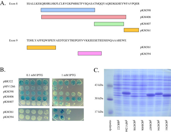
Results: In this report, we use a papillation assay that measures spontaneous mutagenesis of an E. coli mutM mutY strain, a host defective for oxidative DNA repair. Papillation frequencies with this strain are dependent upon a G→T transversion in the lacZ gene (a mutation known to occur as a result of oxidative damage) and are suppressed by in vivo expression of human OXR1. N-terminal, C-terminal and internal deletions of the OXR1 gene were constructed and tested for suppression of the mutagenic phenotype of the mutM mutY strain. We find that the TLDc domain, encoded by the final four exons of the OXR1 gene, is not required for papillation suppression in E. coli. Instead, we show that the protein segment encoded by exon 8 of OXR1 is responsible for the suppression of oxidative damage in E. coli.

Conclusion: The protein segment encoded by OXR1 exon 8 plays an important role in the anti-oxidative function of the human OXR1 protein. This result suggests that the TLDc domain, found in OXR1 exons 12-16 and common in many proteins with nuclear function, has an alternate (undefined) role other than oxidative repair.


Figures

Figure 1
Figure 1
The human OXR1 gene. Top: The genomic structure of OXR1 consists of 16 known exons located on chromosome 8q23. Exons, or portions of exons shown in black are present in an oxidation resistance active form of OXR1 previously described [24], whereas exons in white are known to be dispensable for this activity. Exon 10 is found in a variant that begins with exon 10 and includes exons 12 through 15 [43]. Bottom: The protein sequences encoded by OXR1 exons 7 through 16 are listed. The starting points for the series of OXR1 N-terminal truncations were constructed by placing an ATG at the codon prior to position 203 (shaded lysine) of exon 7, using the methionine codon at position 225 (shaded) in exon 7, or by placing an ATG start codon in front of the codons encoding the first residues listed for exons 8 through 13 (except exon 10). The end points for the series of OXR1 C-terminal truncations are denoted by the arrows. Exon 10 is listed here, but was not found in pMV520; it is shown here for completeness.
Figure 2
Figure 2
(A) Diagram of N-terminal deletions of OXR1. In these assays, the full-length OXR1 construct is represented by both pMV1263, which starts with methionine 225 in exon 7 (the expected start site of OXR1 in pMV520) and pMV1260, which encodes both this species and a slightly larger OXR1 species (by virtue of the presence of an initiating methionine start site inserted prior to codon 200 in exon 7). (B) Papillation assay for full-length OXR1 species (pMV1260 and pMV1263) and three N-terminal deletions starting within exon 8 (pMV1266), exon 9 (pMV1269) and exon 12 (pMV1275). Plasmids were expressed in strain MV4709 (mutM mutY lacZ cc104) and plated on minimal lactose plates containing 100 μg/ml carbenicillin with and without 1 mM IPTG. Aliquots of 10-fold serial dilutions are plated beginning with undiluted cells on the left. No OXR1 controls containing pBR322 showed patterns that were identical to pMV1269 and p1275 (not shown). (C) Papillation assay for full-length OXR1 species (pMV1260 and pMV1263) and three N-terminal deletions starting within exon 8 (pMV1266), exon 9 (pMV1269) and exon 12 (pMV1275). Plasmids were expressed in strain MV4709 (mutM mutY lacZ cc104) containing pMS421 (LacI-producer) and plated on minimal lactose plates containing 100 μg/ml carbenicillin, 40 μg/ml streptomycin, 20 μg/ml spectinomycin and 1 mM IPTG. (D) SDS-PAGE of extracts containing plasmids expressing OXR1 N-terminal deletions. Plasmid pMV1263 makes the full-length OXR1 species encoded by pMV520, while pMV1260 makes both this species and a slightly larger one that starts at position Lys-200 in exon 7. In addition, the arrows denote positions of observed protein bands for pMV1269 and pMV1272. (E) SDS-PAGE comparing full length OXR1 (pMV520) and OXR1 fragment starting at exon 9 (pMV1269). Arrows denote positions of induced protein bands; full length OXR1 is present as a doublet (see text for details).
Figure 3
Figure 3
(A) Diagram of C-terminal deletions of OXR1. (B) Papillation assays with plasmids expressing OXR1 C-terminal deletions in MV4709 containing LacI-producing plasmid pMS421. Dilutions and cell platings were as in Figure 2B using plates containing 0.125 mM and 1.0 mM IPTG on minimal lactose plates containing 100 μg/ml carbenicillin, 40 μg/ml streptomycin and 20 μg/ml spectinomycin. (C) SDS-PAGE of extracts containing plasmids expressing OXR1 C-terminal deletions.
Figure 4
Figure 4
(A) Diagram of internal-terminal deletions of OXR1 (8–13). (B) Papillation assays with plasmids expressing OXR1 internal deletions in MV4709 containing LacI-producing plasmid pMS421. Dilutions and cell platings were as in Figure 2B using plates containing 1.0 mM IPTG on minimal lactose plates containing 100 μg/ml carbenicillin, 40 μg/ml streptomycin and 20 μg/ml spectinomycin. (C) SDS-PAGE of extracts containing plasmids expressing OXR1 internal deletions.
Figure 5
Figure 5
(A) Secondary structural prediction of the polypeptide encoded by OXR1 exon 8 based on the method of Garnieret al[[38]]. Boxed region of protein sequence defines the putative larger helix-turn-helix region of the protein. (B) ClustelW alignment of the protein sequences encoded by OXR1 exon 8 and NCOA7 exon 9.
Figure 6
Figure 6
(A) Diagram of internal deletions of exons 8 and 9 within constructs expressing full length OXR1 proteins (exons 8–16). Bars represent regions of exon 8 that are missing in the OXR1 deletion mutants. (B) Papillation assay for control plasmid (pBR322), an OXR1 encoded by exons 8–16 (pMV1266), and the five internal deletions of OXR1 diagrammed in part A. Plasmids were expressed in strain MV4709 (mutM mutY lacZ cc104) containing LacI-producing plasmid pMS421. Dilutions and cell platings were as in Figure 2B using 0.125 mM and 1.0 mM IPTG on minimal lactose plates containing 100 μg/ml carbenicillin, 40 μg/ml streptomycin and 20 μg/ml spectinomycin. (C) SDS-PAGE comparing full length OXR1 (pMV1266) and OXR1 deletion mutants described in part A. The expected sizes of the protein fragments from these plasmid are as follows: pMV1266 (38.2 kDa); pKM398 (35.2 kDa); pKM406 (33.1 kDa); pKM407 (36.5 kDa) pKM394 (36.8 kDa); pKM361 (35.5 kDa).
Figure 7
Figure 7
(A) Lac + reversion frequencies of MV6543 (MV4709 with pMS421) containing various OXR1 fragment-producing plasmids. Cells containing plasmids were grown overnight as described in the Methods section. All cultures contained 1 mM IPTG, except ones that contained pMV1266 and pKM407, which contained 0.1 mM IPTG (to prevent lethality that results from higher levels of expression of these OXR1 proteins). All cultures grew to saturation (2–4 x 10⁹ cells/ml). The results are reported as the mean of 3–6 determinations for each strain; error bars report the standard error. A t-test analysis of the values reported for pKM398 and pKM406 determined that there is no significant difference between the two frequencies reported for these plasmids. (B) Diagram of the OXR1 fragment-producing plasmids used in the Lac + reversion assay.

Similar articles

Cited by

References

    1. Cadet J, Douki T, Ravanat JL. Oxidatively generated base damage to cellular DNA. Free Radic Biol Med. 2010;49(1):9–21. doi: 10.1016/j.freeradbiomed.2010.03.025. - DOI - PubMed
    1. Negre-Salvayre A, Coatrieux C, Ingueneau C, Salvayre R. Advanced lipid peroxidation end products in oxidative damage to proteins Potential role in diseases and therapeutic prospects for the inhibitors. Br J Pharmacol. 2008;153(1):6–20. doi: 10.1038/sj.bjp.0707395. - DOI - PMC - PubMed
    1. Sedelnikova OA, Redon CE, Dickey JS, Nakamura AJ, Georgakilas AG, Bonner WM. Role of oxidatively induced DNA lesions in human pathogenesis. Mutat Res. 2010;704(1–3):152–159. - PMC - PubMed
    1. Liochev SI, Fridovich I. The Haber-Weiss cycle -- 70 years later: an alternative view. Redox Rep. 2002;7(1):55–57. doi: 10.1179/135100002125000190. author reply 59–60. - DOI - PubMed
    1. Hancock JT, Desikan R, Neill SJ. Role of reactive oxygen species in cell signalling pathways. Biochem Soc Trans. 2001;29(Pt 2):345–350. - PubMed

Publication types