

Recent advances in optical cancer imaging of EGF receptors
- PMID: 22873662
- PMCID: PMC6330113
- DOI: 10.2174/092986712803341584
Recent advances in optical cancer imaging of EGF receptors
Abstract
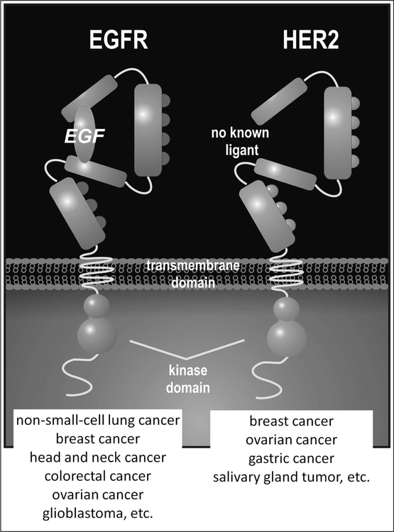
Epidermal growth factor (EGF) receptors are commonly expressed on the cell membrane of cancer cells and activity of these receptors results in accelerated cell growth and carcinogenesis. A variety of targeted molecules have been developed to block ligand binding and/or inhibit the function of these receptor tyrosine kinases, and several have proven therapeutic benefits. Along with the advent of new therapeutic agents comes a need for non-invasive tools to diagnose, characterize, and monitor tumor responsiveness to therapy. Imaging EGF receptors with radionuclides has been performed for decades. However, recently this area has advanced considerably with the development of EGF receptor-targeted optical imaging probes. Herein, we review recent advances in molecular imaging of the EGF receptor family, focusing specifically on optical imaging. Such agents provide the opportunity for earlier diagnosis, improved tumor characterization, and the ability to measure and monitor tumor responsiveness to anti-EGF receptor treatment strategies.
Conflict of interest statement
CONFLICT OF INTEREST
The authors do not have any conflict of interests for writing this article.
Figures
Similar articles
-
Quantitative assays for the measurement of HER1-HER2 heterodimerization and phosphorylation in cell lines and breast tumors: applications for diagnostics and targeted drug mechanism of action.Breast Cancer Res. 2011 Apr 15;13(2):R44. doi: 10.1186/bcr2866. Breast Cancer Res. 2011. PMID: 21496232 Free PMC article.
-
Bioorthogonal reaction pairs enable simultaneous, selective, multi-target imaging.Angew Chem Int Ed Engl. 2012 Jan 23;51(4):920-2. doi: 10.1002/anie.201104389. Epub 2011 Dec 12. Angew Chem Int Ed Engl. 2012. PMID: 22162316 Free PMC article.
-
Epidermal growth factor-related peptides activate distinct subsets of ErbB receptors and differ in their biological activities.J Biol Chem. 1996 Mar 15;271(11):6071-6. doi: 10.1074/jbc.271.11.6071. J Biol Chem. 1996. PMID: 8626392
-
Recent advances in receptor-targeted fluorescent probes for in vivo cancer imaging.Curr Med Chem. 2012;19(28):4742-58. doi: 10.2174/092986712803341467. Curr Med Chem. 2012. PMID: 22873663 Review.
-
Clinical implications of the EGF receptor/ligand system for tumor progression and survival in gastrointestinal carcinomas: evidence for new therapeutic options.Recent Results Cancer Res. 2003;162:115-32. doi: 10.1007/978-3-642-59349-9_10. Recent Results Cancer Res. 2003. PMID: 12790326 Review.
Cited by
-
The status of contemporary image-guided modalities in oncologic surgery.Ann Surg. 2015 Jan;261(1):46-55. doi: 10.1097/SLA.0000000000000622. Ann Surg. 2015. PMID: 25599326 Free PMC article. Review.
-
Molecular Targeting of Epidermal Growth Factor Receptor (EGFR) and Vascular Endothelial Growth Factor Receptor (VEGFR).Molecules. 2021 Feb 18;26(4):1076. doi: 10.3390/molecules26041076. Molecules. 2021. PMID: 33670650 Free PMC article. Review.
-
Molecular imaging of EGFR/HER2 cancer biomarkers by protein MRI contrast agents.J Biol Inorg Chem. 2014 Feb;19(2):259-70. doi: 10.1007/s00775-013-1076-3. Epub 2013 Dec 24. J Biol Inorg Chem. 2014. PMID: 24366655 Free PMC article. Review.
-
New whole-body multimodality imaging of gastric cancer peritoneal metastasis combining fluorescence imaging with ICG-labeled antibody and MRI in mice.Gastric Cancer. 2014;17(3):497-507. doi: 10.1007/s10120-013-0316-0. Epub 2013 Nov 28. Gastric Cancer. 2014. PMID: 24288123
-
Molecular and functional imaging in cancer-targeted therapy: current applications and future directions.Signal Transduct Target Ther. 2023 Feb 27;8(1):89. doi: 10.1038/s41392-023-01366-y. Signal Transduct Target Ther. 2023. PMID: 36849435 Free PMC article. Review.
References
-
- Ciardiello F; Tortora G, EGFR antagonists in cancer treatment. N Engl J Med, 2008, 358, 1160–1174. - PubMed
-
- Jones HE; Gee JM; Taylor KM; Barrow D; Williams HD; Rubini M; Nicholson RI, Development of strategies for the use of anti-growth factor treatments. Endocr Relat Cancer, 2005, 12 Suppl 1, S173–182. - PubMed
-
- Lurje G; Lenz HJ, EGFR signaling and drug discovery. Oncology, 2009, 77, 400–410. - PubMed
-
- Cai W; Niu G; Chen X, Multimodality imaging of the HER-kinase axis in cancer. Eur J Nucl Med Mol Imaging, 2008, 35, 186–208. - PubMed
Publication types
MeSH terms
Substances
Grants and funding
LinkOut - more resources
Full Text Sources
