. 2012 Dec 10;29(18):2805-15.
doi: 10.1089/neu.2011.2298. Epub 2012 Nov 19.

Influence of breaching the connective sheaths of the donor nerve on its myelinated sensory axons and on their sprouting into the end-to-side coapted nerve in the rat

Affiliations

Influence of breaching the connective sheaths of the donor nerve on its myelinated sensory axons and on their sprouting into the end-to-side coapted nerve in the rat

Uroš Kovačič et al. J Neurotrauma. .

Abstract

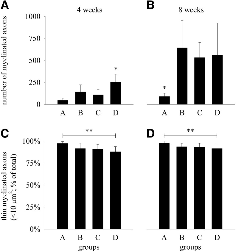
The influence of breaching the connective sheaths of the donor sural nerve on axonal sprouting into the end-to-side coapted peroneal nerve was examined in the rat. In parallel, the effect of these procedures on the donor nerve was assessed. The sheaths of the donor nerve at the coaptation site were either left completely intact (group A) or they were breached by epineurial sutures (group B), an epineurial window (group C), or a perineurial window (group D). In group A, the compound action potential (CAP) of sensory axons was detected in ~10% and 40% of the recipient nerves at 4 and 8 weeks, respectively, which was significantly less frequently than in group D at both recovery periods. In addition, the number of myelinated axons in the recipient nerve was significantly larger in group D than in other groups at 4 weeks. At 8 weeks, the number of axons in group A was only ~15% of the axon numbers in other groups (p<0.05). Focal subepineurial degenerative changes in the donor nerves were only seen after 4 weeks, but not later. The average CAP area and the total number of myelinated axons in the donor nerves were not different among the experimental groups. In conclusion, myelinated sensory axons are able to penetrate the epiperineurium of donor nerves after end-to-side nerve coaption; however, their ingrowth into recipient nerves is significantly enhanced by breaching the epiperineurial sheets at the coaptation site. Breaching does not cause permanent injury to the donor nerve.


Figures

FIG. 1.
FIG. 1.
Schematic drawing of the surgical procedures. The sciatic nerve and its branches were exposed in the right thigh of the rat. The peroneal and tibial nerves as well as the saphenous nerve, which is not shown in the figure, were transected, and their proximal stumps were ligated and implanted in nearby muscles. In addition, the segment of the recipient peroneal nerve was coapted to the side of the ipsilateral donor sural nerve (end-to-side coaptation). In four experimental groups of rats, the connective tissue sheaths of the donor sural nerve at the coaptation site (enlarged frame in lower row) were either left completely intact (group A) or breached by epineurial sutures (group B), epineurial window (group C), or perineurial window (group D).
FIG. 2.
FIG. 2.
Representative light photomicrographs of the recipient peroneal nerve from groups A, B, and D 4 weeks (left column – 4w) and 8 weeks (right column – 8w) after its proximal end was coapted to the side of donor sural nerve. Cross-sections were taken ∼4 mm distal from the site of coaptation and were stained with azure blue. Thin myelinated axons are shown by arrows.
FIG. 3.
FIG. 3.
Total number of myelinated axons (diagrams A and B) and percentages of myelinated axons with their cross-sectional area <10 μm2 (diagrams C and D) in the recipient peroneal nerve segment 4 weeks (diagrams A and C) and 8 weeks (diagrams B and D) after the end-to-side nerve coaptation in rats in which the connective tissue sheaths of the donor sural nerve at the coaptation site were either left completely intact (group A) or breached by epineurial sutures (group B), epineurial window (group C), or perineurial window (group D). Data are shown as mean values and SD (4 weeks: n=8 in each group; 8 weeks: n=12 in each group). For comparison, the total number of myelinated axons in normal contralateral peroneal nerve was 1935±108 (n=4). *Statistically significantly different from the other experimental groups at the corresponding time point (p<0.05; 1W ANOVA and Bonferroni's test). **Statistically significantly different between groups A and D at the corresponding time point (p<0.05; 1W ANOVA and Bonferroni's test).
FIG. 4.
FIG. 4.
Light photomicrographs of the representative donor sural nerves 4 weeks (left column – 4w) and 8 weeks (right column – 8w) after the end-to-side nerve coaptation was performed completely without injury to the epineurium (group A), with sutures to the epineurium (group B), with an epineurial window (group C), and with a perineurial window (group D). The cross-sections of the donor nerves were obtained 4 mm distal from the site of coaptation and stained with azure blue. Note the presence of degenerating fiber profiles focally distributed in the subperineurial region of the donor nerves (shown by arrows) from the groups 4 weeks (left column) after the surgery in contrast to the nerves obtained from animals that recovered for 8 weeks (right column).
FIG. 5.
FIG. 5.
Comparisons of the frequency histograms of cross-sectional areas of myelinated fibers in donor and contralateral sural nerves. Samples of the donor sural nerves were obtained 4 mm distal from the site of coaptation from the animals that were left to survive for 4 weeks (left column) and 8 weeks (right column) after the surgery. Samples of the contralateral intact sural nerve were obtained at the corresponding sites and served as controls. Data are shown as mean values and SD (n=8 in each group). At 4 weeks, the bimodality coefficients for groups A, B, C, D, and the control group were 0.81, 0.69, 0.67, 0.72, and 0.64, respectively. At 8 weeks, the bimodality coefficients for groups A, B, C, D, and the control group were 0.72, 0.73, 0.61, 0.69 and 0.71, respectively. Notably, bimodality coefficient >0.555 indicates that a distribution is bimodal (but see Jackson, P.R., Tucker, G.T., and Woods, H.F. (1989). Testing for bimodality in frequency distributions of data suggesting polymorphisms of drug metabolism – hypothesis testing. Br. J. Clin. Pharmacol. 28, 655–662.) Note the tendency for a greater proportion of thin fibers in donor sural nerves from group A than in other groups at 4 weeks after surgery, and a smaller proportion of thin fibers in donor nerves from group A than in other groups at 8 weeks after surgery (see also Figure 6).
FIG. 6.
FIG. 6.
Total number of myelinated axons (diagrams A and B) and percentages of thin myelinated axons with their cross-sectional area <20 μm2 (diagrams C and D) in the donor and contralateral (control) sural nerves from the animals that were left to survive for 4 weeks (diagrams A and C) and 8 weeks (diagrams B and D) after the surgery. Samples of the donor sural nerves were obtained 4 mm distal from the site of coaptation and samples of the control nerves were obtained at the corresponding sites on the contralateral side. The end-to-side nerve coaptation was performed completely without injury to the epineurium (group A), with sutures to the epineurium (group B), with an epineurial window (group C), and with a perineurial window (group D). *Statistically significantly different from the contralateral sural nerves at 4 weeks and from the group B at 8 weeks (p<0.05, 1W ANOVA followed by Bonferroni's test). The differences in all other pairwise comparisons between the groups were not statistically significant (p>0.05, n=8 in each group).

Similar articles

Cited by

References

    1. Beris A. Lykissas M. Korompilias A. Mitsionis G. End-to-side nerve repair in peripheral nerve injury. J. Neurotrauma. 2007;24:909–916. - PubMed
    1. Dvali L.T. Myckatyn T.M. End-to-side nerve repair: review of the literature and clinical indications. Hand Clin. 2008;24:455–460. - PubMed
    1. Geuna S. Papalia I. Tos P. End-to-side (terminolateral) nerve regeneration: a challenge for neuroscientists coming from an intriguing nerve repair concept. Brain. Res. Rev. 2006;52:381–388. - PubMed
    1. Pannucci C. Myckatyn T.M. Mackinnon S.E. Hayashi A. End-to-side nerve repair: review of the literature. Restor. Neurol. Neurosci. 2007;25:45–63. - PubMed
    1. Tos P. Geuna S. Papalia I. Conforti L.G. Artiaco S. Battiston B. Experimental and clinical employment of end-to-side coaptation: our experience. Acta Neurochir. 2011;(Suppl. 108):241–245. - PubMed

Publication types

LinkOut - more resources