. 2012 Aug 8:12:345.
doi: 10.1186/1471-2407-12-345.

Immediate in vivo target-specific cancer cell death after near infrared photoimmunotherapy

Affiliations

Immediate in vivo target-specific cancer cell death after near infrared photoimmunotherapy

Makoto Mitsunaga et al. BMC Cancer. .

Abstract

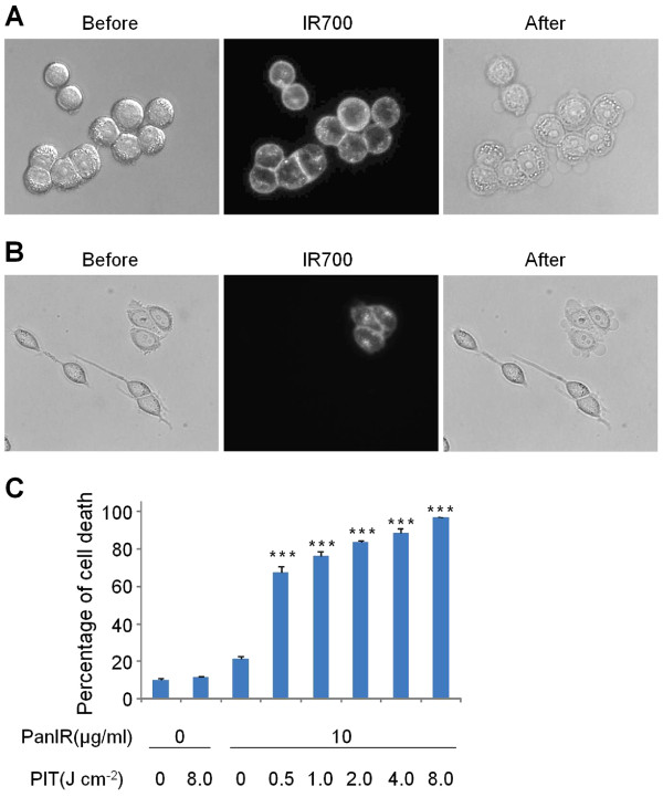
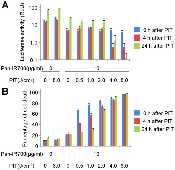
Background: Near infrared (NIR) photoimmunotherapy (PIT) is a new type of cancer treatment based on a monoclonal antibody (mAb)-NIR phthalocyanine dye, (IR700) conjugate. In vitro cancer-specific cell death occurs during NIR light exposure in cells previously incubated with mAb-IR700 conjugates. However, documenting rapid cell death in vivo is more difficult.

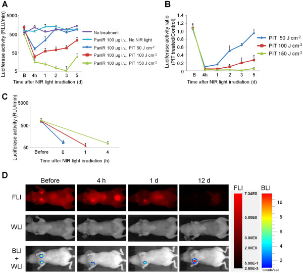
Methods: A luciferase-transfected breast cancer cell (epidermal growth factor receptor+, MDA-MB-468luc cells) was produced and used for both in vitro and in vivo experiments for monitoring the cell killing effect of PIT. After validation of cytotoxicity with NIR exposure up to 8 J/cm2in vitro, we employed an orthotopic breast cancer model of bilateral MDA-MB-468luc tumors in female athymic mice, which subsequently received a panitumumab-IR700 conjugate in vivo. One side was used as a control, while the other was treated with NIR light of dose ranging from 50 to 150 J/cm2. Bioluminescence imaging (BLI) was performed before and after PIT.

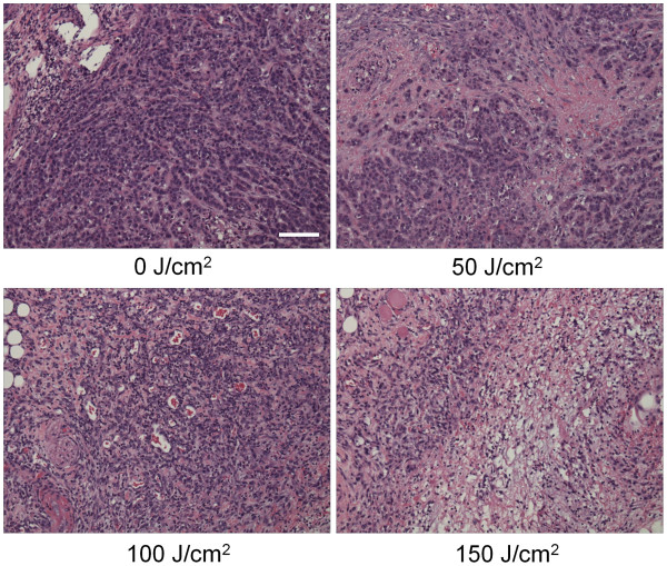
Results: Dose-dependent cell killing and regrowth was successfully monitored by the BLI signal in vitro. Although tumor sizes were unchanged, BLI signals decreased by >95% immediately after PIT in vivo when light intensity was high (>100 J/cm2), however, in mice receiving lower intensity NIR (50 J/cm2), tumors recurred with gradually increasing BLI signal.

Conclusion: PIT induced massive cell death of targeted tumor cells immediately after exposure of NIR light that was demonstrated with BLI in vivo.


Figures

Figure 1
Figure 1
EGFR-specific PIT in vitro. (A) MDA-MB-468luc cells were treated with Pan-IR700 and observed with microscopy (Before and after Pan-IR700). Necrotic cell death was observed upon excitation with NIR light (After). (B) Both EGFR positive (MDA-MB-468luc) and negative (Balb/3T3) cells were co-cultured and treated with Pan-IR700 (Before and after Pan-IR700). EGFR-specific necrotic cell death was observed (After). (C) NIR light irradiation results in dose dependent cell death as determined by the cytotoxicity assay. (n = 3, *** P<0.001 vs. non treatment control, Student’s t test).
Figure 2
Figure 2
Effect of phototoxicity in response to Pan-IR700 mediated PIT. Cell viability was analyzed with (A) bioluminescence and (B) fluorescence LIVE/DEAD cytotoxicity assay after NIR light irradiation in MDA-MB-468luc cells.
Figure 3
Figure 3
In vivo fluorescence and bioluminescence imaging of orthotopic breast tumors in response to Pan-IR700 mediated PIT. (A) Monitoring bioluminescence activity in response to Pan-IR700 mediated PIT (n = 5 each). B: just before NIR light treatment. Bioluminescence signals of tumors in 100 and 150 J/cm2 treated groups showed significant difference with that in control or 50 J/cm2 treated group at all time points examined after PIT (P<0.001). (B) Monitoring bioluminescence activity ratio of treated tumor to internal control (n = 5 each). B: just before NIR light treatment. Bioluminescence signals of tumors in 100 and 150 J/cm2 treated groups showed significant difference with that in control or 50 J/cm2 treated group at all time points examined after PIT (P<0.001). (C) Rapid changes of bioluminescence activity in response to Pan-IR700 mediated PIT (n = 5 each). Before: 1 d before NIR light treatment. (D) IR700 fluorescence and bioluminescence imaging in response to Pan-IR700 mediated PIT. Representative images are shown. Before: just before NIR light treatment, FLI: IR700 fluorescence imaging, WLI: white light imaging, BLI: bioluminescence imaging.
Figure 4
Figure 4
Histological findings immediately after PIT with various NIR exposures. Histological specimens of MDA-MB-468luc tumors, which were treated with PIT at 0, 50, 100, and 150 J/cm2, are shown. All specimens are stained with Hematoxylin and Eosin. A few scattered clusters of damaged tumor cells are seen within a background of diffuse cellular necrosis and micro-hemorrhage immediately after PIT with 50 J/cm2. Necrotic damage was more intense when higher intensity NIR light was administered. Scale indicates 50μm.

References

    1. Waldmann TA. Immunotherapy: past, present and future. Nat Med. 2003;9:269–277. doi: 10.1038/nm0303-269. - DOI - PubMed
    1. Pastan I, Hassan R, Fitzgerald DJ, Kreitman RJ. Immunotoxin therapy of cancer. Nat Rev Cancer. 2006;6:559–565. doi: 10.1038/nrc1891. - DOI - PubMed
    1. Mitsunaga M, Ogawa M, Kosaka N, Rosenblum LT, Choyke PL, Kobayashi H. Cancer cell-selective in vivo near infrared photoimmunotherapy targeting specific membrane molecules. Nat Med. 2011;17:1685–1691. doi: 10.1038/nm.2554. - DOI - PMC - PubMed
    1. Mitsunaga M, Nakajima T, Sano K, Choyke PL, Kobayashi H. Near-infrared theranostic photoimmunotherapy (PIT): repeated exposure of light enhances the effect of immunoconjugate. Bioconjug Chem. 2012;23:604–609. doi: 10.1021/bc200648m. - DOI - PMC - PubMed
    1. Contag PR, Olomu IN, Stevenson DK, Contag CH. Bioluminescent indicators in living mammals. Nat Med. 1998;4:245–247. doi: 10.1038/nm0298-245. - DOI - PubMed

Publication types

MeSH terms