. 2012 Nov;9(4):239-49.
doi: 10.2174/156720212803530618.

Wnt1 inducible signaling pathway protein 1 (WISP1) targets PRAS40 to govern β-amyloid apoptotic injury of microglia

Affiliations

Wnt1 inducible signaling pathway protein 1 (WISP1) targets PRAS40 to govern β-amyloid apoptotic injury of microglia

Yan Chen Shang et al. Curr Neurovasc Res. 2012 Nov.

Abstract

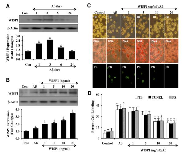
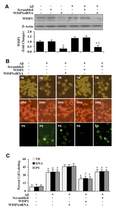
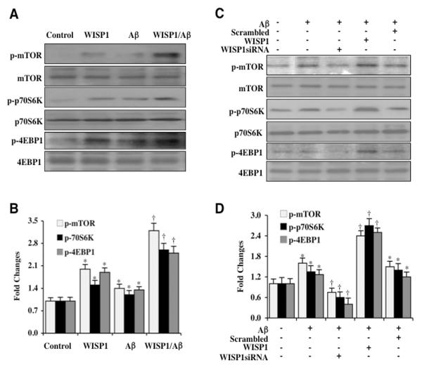
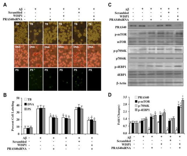
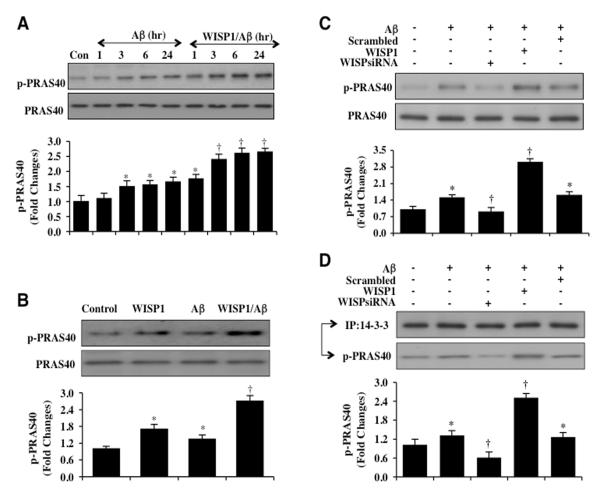
Given the present challenges to attain effective treatment for β-amyloid (Aβ) toxicity in neurodegenerative disorders such as Alzheimer's disease, development of novel cytoprotective pathways that can assist immune mediated therapies through the preservation of central nervous system microglia could offer significant promise. We show that the CCN4 protein, Wnt1 inducible signaling pathway protein 1 (WISP1), is initially up-regulated by Aβ and can modulate its endogenous expression for the protection of microglia during Aβ mediated apoptosis. WISP1 activates mTOR and phosphorylates p70S6K and 4EBP1 through the control of the regulatory mTOR component PRAS40. Loss of PRAS40 through gene reduction or inhibition by WISP1 is cytoprotective. WISP1 ultimately governs PRAS40 by sequestering PRAS40 intracellularly through post-translational phosphorylation and binding to protein 14-3-3. Our work identifies WISP1, mTOR signaling, and PRAS40 as targets for new strategies directed against Alzheimer's disease and related disorders.


Figures

Figure 1
Figure 1. WISP1 increases endogenous expression and blocks apoptotic injury during Aβ exposure
(A) Microglial cultures were exposed to Aβ at the concentration of 10 μM for the time period (hr=hour) as indicated and the expression of WISP1 was determined by western blot analysis (*P <0.01 vs. Control). Con = Control = untreated microglia. Each data point represents the mean and SEM from 3 experiments. (B) Exogenous WISP1 (1, 5, 10, and 20 ng/ml) was applied 1 hour prior to Aβ (10 μM) exposure, maintained for 6 hours and then removed through 3 media exchanges. Subsequent expression of WISP1 was determined by western blot analysis (*P<0.01 vs. untreated control; P <0.01 vs. Aβ). Each data point represents the mean and SEM from 3 experiments. Con = control = untreated microglia. (C and D) WISP1 (1, 5, 10, and 20 ng/ml) was applied to cultures of microglia 1 hour prior to the administration of Aβ (10 μM) and cell survival, DNA fragmentation, and membrane PS exposure were determined 24 hours later through trypan blue dye exclusion (TB, for cell survival), TUNEL (DNA fragmentation), and annexin-V labeling (PS exposure) respectively. Representative images (C) and quantitative analysis (D) demonstrate that Aβ leads to a significant increase in trypan blue dye, TUNEL, and annexin-V labeling in microglia 24 hours after Aβ exposure when compared to untreated control cultures. In contrast, WISP1 (10 or 20 ng/ml) given 1 hour prior to Aβ exposure significantly reduced trypan blue dye, TUNEL, and annexin-V labeling (*P < 0.01 vs. Control; P <0.01 vs. Aβ treated alone). Each data point represents the mean and SEM from 6 experiments.
Figure 2
Figure 2. Endogenous WISP1 is a necessary for microglial cell survival against Aβ
(A) Gene knockdown of WISP1 was performed with transfection of WISP1 siRNA prior to Aβ (10 μM) exposure in microglial cells. The expression of WISP1 was determined 3 hours following Aβ exposure by western blot analysis (*P <0.01 vs. Control; P <0.01 vs. Aβ). Control = untreated microglia. Each data point represents the mean and SEM from 3 experiments. Transfection with scrambled siRNA did not change WISP1 expression following Aβ exposure when compared to Aβ exposure alone (*P < 0.01 vs. Control; P <0.01 vs. Aβ treated alone). (B) Gene knockdown of WISP1 was performed with transfection of WISP1 siRNA prior to Aβ (10 μM) exposure in microglia cells and cell survival, DNA fragmentation, and membrane PS exposure were determined 24 hours later through trypan blue dye exclusion (TB), TUNEL (DNA), and annexin-V labeling (PS) respectively. Representative images demonstrate that Aβ results in a significant increase in trypan blue dye staining, DNA fragmentation, and membrane PS exposure in microglia 24 hours after Aβ exposure. WISP1 siRNA transfection further increased trypan blue dye staining, DNA fragmentation, and membrane PS exposure labeling following Aβ exposure, but not with statistical significance.. (C) Quantitative analysis demonstrates that transfection with WISP1 siRNA increased percent cell labeling of trypan blue dye staining, DNA fragmentation, and membrane PS exposure following Aβ exposure. Transfection with scrambled siRNA did not change the percent trypan blue dye staining, DNA fragmentation, and membrane PS exposure labeling following Aβ exposure when compared to Aβ exposure alone (*P < 0.01 vs. Control; P <0.01 vs. Aβ treated alone). Each data point represents the mean and SEM from 3 experiments.
Figure 3
Figure 3. WISP1 activates mTOR and phosphorylates p70S6K and 4EBP1
(A) WISP1 (10 ng/ml) was applied directly to microglial cultures and the expression of phosphorylated (p)-mTOR (Ser2448), p-p70S6K (Thr389), and p-4EBP1 (Ser65/Thr70) was determined at 3 hours later by western blot analysis. WISP1 significantly increased the expression of p-mTOR, p-p70S6K, and p-4EBP1 in microglia. Following WISP1 (10 ng/ml) application to microglia 1 hour prior to Aβ (10 μM) administration, the expression of p-mTOR, p-p70S6K, and p-4EBP1 was significantly increased when compared with Aβ cultures treated alone. (B) Quantitative results illustrate that WISP1 during Aβ exposure significantly increased the expression of p-mTOR, p-p70S6K, and p-4EBP1 in microglia (*P < 0.01 vs. Control; †P<0.01 vs. Aβ treated alone). (C) WISP1 siRNA was transfected into microglia and cell protein extracts (50 μg/lane) were immunoblotted with phosphorylated (p)-mTOR (Ser2448), p-p70S6K (Thr389), and p-4EBP1 (Ser 65/Thr70) antibodies at 3 hours following administration of Aβ (10 μM). Aβ resulted in a mild increase in the expression of p-mTOR, p-p70S6K, and p-4EBP1 that was further increased by WISP1 (10 ng/ml) administration 1 hour prior to Aβ exposure. WISP1 siRNA transfection significantly limited the expression of p-mTOR, p-p70S6K, and p-4EBP1 3 hours following Aβ exposure. (D) Quantitative results demonstrate that gene knockdown of WISP1 significantly limited the expression of p-mTOR, p-p70S6K, and p-4EBP1 3 hours following Aβ exposure (*P <0.01 vs. Control; †P<0.01 vs. Aβ treated alone). Scrambled siRNA transfection did not alter the expression of p-mTOR, p-p70S6K, and p-4EBP1 following Aβ exposure. Control=untreated microglia. In all cases, each data point represents the mean and SEM from 3 experiments.
Figure 4
Figure 4. PRAS40 controls apoptotic injury and the WISP1 phosphorylation of mTOR, p70S6K, and 4EBP1
(A) PRAS40 siRNA was transfected into microglial cultures prior to administration of Aβ (10 μM) and cell injury was determined 24 hours following Aβ exposure through trypan blue dye exclusion (TB, for cell survival), TUNEL (DNA fragmentation), and annexin-V labeling (PS exposure) respectively. Representative images of trypan blue staining, TUNEL, and PS exposure following Aβ exposure in microglia show that PRAS40 siRNA transfection reduced trypan blue staining, DNA fragmentation, and PS exposure and tended to increase cytoprotection of WISP1 (10 ng/ml) without statistical significance. (B) The quantitative results of trypan blue dye exclusion, DNA fragmentation, and membrane PS exposure demonstrated that percent trypan blue staining, DNA fragmentation, and PS exposure was significantly decreased by PRAS40 siRNA transfection or WISP1 (10 ng/ml) administration prior to Aβ exposure (*P<0.01 vs. Aβ treated alone). Scrambled siRNA transfection did not alter trypan blue dye exclusion, DNA fragmentation, and membrane PS exposure following Aβ exposure when compared to Aβ exposure alone. Each data point represents the mean and SEM from 3 experiments. (C) Gene knockdown of PRAS40 was performed with transfection of PRAS40 siRNA prior to Aβ (10 μM) exposure in microglia and the expression of PRAS40, phosphorylated (p) p-mTOR (Ser2448), p-p70S6K (Thr389), and p-4EBP1 (Ser65/Thr70) was determined 3 hours following Aβ exposure. WISP1 (10 ng/ml) applied 1 hour prior to Aβ exposure significantly increased the expression of p-mTOR, p-p70S6K, and p-4EBP1. Transfection with PRAS40 siRNA significantly limited the expression of PRAS40 and significantly increased the expression of p-mTOR, p-p70S6K, and p-4EBP1 in microglia following a 3 hour period of Aβ exposure or in cells treated with WISP1 (10 ng/ml). Scrambled siRNA transfection did not alter the expression of PRAS40, p-mTOR, p-p70S6K, and p-4EBP1 following Aβ exposure. (D) Quantitative results of western blot band density in (C) illustrate that PRAS40 gene reduction leads to significantly increased expression of p-mTOR, p-p70S6K, and p-4EBP1 in microglia during Aβ exposure alone or during Aβ exposure with WISP1 (10 ng/ml) application (*P < 0.01 vs. Aβ treated alone; P<0.01 vs. WISP1/Aβ).
Figure 5
Figure 5. WISP1 leads to the phosphorylation and binding of PRAS40 to protein 14-3-3 during Aβ exposure
(A) Western blot was performed for phosphorylated-PRAS40 (p-PRAS40, Thr246) in microglia at 1, 3, 6, or 24 hours (hr) following Aβ (10 μM) exposure. Aβ resulted in a mild increase in the expression of p-PRAS40 after 3 hours following of Aβ (*P<0.01 vs. Control). Application of WISP1 (10 ng/ml) 1 hour prior to Aβ administration significantly increased the expression of p-PRAS40 at 3, 6, and 24 hours after Aβ exposure (P<0.01 vs. Aβ at the corresponding time points). (B) Application of WISP1 (10 ng/ml) microglial cultures significantly increased the expression of p-PRAS40 3 hours later. WISP1 (10 ng/ml) treatment prior to Aβ (10 μM) administration also significantly increased the expression of p-PRAS40 when compared to Aβ treated alone 3 hours following Aβ exposure (*P < 0.01 vs. untreated Control; P<0.01 vs. Aβ treated alone). (C) WISP1 siRNA was transfected into microglial cultures prior to the administration of Aβ (10 μM) and expression of p-PRAS40 was determined 3 hours following Aβ exposure by western blot analysis. Transfection of WISP1 siRNA reduced the expression of p-PRAS40 3 hours following Aβ exposure. Non-specific scrambled siRNA did not alter the expression of p-PRAS40 during Aβ exposure (*P < 0.01 vs. untreated Control; P<0.01 vs. Aβ treated alone). In all cases, each data point represents the mean and SEM from three experiments. (D) WISP1 siRNA was transfected into microglial cultures prior to the administration of Aβ (10 μM) and cell extracts were immunoprecipitated by antibodies against protein 14-3-3 three hours later. Western blot analysis was performed to detect the expression p-PRAS40 and 14-3-3 in the precipitate. Application of WISP1 (10 ng/ml) increased the expression of p-PRAS40 in the precipitate. In contrast, WISP1 siRNA significantly reduced expression of p-PRAS40 in the precipitate following Aβ exposure (*P<0.01 vs. untreated control; P< 0.01 vs. Aβ treated alone).

References

    1. Yeger H, Perbal B. The CCN family of genes: a perspective on CCN biology and therapeutic potential. J Cell Commun Signal. 2007 Dec;1(3-4):159–64. - PMC - PubMed
    1. Su F, Overholtzer M, Besser D, Levine AJ. WISP-1 attenuates p53-mediated apoptosis in response to DNA damage through activation of the Akt kinase. Genes Dev. 2002 Jan 1;16(1):46–57. - PMC - PubMed
    1. French DM, Kaul RJ, D’Souza AL, Crowley CW, Bao M, Frantz GD, et al. WISP-1 is an osteoblastic regulator expressed during skeletal development and fracture repair. Am J Pathol. 2004 Sep;165(3):855–67. - PMC - PubMed
    1. Macsai CE, Georgiou KR, Foster BK, Zannettino AC, Xian CJ. Microarray expression analysis of genes and pathways involved in growth plate cartilage injury responses and bony repair. Bone. 2012 May;50(5):1081–91. - PubMed
    1. Colston JT, de la Rosa SD, Koehler M, Gonzales K, Mestril R, Freeman GL, et al. Wnt-induced secreted protein-1 is a prohypertrophic and profibrotic growth factor. Am J Physiol Heart Circ Physiol. 2007 Sep;293(3):H1839–46. - PubMed

Publication types

MeSH terms

Substances