. 2012 Aug 21;51(33):6511-8.
doi: 10.1021/bi3002538. Epub 2012 Aug 8.

Adhesion of mussel foot protein Mefp-5 to mica: an underwater superglue

Affiliations

Adhesion of mussel foot protein Mefp-5 to mica: an underwater superglue

Eric W Danner et al. Biochemistry. .

Abstract

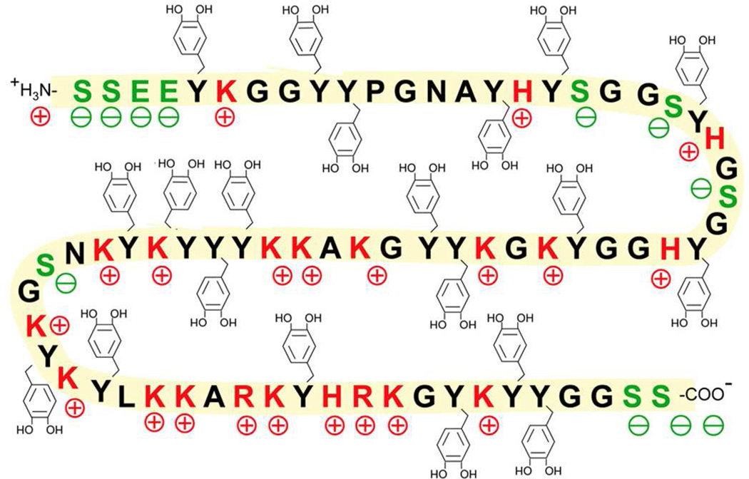
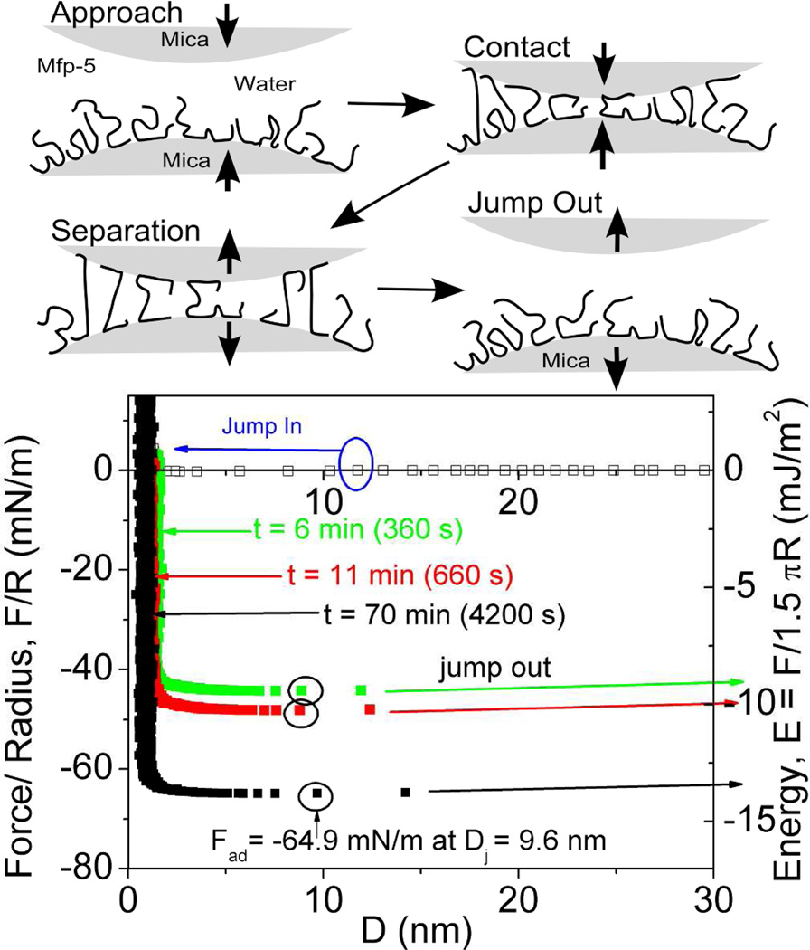
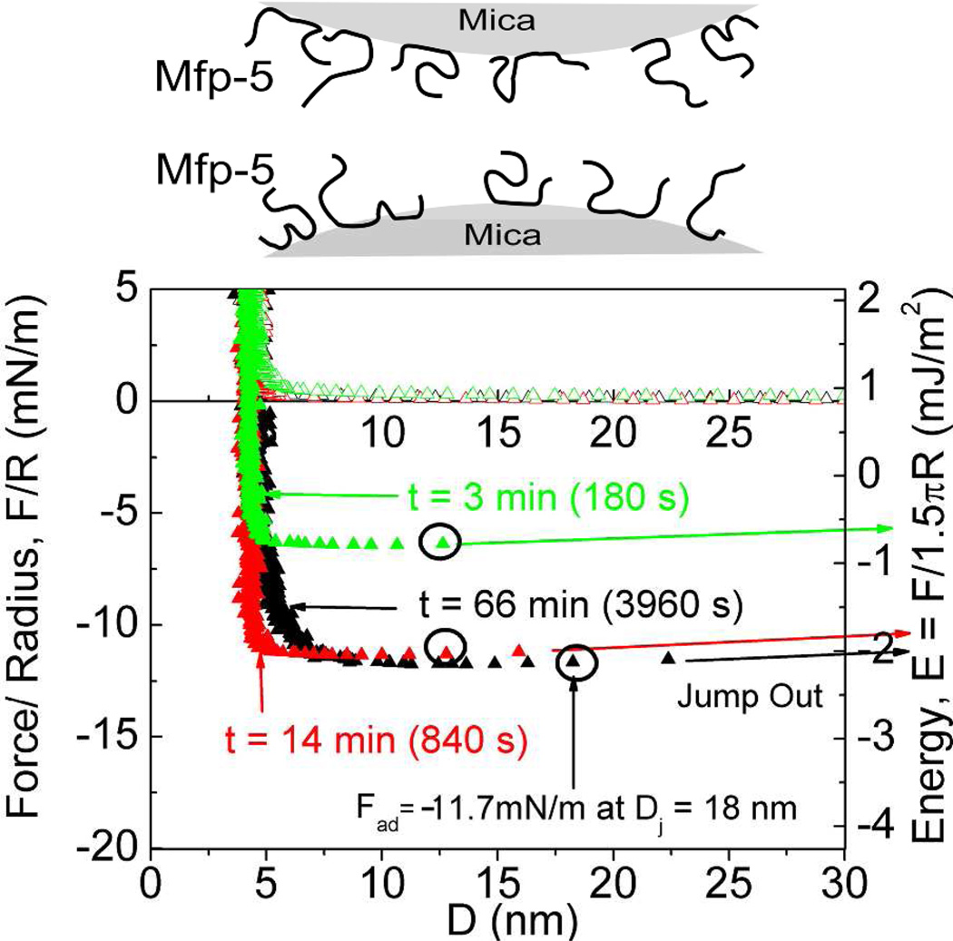
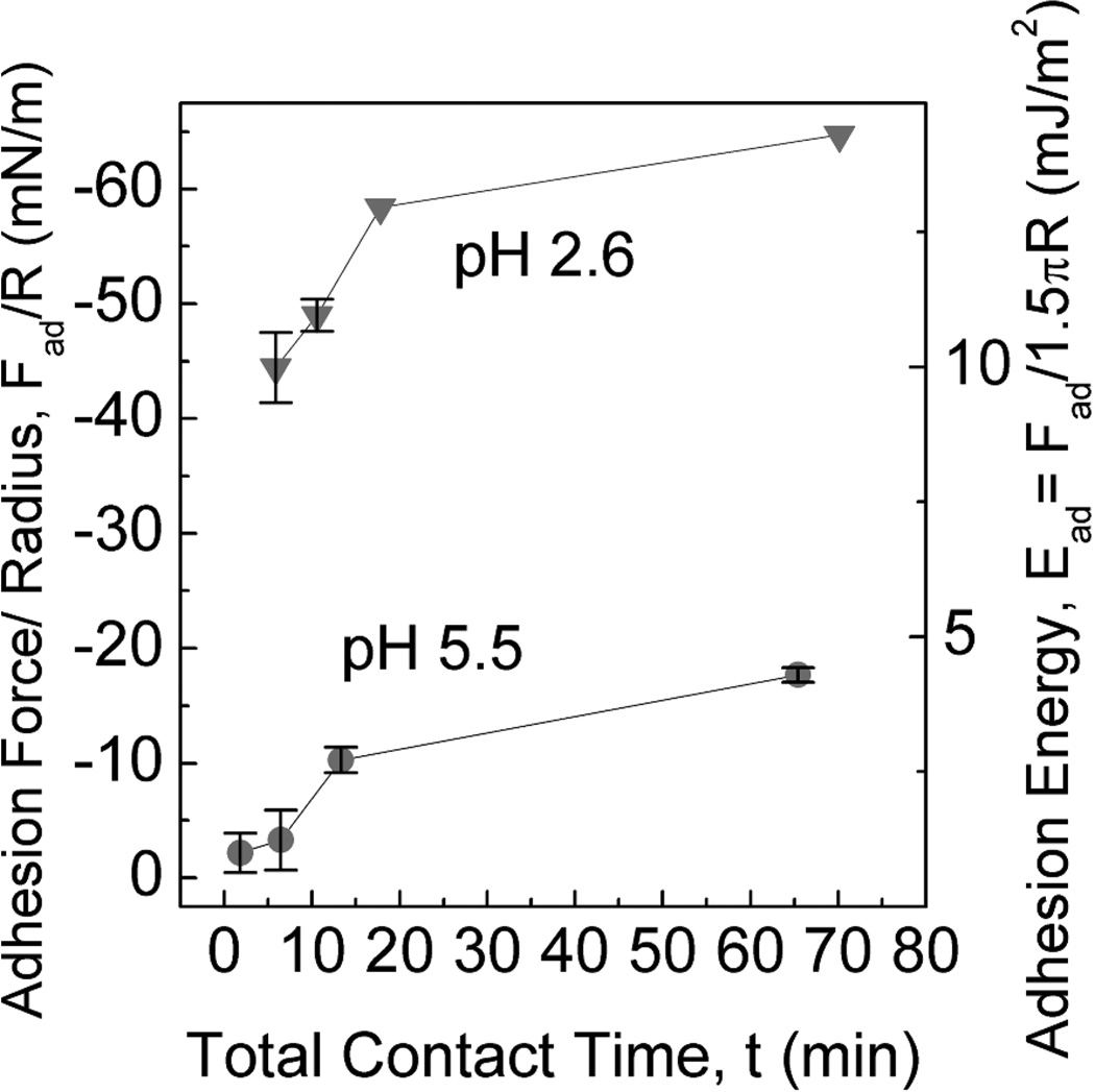
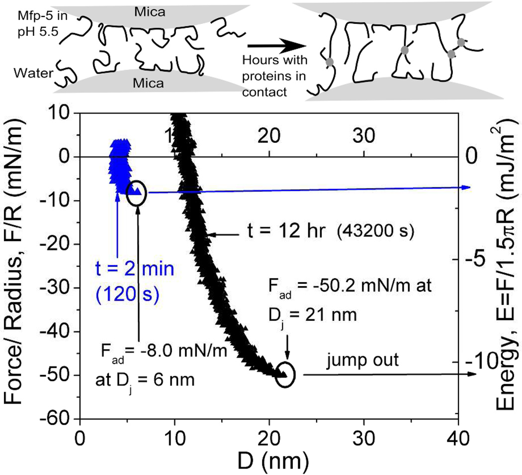
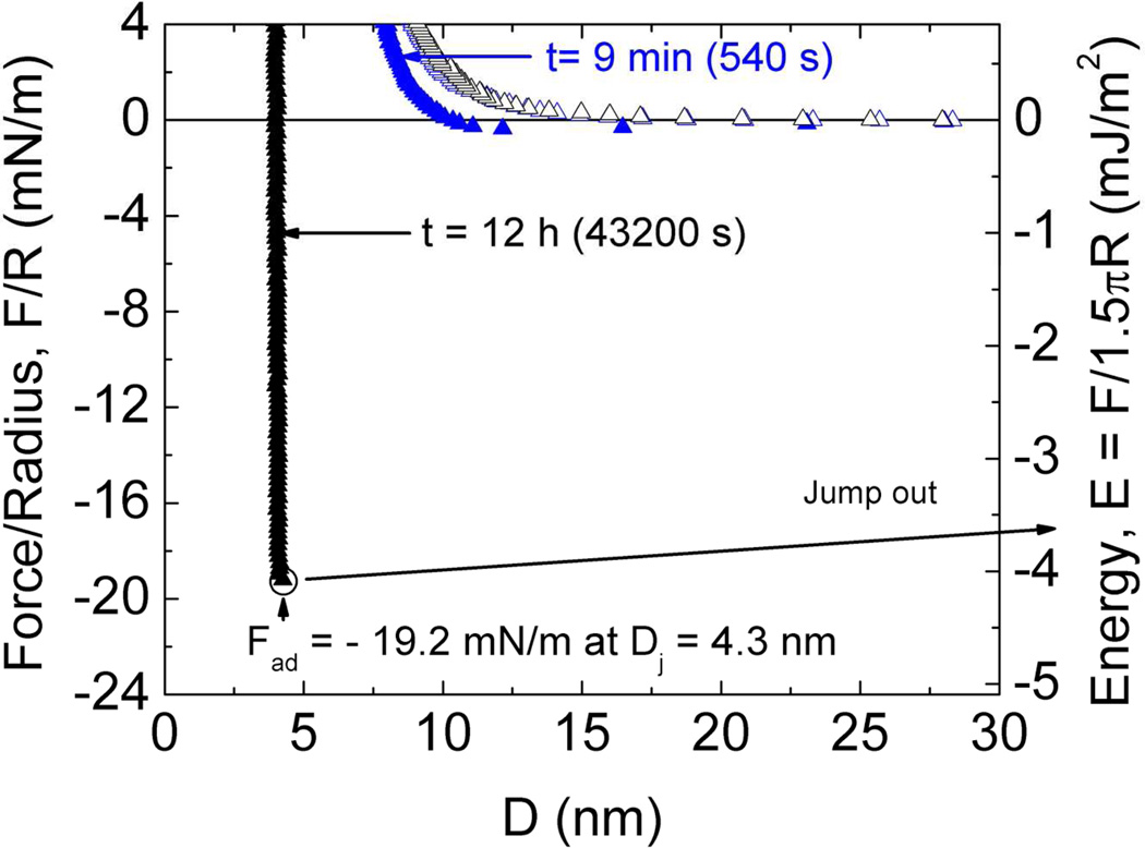
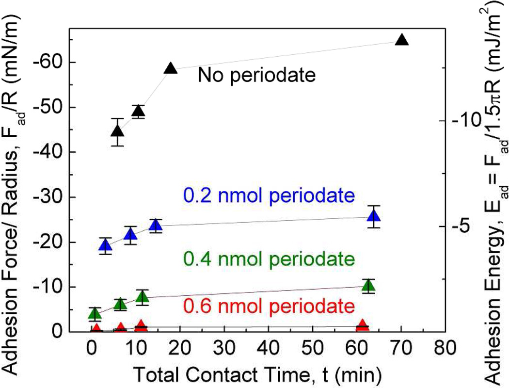
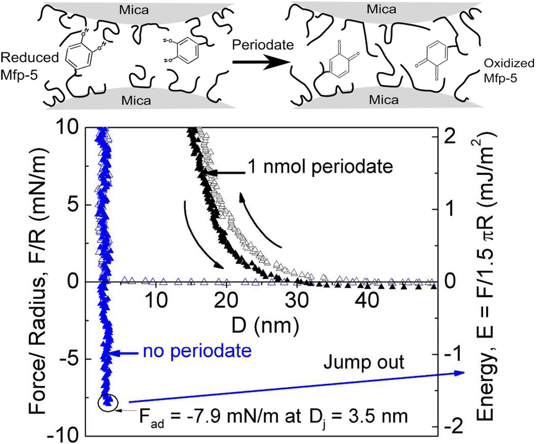
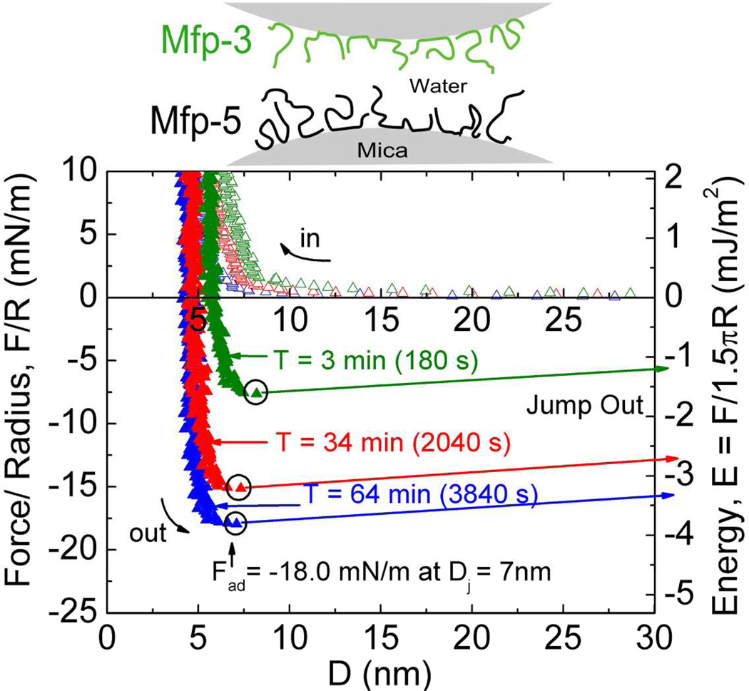
Mussels have a remarkable ability to attach their holdfast, or byssus, opportunistically to a variety of substrata that are wet, saline, corroded, and/or fouled by biofilms. Mytilus edulis foot protein-5 (Mefp-5) is one of several proteins in the byssal adhesive plaque of the mussel M. edulis. The high content of 3,4-dihydroxyphenylalanine (Dopa) (~30 mol %) and its localization near the plaque-substrate interface have often prompted speculation that Mefp-5 plays a key role in adhesion. Using the surface forces apparatus, we show that on mica surfaces Mefp-5 achieves an adhesion energy approaching E(ad) = ~-14 mJ/m(2). This exceeds the adhesion energy of another interfacial protein, Mefp-3, by a factor of 4-5 and is greater than the adhesion between highly oriented monolayers of biotin and streptavidin. The adhesion to mica is notable for its dependence on Dopa, which is most stable under reducing conditions and acidic pH. Mefp-5 also exhibits strong protein-protein interactions with itself as well as with Mefp-3 from M. edulis.


Figures

Figure 1
Figure 1
Sequence of Mefp-5. Red residues are positively charged, green residues are negatively charged and Dopa modifications (Y) are depicted with catecholic side chains. S denotes the location of phospho-Serine. Whereas the conversion of tyrosine to Dopa is complete in purified Mefp-5, the conversion of serine to phosphoserine is variable.
Figure 2
Figure 2
Mefp-5 interactions with mica as measured by the surface forces apparatus (SFA) with variable total contact times. Upper panels show how an asymmetric configuration with a Mefp-5 monolayer deposited on the lower mica surface and freshly cleaved mica on the upper surface was used to measure adhesion. Blue circle in the lower plot indicates beginning of jump-in; black circles denote Fad for each run. Solution I=100mM, pH 2.6.
Figure 3
Figure 3
Mefp-5/Mefp-5 symmetric configuration with 20 µl of 60µg/ml protein deposited on both surfaces (schematic above graph) with 3× higher protein concentration during deposition than in other experiments. The total contact times are denoted with (t). Circled points indicate Fad of the run. Solution I=100 mM, pH 2.6.
Figure 4
Figure 4
Effect of pH on peak adhesive force Fad and adhesion energy Ead between Mefp-5 and freshly cleaved mica surface (upper panel schematic). Results are plotted at pH 2.6 and 5.5 as a function of total contact times. Ionic strength is 100 mM. Error bars indicate standard deviation of at least two measurements.
Figure 5
Figure 5
Effect of auto-oxidation on cohesion in symmetrically deposited Mefp-5 in at pH 5.5 and I=350 mM. After an initial short cohesion (blue), protein covered surfaces were left in contact in the auto-oxidizing solution for 12 hrs before separation (black). Upper panel depicts cross-link formation (dots) during contact following exposure to pH 5.5.
Figure 6
Figure 6
Effect of auto-oxidation on cohesion in symmetrically deposited Mefp-5 in at pH 7.5 and I=234 mM. After the short-term contact (blue), protein covered surfaces were left in contact in the auto-oxidizing solution for 12 hrs before separation (black).
Figure 7
Figure 7
Effect of Dopa oxidation by periodate treatment (upper panel) on peak Fad and Ead between Mefp-5 and mica at different total contact times before separation (lower panel). All runs were done at pH 2.6 and I=100 mM. Error bars indicate standard deviation of at least two measurements.
Figure 8
Figure 8
Effect of periodate treatment on the cohesive interaction between symmetrically deposited Mefp-5 films taken after 12 minute (760 s) total contact times. Oxidation of Dopa to Dopaquinone by periodate treatment (upper panel) while protein films are separated abolishes cohesion (black), evident in the untreated controls (blue). pH 2.6 and I= 100 mM.
Figure 9
Figure 9
Interaction between two interfacial plaque proteins, Mefp-3 and Mefp-5 with different total contact times. Mefp-3 and Mefp-5 monolayers were deposited asymmetrically on opposing mica surfaces (upper panel). Circled portions indicate Fad for run. Solution pH 2.6 and I=100 mM.

References

    1. Rubin DJ, Miserez A, Waite JH. Diverse strategies of protein sclerotization in marine invertebrate structure–property relationships in natural biomaterials. Adv. Insect Physiol. 2010;38:75–133.
    1. Lee H, Scherer NF, Messersmith PB. Single-molecule mechanics of mussel adhesion. Proc. Natl. Acad. Sci. 2006;103:12999–13003. - PMC - PubMed
    1. Anderson TH, Yu J, Estrada A, Hammer MU, Waite JH, Israelachvili JN. The contribution of Dopa to substrate-peptide adhesion and internal cohesion of mussel-inspired synthetic peptide films. Adv. Funct. Mater. 2010;20:4196–4205. - PMC - PubMed
    1. Wang J, Tahir MN, Kappl M, Tremel W, Metz N, Barz M, Theato P, Butt HJ. Influence of binding-site density in wet bioadhesion. Adv. Mater. 2008;20:3872–3876.
    1. Sivasankar S, Brieher W, Lavrik N, Gumbiner B, Leckband D. Direct molecular force measurements of multiple adhesive interactions between cadherin ectodomains. Proc. Natl. Acad. Sci. 1999;96:11820–11824. - PMC - PubMed

Publication types