Review
. 2012 Nov 7;9(76):2749-66.
doi: 10.1098/rsif.2012.0341. Epub 2012 Aug 8.

The hierarchical structure and mechanics of plant materials

Affiliations
Review

The hierarchical structure and mechanics of plant materials

Lorna J Gibson. J R Soc Interface. .

Abstract

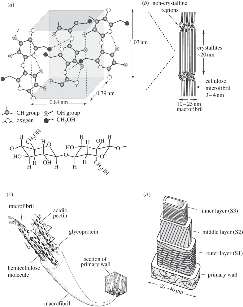
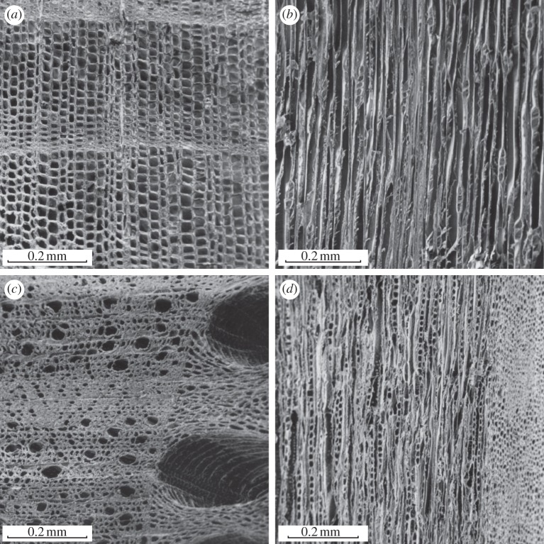
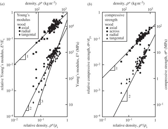
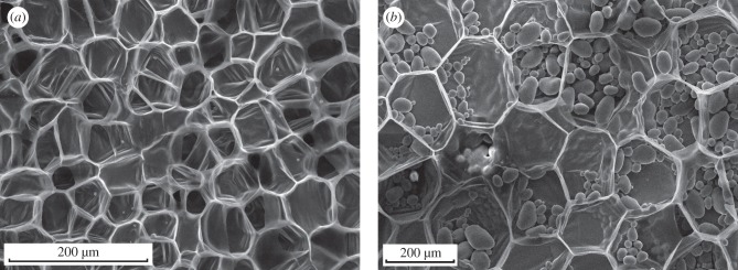
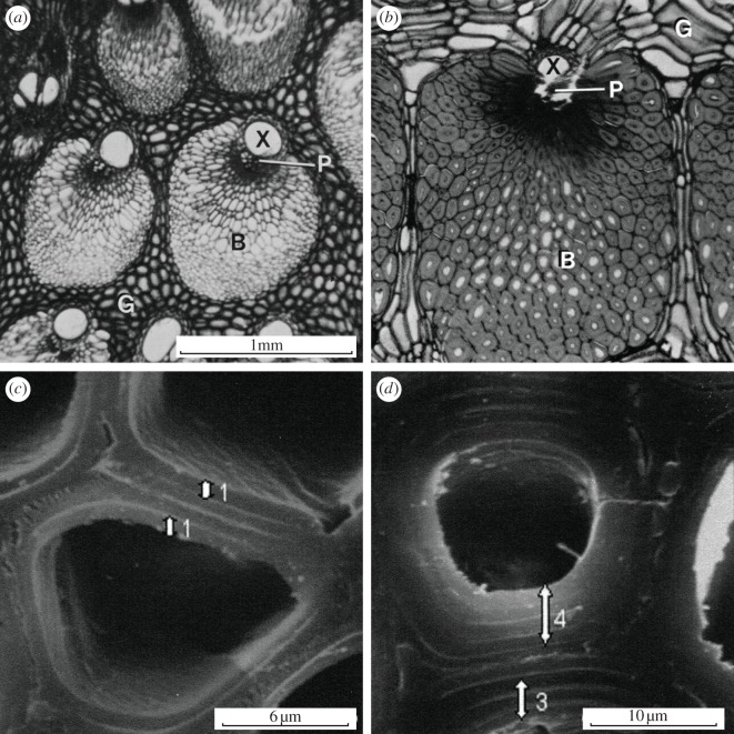
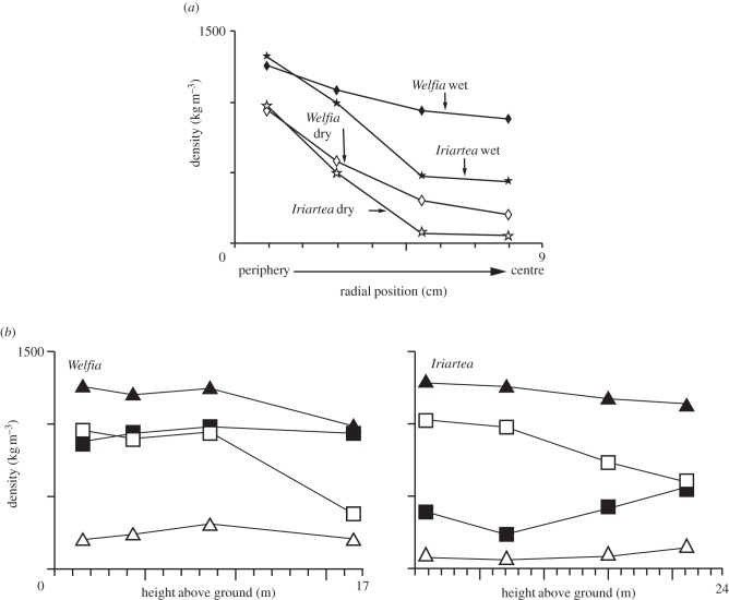
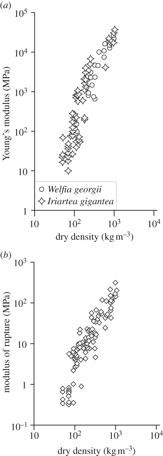
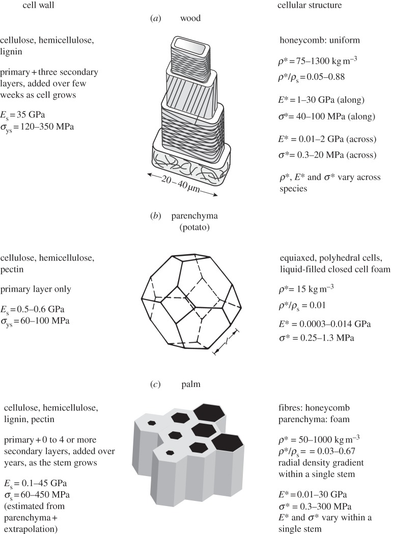
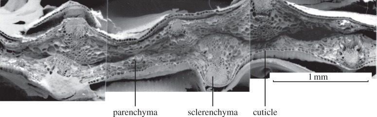
The cell walls in plants are made up of just four basic building blocks: cellulose (the main structural fibre of the plant kingdom) hemicellulose, lignin and pectin. Although the microstructure of plant cell walls varies in different types of plants, broadly speaking, cellulose fibres reinforce a matrix of hemicellulose and either pectin or lignin. The cellular structure of plants varies too, from the largely honeycomb-like cells of wood to the closed-cell, liquid-filled foam-like parenchyma cells of apples and potatoes and to composites of these two cellular structures, as in arborescent palm stems. The arrangement of the four basic building blocks in plant cell walls and the variations in cellular structure give rise to a remarkably wide range of mechanical properties: Young's modulus varies from 0.3 MPa in parenchyma to 30 GPa in the densest palm, while the compressive strength varies from 0.3 MPa in parenchyma to over 300 MPa in dense palm. The moduli and compressive strength of plant materials span this entire range. This study reviews the composition and microstructure of the cell wall as well as the cellular structure in three plant materials (wood, parenchyma and arborescent palm stems) to explain the wide range in mechanical properties in plants as well as their remarkable mechanical efficiency.


Figures

Figure 1.
Figure 1.
Strength plotted against Young's modulus for selected plant materials. Note the large range in properties produced by varying the arrangement of the four building blocks (cellulose, lignin, hemicellulose and pectin) in the cell wall as well as the cellular structure. The properties of the cellulose and lignin are indicated in red. Adapted from Gibson et al. [1] with kind permission from Cambridge University Press. (Online version in colour.)
Figure 2.
Figure 2.
The hierarchical structure of plant cell walls showing: (a) the molecular structure of cellulose, with glucose molecules alternately rotated 180° (solid line, covalent bonds; dashed line, hydrogen bonds); (b) cellulose microfibrils, with both crystalline and non-crystalline regions, aggregated into a macrofibril; (c) a macrofibril from a primary cell wall and (d) the cell wall of wood, made up of a primary layer and three secondary layers (S1, S2 and S3), with the cellulose microfibrils arranged in different orientations in each layer. Neighbouring cells are attached to each other by the middle lamella (not indicated) (a,b,d adapted from Gibson et al. [1], with kind permission from Cambridge University Press; based on Dinwoodie [2]; figure 2c is reproduced from Niklas [3] with kind permission from the University of Chicago Press).
Figure 3.
Figure 3.
Scanning electron micrographs of woods: (a) cedar, cross section; (b) cedar, longitudinal section; (c) oak, cross section; (d) oak, longitudinal section. Adapted from Gibson et al. [1] with kind permission from Cambridge University Press.
Figure 4.
Figure 4.
(a) Young's modulus and (b) compressive strength of wood plotted against density. Data for loading across the grain are for loading in the radial or tangential directions; the direction of loading is not specified. Data from Goodman & Bodig [48,49] Bodig & Goodman [50]; Wood Handbook [51]; Dinwoodie [2]; Bodig & Jayne [9] and Easterling et al. [52]. Adapted from Gibson et al. [1], with kind permission from Cambridge University Press.
Figure 5.
Figure 5.
(a) Young's modulus and (b) strength plotted against density for woods and their constituents. (a) Adapted from Gibson et al. [1] with kind permission from Cambridge University Press.
Figure 6.
Figure 6.
Scanning electron micrographs of (a) carrot and (b) potato showing the relatively thin-walled cells. The ellipsoidal objects within the potato tissue are starch granules. Adapted from Gibson et al. [1], with kind permission from Cambridge University Press.
Figure 7.
Figure 7.
(a,b) Optical micrographs of cross sections of Iriartea gigantea showing the peripheral stem tissue of (a) a young individual and (b) an older individual. B denotes vascular bundle of honeycomb-like cells, including xylem (X) and phloem (P), which conduct water and sap; G denotes ground tissue, made up of polyhedral parenchyma cells, similar to a closed-cell foam. (c,d) Scanning electron micrographs of cross sections of coconut palm Cocus nucifera, showing cells near (c) the centre of the stem, with a primary layer and one secondary layer and (d) the periphery of the stem, with a primary cell wall and three or four secondary layers. (a,b) Adapted from Rich [, figs 22 and 23], with kind permission of the Botanical Society of America Inc.; (c,d) Reprinted from Kuo-Huang et al. [66], fig. 1e,f, with kind permission of the International Association of Wood Anatomists (IAWA).
Figure 8.
Figure 8.
Density plotted against (a) radial position (at breast height) and (b) against height above ground in the stem for a 19 m tall Welfia georgii and for a 17 m tall Iriartea gigantea. All adapted from Rich [65]. Filled triangles, peripheral wet; filled squares, central wet; open squares, peripheral dry; open triangles, central dry.
Figure 9.
Figure 9.
(a) Young's modulus and (b) modulus of rupture plotted against dry density for two species of palm, Welfia georgii and Iriartea gigantea. (b) Modulus of rupture plotted against dry density for six species of palm: W. georgii, I. gigantea, Socratea durissima, Euterpe macrospadix, Prestoea decurrens and Cryosophila albida. Adapted from Rich [65].
Figure 10.
Figure 10.
A comparison of the cell wall and cellular structure of wood, parenchyma and arborescent palm stem. Left: the cell wall composition, microstructure, modulus and strength. Centre: schematic showing the structure of each plant material. Right: plant cellular microstructure, and ranges of density, relative density, modulus and strength. Top and centre schematics from Gibson et al. [1], with kind permission from Cambridge University Press.
Figure 11.
Figure 11.
(a) Young's modulus–density chart for engineering materials, including woods. The performance index E1/2/ρ gives the performance of a material for resisting bending deflections: every point on a single line E1/2/ρ has the same value of E1/2/ρ. As the line moves to the upper left, the value of E1/2/ρ increases. Woods loaded along the grain have comparable values to the best engineering materials, fibre composites (adapted from figure from MF Ashby, with permission). (b) Strength–density chart for engineering materials, including woods. The performance index σ2/3/ρ gives the performance of a material for resisting bending stresses: every point on a single line σ2/3/ρ has the same value of σ2/3/ρ . As the line moves to the upper left, the value of σ2/3/ρ  increases. Woods loaded along the grain have comparable values to the best engineering materials, fibre composites. Adapted from figure from Ashby, with permission. (Online version in colour.)
Figure 12.
Figure 12.
Scanning electron micrograph of the iris leaf, showing the core of foam-like parenchyma tissue and the outer faces of dense sclerenchyma fibres in a matrix of cuticle (reproduced from Gibson et al. [71], with kind permission of Springer).
Figure 13.
Figure 13.
The distribution of bending stress, σ (dashed line) and bending strength, σ* (solid line) in the arborescent palm stem, Iriartea gigantea, with its radial density gradient. The strength of the palm tissue closely matches the stress distribution (adapted from Gibson et al. [1] with kind permission from Cambridge University Press).

Similar articles

Cited by

References

    1. Gibson L. J., Ashby M. F., Harley B. A. 2010. Cellular materials in nature and medicine. Cambridge, UK: Cambridge University Press
    1. Dinwoodie J. M. 1981. Timber: its nature and behaviour. New York, NY: Van Nostrand Reinhold
    1. Niklas K. J. 1992. Plant biomechanics. Chicago, IL: University of Chicago Press
    1. Hori R., Muller M., Watanabe U., Lichtenegger H. C., Fratzl P., Sugiyama J. 2002. The importance of seasonal differences in the cellulose microfibril angle in softwoods in determining acoustic properties. J. Mater. Sci. 37, 4279–428410.1023/A:1020688132345 (doi:10.1023/A:1020688132345) - DOI - DOI
    1. Peura M., Muller M., Vainio U., Saren M.-P., Saranpaa P., Serimaa R. 2008. X-ray microdiffraction reveals the orientation of cellulose microfibrils and the size of cellulose crystallites in single Norway spruce tracheids. Trees 22, 49–6110.1007/s00468-007-0168-5 (doi:10.1007/s00468-007-0168-5) - DOI - DOI

Publication types

LinkOut - more resources