. 2012 Oct 15;303(8):R834-42.
doi: 10.1152/ajpregu.00260.2012. Epub 2012 Aug 8.

Intrinsic properties of the sodium sensor neurons in the rat median preoptic nucleus

Affiliations

Intrinsic properties of the sodium sensor neurons in the rat median preoptic nucleus

Aurore N Voisin et al. Am J Physiol Regul Integr Comp Physiol. .

Abstract

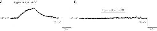
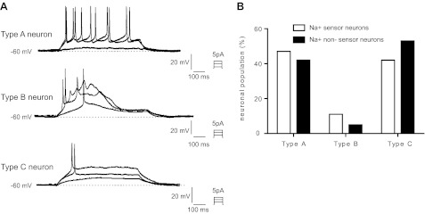
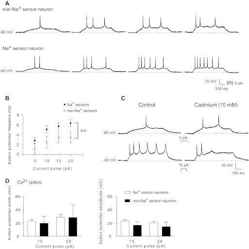
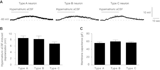
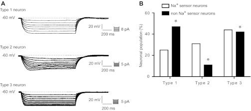
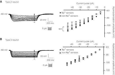
The essential role of the median preoptic nucleus (MnPO) in the integration of chemosensory information associated with the hydromineral state of the rat relies on the presence of a unique population of sodium (Na+) sensor neurons. Little is known about the intrinsic properties of these neurons; therefore, we used whole cell recordings in acute brain slices to determine the electrical fingerprints of this specific neural population of rat MnPO. The data collected from a large sample of neurons (115) indicated that the Na+ sensor neurons represent a majority of the MnPO neurons in situ (83%). These neurons displayed great diversity in both firing patterns induced by transient depolarizing current steps and rectifying properties activated by hyperpolarizing current steps. This diversity of electrical properties was also present in non-Na+ sensor neurons. Subpopulations of Na+ sensor neurons could be distinguished, however, from the non-Na+ sensor neurons. The firing frequency was higher in Na+ sensor neurons, showing irregular spike discharges, and the amplitude of the time-dependent rectification was weaker in the Na+ sensor neurons than in non-Na+ sensor neurons. The diversity among the electrical properties of the MnPO neurons contrasts with the relative function homogeneity (Na+ sensing). However, this diversity might be correlated with a variety of direct synaptic connections linking the MnPO to different brain areas involved in various aspects of the restoration and conservation of the body fluid homeostasis.


Figures

Fig. 1.
Fig. 1.
Functional identification of the Na+ sensor neurons in the ventral region of median preoptic nucleus (vMnPO). Transient application of hypernatriuric artifical cerebrospinal fluid (aCSF) (170 mM) induced either a membrane depolarization (A) or no change in the membrane potential (B). The reversible membrane depolarization triggered by the application of hypernatriuric aCSF identifies the Na+ sensor neurons.
Fig. 2.
Fig. 2.
The Na+ sensor neurons display distinct firing patterns in acute brain slices. A: typical illustration of the three distinct firing patterns observed in the vMnPO neurons in response to depolarizing current steps of 800 ms. Type A neurons were characterized by irregular spike discharges (top trace). Type B neurons displayed a robust Ca2+ spike (middle trace) and type C neurons showed a strong spike adaptation (bottom trace). B: bar graph illustrating the distribution of the firing patterns in the Na+ sensor and non-Na+ sensor neurons.
Fig. 3.
Fig. 3.
Type A Na+ sensor neurons fire at higher frequency than the non-Na+ sensor neurons. A: illustration of the firing pattern of the type A neurons in the two populations of MnPO neurons. B: intensity-to-firing frequency plot indicates that the Na+ sensor neurons discharge at higher frequency that the non-Na+ sensor neurons. **Statistical significance at P < 0.01. C: typical illustration of high-threshold Ca2+ spikes in MnPO neurons. Local application of cadmium (10 mM) abolished the high threshold spikes elicited by depolarizing current steps (>10 pA). D: bar graph histograms indicating that the Ca2+ spike characteristics were identical in the Na+ sensor and in the non-Na+ sensor neurons. The duration of each depolarizing current step was 800 ms. Results were expressed as means ± SE.
Fig. 4.
Fig. 4.
Amplitude of the Na+-evoked depolarization is not a valid criteria to distinguish between the populations of Na+ sensor neurons. A: typical illustration of the depolarization triggered by hypernatriuric aCSF (170 mM) recorded in individual Na+-sensor neurons characterized by a distinct firing pattern (type A, B, and C neuron). Neurons were held around −60 mV and four depolarizing steps of 800 ms were applied (5 pA increment). B: bar graph histogram indicating that the Na+-evoked depolarization was similar in the three populations of Na+ sensor neurons. C: bar graph histogram indicating that membrane capacitance (neuronal size indicator) was identical in the three populations of Na+ sensor neurons.
Fig. 5.
Fig. 5.
The Na+ sensor neurons display various patterns of membrane rectification in acute brain slice. A: two distinct rectifying patterns were observed in the MnPO neurons in response to hyperpolarizing current steps: neurons showing a time-dependent membrane rectification (type 2 neuron) and neurons displaying a time-independent membrane rectification (type 3 neuron). In addition, neurons without evoked membrane rectification were also present in the MnPO (type 1 neuron). B: bar graph histogram representing the distribution of the three types of neurons in the Na+ sensor and in the non-Na+ sensor neurons. The hyperpolarizing current steps were applied during 1,200 ms. Results were expressed as means ± SE. *Statistical significance at P < 0.05.
Fig. 6.
Fig. 6.
Type 2 Na+ sensor neurons are characterized by a weaker membrane rectification than the non-Na+ sensor neurons. A: amplitude of the time-dependent hyperpolarization evoked by negative current steps of 1,200 ms duration was measured at a steady-state membrane potential (a). The current-voltage relationship (I/V curve) indicated that the amplitude of the hyperpolarization was higher in the Na+ sensor neurons than in the non-Na+ sensor neurons. **Statistical significance at P < 0.01. B: amplitude of the time independent hyperpolarization was similar in the Na+ sensor and in the non-Na+ sensor neurons. Results were expressed as means ± SE.

Similar articles

Cited by

References

    1. Aradachi H, Honda K, Negoro H, Kubota T. Median preoptic neurones projecting to the supraoptic nucleus are sensitive to haemodynamic changes as well as to rise in plasma osmolality in rats. J Neuroendocrinol 8: 35–43, 1996 - PubMed
    1. Bai D, Renaud LP. ANG II AT1 receptors induce depolarization and inward current in rat median preoptic neurons in vitro. Am J Physiol Regul Integr Comp Physiol 275: R632–R639, 1998 - PubMed
    1. Blatz AL, Magleby KL. Single apamin-blocked Ca-activated K+ channels of small conductance in cultured rat skeletal muscle. Nature 323: 718–720, 1986 - PubMed
    1. Bodnar RJ, Glass MJ, Ragnauth A, Cooper ML. General, mu and kappa opioid antagonists in the nucleus accumbens alter food intake under deprivation, glucoprivic and palatable conditions. Brain Res 66: 205–212, 1995 - PubMed
    1. Brown DR, Holtzman SG. Opiate antagonists: central sites of action in suppressing water intake of the rat. Brain Res 221: 432–436, 1981 - PubMed

Publication types