. 2012 Aug 15;11(16):3019-35.
doi: 10.4161/cc.21384. Epub 2012 Aug 9.

Metabolic reprogramming of cancer-associated fibroblasts by TGF-β drives tumor growth: connecting TGF-β signaling with "Warburg-like" cancer metabolism and L-lactate production

Affiliations

Metabolic reprogramming of cancer-associated fibroblasts by TGF-β drives tumor growth: connecting TGF-β signaling with "Warburg-like" cancer metabolism and L-lactate production

Carmela Guido et al. Cell Cycle. .

Abstract

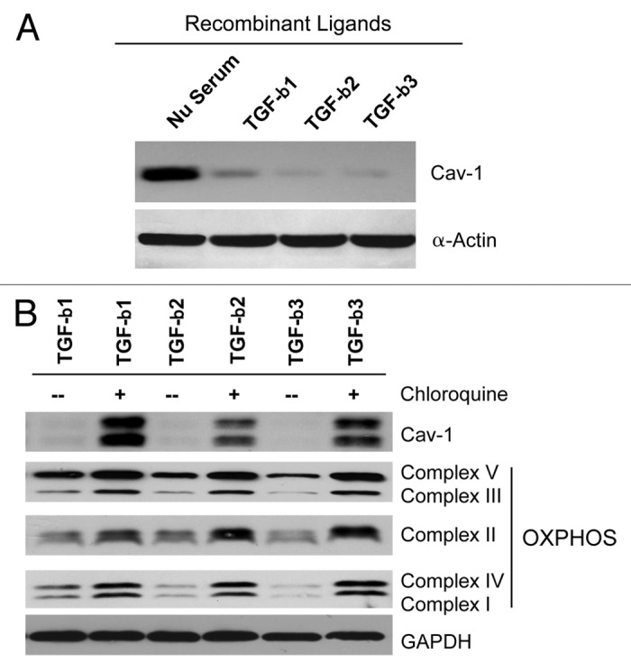
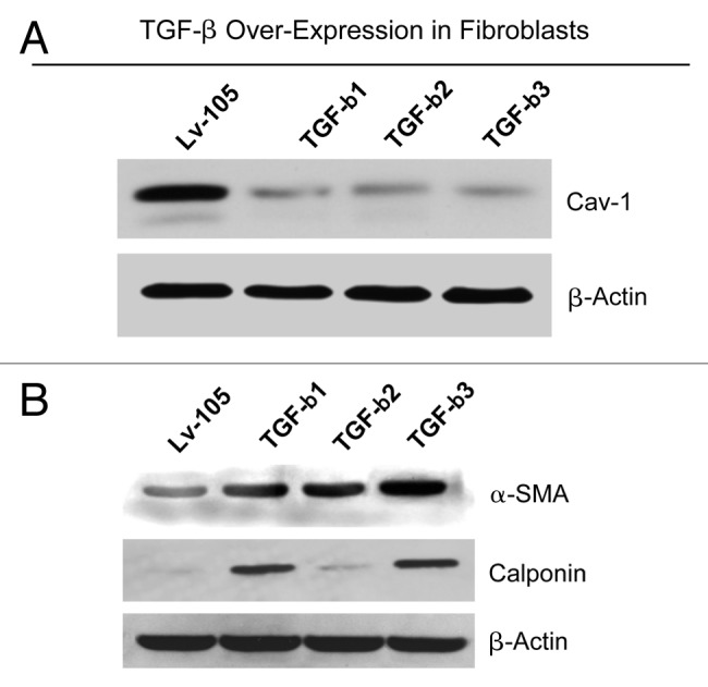
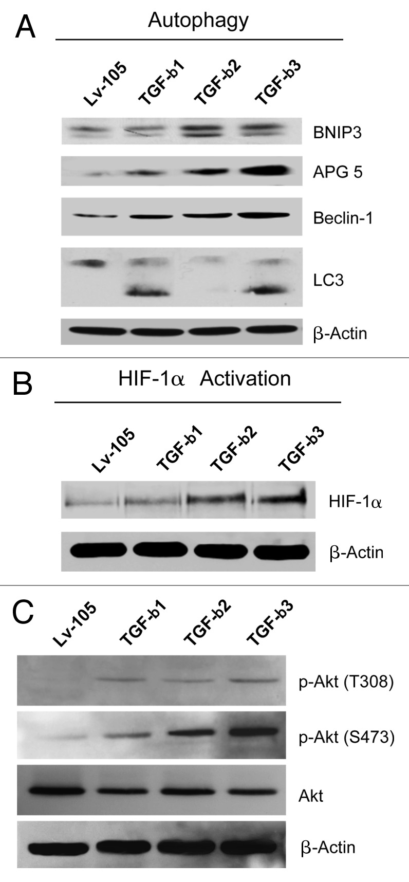
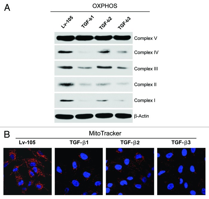
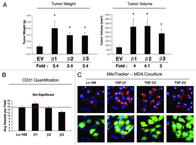
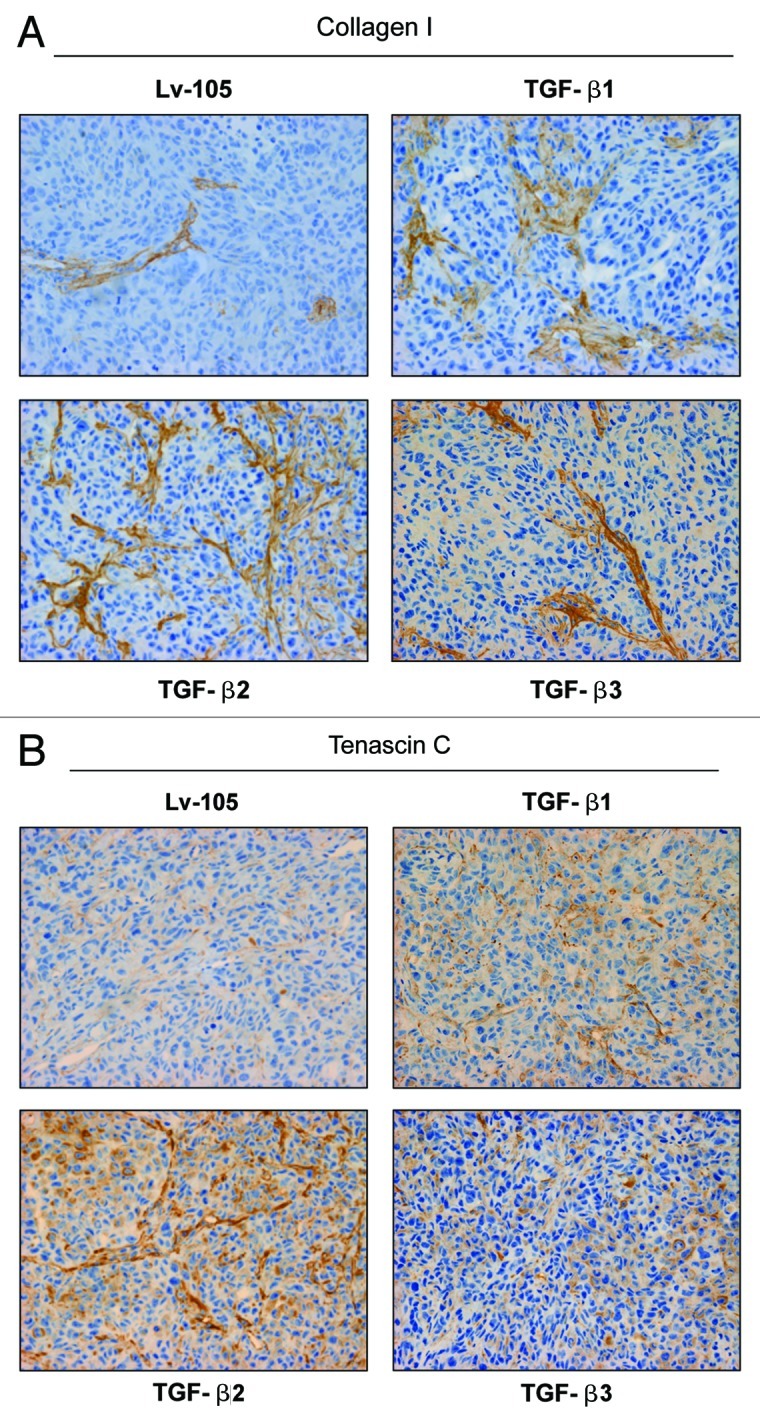
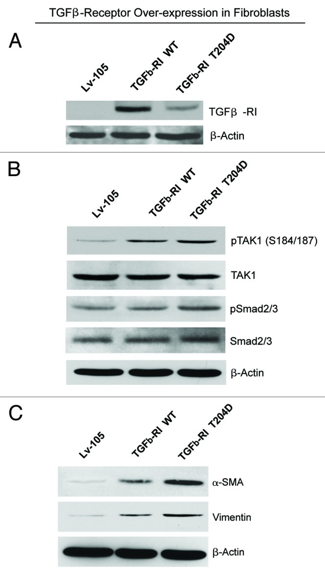
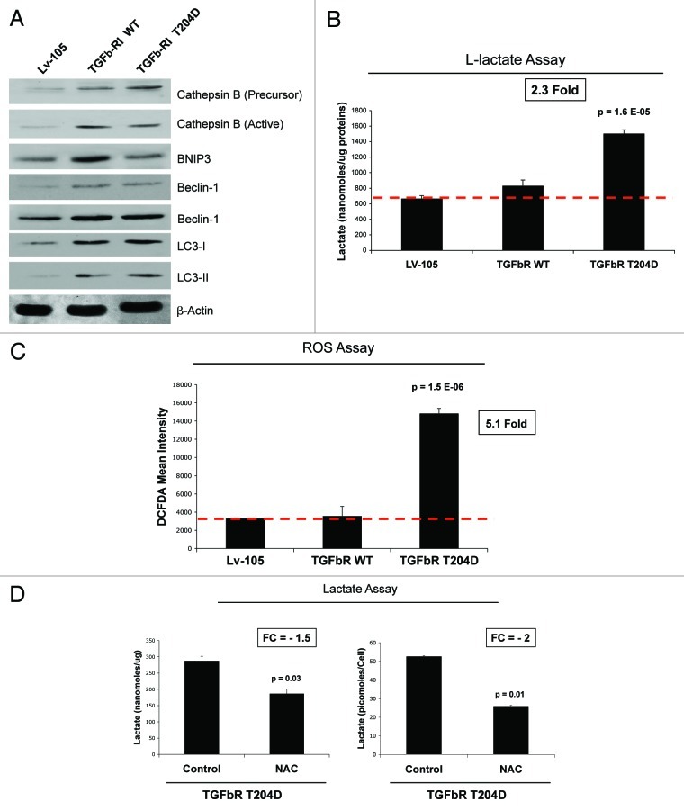
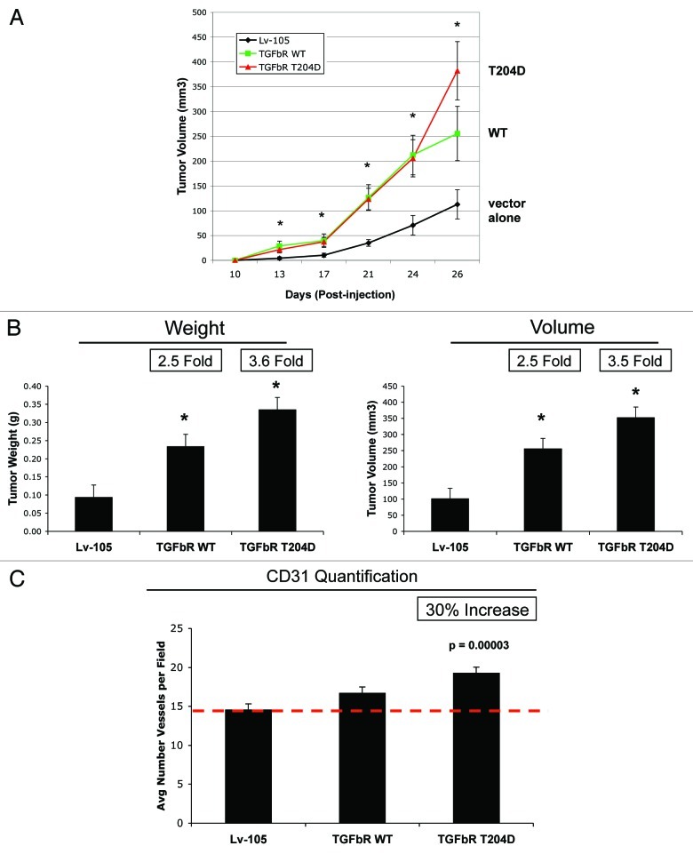
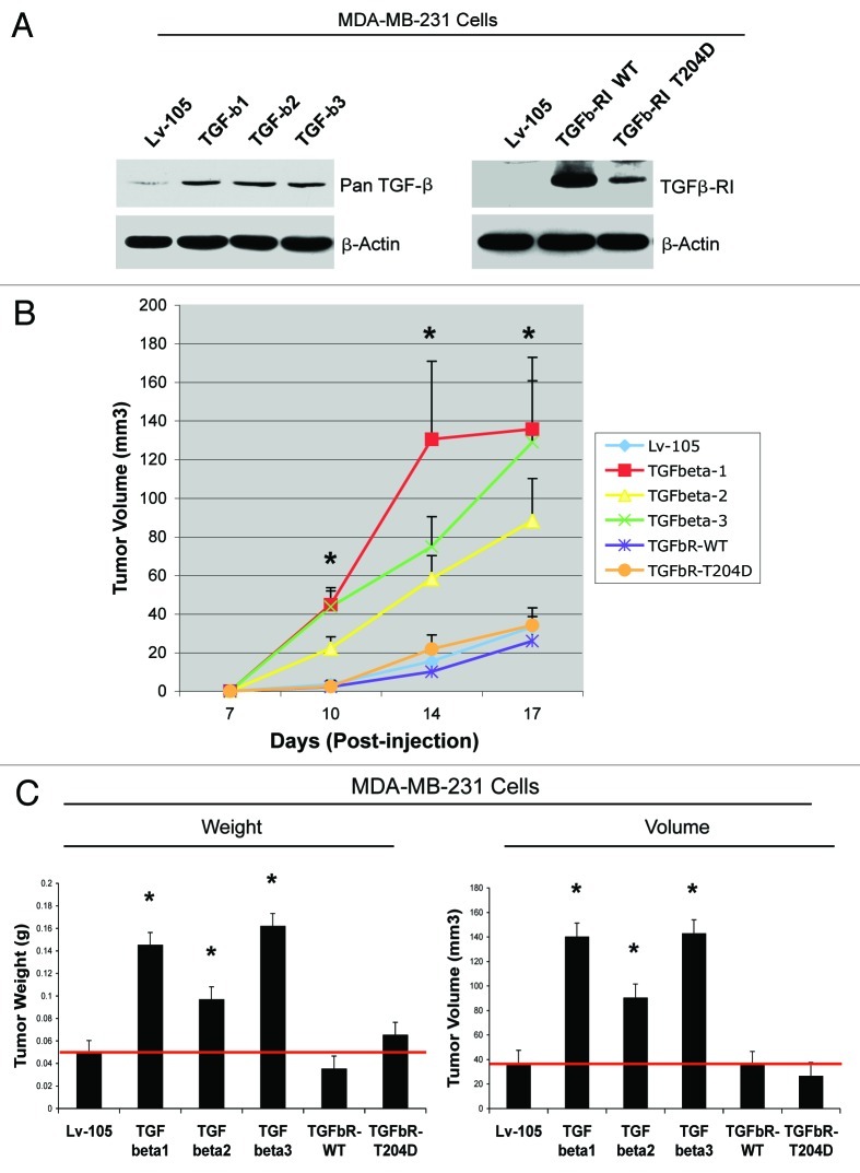
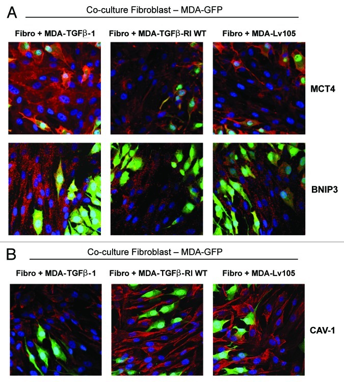
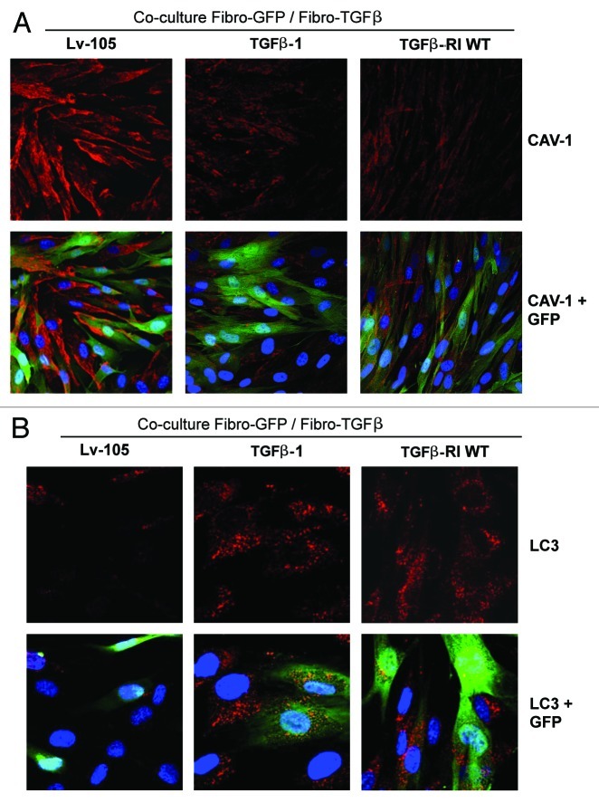
We have previously shown that a loss of stromal Cav-1 is a biomarker of poor prognosis in breast cancers. Mechanistically, a loss of Cav-1 induces the metabolic reprogramming of stromal cells, with increased autophagy/mitophagy, mitochondrial dysfunction and aerobic glycolysis. As a consequence, Cav-1-low CAFs generate nutrients (such as L-lactate) and chemical building blocks that fuel mitochondrial metabolism and the anabolic growth of adjacent breast cancer cells. It is also known that a loss of Cav-1 is associated with hyperactive TGF-β signaling. However, it remains unknown whether hyperactivation of the TGF-β signaling pathway contributes to the metabolic reprogramming of Cav-1-low CAFs. To address these issues, we overexpressed TGF-β ligands and the TGF-β receptor I (TGFβ-RI) in stromal fibroblasts and breast cancer cells. Here, we show that the role of TGF-β in tumorigenesis is compartment-specific, and that TGF-β promotes tumorigenesis by shifting cancer-associated fibroblasts toward catabolic metabolism. Importantly, the tumor-promoting effects of TGF-β are independent of the cell type generating TGF-β. Thus, stromal-derived TGF-β activates signaling in stromal cells in an autocrine fashion, leading to fibroblast activation, as judged by increased expression of myofibroblast markers, and metabolic reprogramming, with a shift toward catabolic metabolism and oxidative stress. We also show that TGF-β-activated fibroblasts promote the mitochondrial activity of adjacent cancer cells, and in a xenograft model, enhancing the growth of breast cancer cells, independently of angiogenesis. Conversely, activation of the TGF-β pathway in cancer cells does not influence tumor growth, but cancer cell-derived-TGF-β ligands affect stromal cells in a paracrine fashion, leading to fibroblast activation and enhanced tumor growth. In conclusion, ligand-dependent or cell-autonomous activation of the TGF-β pathway in stromal cells induces their metabolic reprogramming, with increased oxidative stress, autophagy/mitophagy and glycolysis, and downregulation of Cav-1. These metabolic alterations can spread among neighboring fibroblasts and greatly sustain the growth of breast cancer cells. Our data provide novel insights into the role of the TGF-β pathway in breast tumorigenesis, and establish a clear causative link between the tumor-promoting effects of TGF-β signaling and the metabolic reprogramming of the tumor microenvironment.


Figures

None
Figure 1. TGF-β treatment induces the autophagy-mediated downregulation of Cav-1 in fibroblasts. (A) hTERT-immortalized human fibroblasts were treated with TGF-β1, TGF-β2 or TGF-β3 (10 ng/ml) for 48 h. Then, cells were analyzed by immunoblotting with anti-Cav-1 antibodies. Note that TGF-β1, TGF-β2 and TGF-β3 ligand treatment induces Cav-1 downregulation. β-actin was used as an equal loading control. (B) Chloroquine treatment rescues the expression of Cav-1 and OXPHOS markers. Fibroblasts treated with the three TGF-β ligands were incubated with the autophagy inhibitor chloroquine (25 μM) for 24 h, and then subjected to immunoblot analysis with antibodies directed against Cav-1 and OXPHOS. Note that treatment with chloroquine rescues the expression of Cav-1 and greatly augments the levels of OXPHOS markers. Immunoblotting with GAPDH is shown as a control for equal loading.
None
Figure 2. Fibroblasts overexpressing TGF-β1, TGF-β2 or TGF-β3 show a fibroblast to myofibroblast conversion, with Cav-1 downregulation. (A) hTERT-immortalized human fibroblasts, stably expressing TGF-β1, TGF-β2, TGF-β3 or the empty vector (Lv-105) control, were generated using a lentiviral approach. After selection with puromycin, the cells were analyzed by immunoblotting with anti-Cav-1 antibodies. Note that the stable overexpression of TGF-β ligands downregulates Cav-1 protein expression. β-actin was used as an equal loading control. (B) Cells were subjected to immunoblot analysis with antibodies directed against myofibroblast markers. Note that α-SMA and calponin levels are upregulated. β-actin was used as an equal loading control.
None
Figure 3. Fibroblasts overexpressing TGF-β1, TGF-β2 or TGF-β3 show increased autophagy, with HIF-1α activation. (A) TGF-β-induced autophagy/mitophagy was evaluated by immunoblot analysis. TGF-β1-, TGF-β2- and TGF-β3-overexpressing fibroblasts display a significant increase of the expression of the mitophagy (BNIP3) and autophagy markers [APG5, Beclin-1, and LC3II (lower band)]. Note that TGF-β2 is able to induce upregulation of APG5 and Beclin-1, but it does not affect LC3 activation. β-actin was used as loading control. (B and C) To evaluate if TGF-β induces activation of HIF-1α and the Akt pathway, cells were analyzed by immunoblotting with antibodies directed against HIF-1α or phospho-Akt. β-actin was used as loading control. Note that TGF-β1-, TGF-β2- and TGF-β3-overexpressing fibroblasts display a significant increase of HIF-1α expression (B), and increased phospho-Akt, with decreased total Akt levels (C).
None
Figure 4. Fibroblasts overexpressing TGF-β1, TGF-β2 or TGF-β3 show impaired mitochondrial function. (A) Cell lysates from TGF-β1, TGF-β2, TGF-β3 and Lv-105 control fibroblasts were subjected to immunoblot analysis with a panel of antibodies against OXPHOS subunits. Note that all three TGF-β isoforms induce a decrease in the expression of key subunits of mitochondrial OXPHOS complexes, relative to control cells (complex IV, COXII; complex III, Core 2 subunit; complex II, 30KDa FeS subunit; complex I, 20KDa subunit). However, TGF-β2 fibroblasts do not show a strong downregulation of complex III. β-actin was used as loading control. (B) Decreased mitochondrial activity of TGF-β-fibroblasts was independently confirmed by MitoTracker staining (red). Nuclei were stained with DAPI (blue). MitoTracker labels only “healthy” mitochondria, with an active membrane potential. Original magnification, 60×.
None
Figure 5. Fibroblasts overexpressing TGF-β1, TGF-β2 or TGF-β3 promote tumor growth, in an angiogenesis-independent manner. (A) Tumor Growth. To evaluate the tumor-promoting properties of TGF-β-fibroblasts, we used a xenograft model. TGF-β1, TGF-β2, TGF-β3 or Lv-105 control fibroblasts were co-injected with MDA-MB-231 breast cancer cells into the flanks of athymic nude mice. After 4 weeks tumors were analyzed. Note that all three TGF-β isoforms induce an increase in tumor weight and volume, compared with the Lv-105 empty vector control. Fold increases are as indicated. n = 10. All (*) p-values were < 0.05. (B) Tumor Angiogenesis. To evaluate if fibroblast-derived TGF-β promote tumor growth by increasing tumor angiogenesis, frozen tumor sections were analyzed by CD31 immunostaining. Quantification of the number of CD31 (+) vessels per field indicates that angiogenesis is not modified in the three TGF-β-xenograft groups, relative to control tumors, indicating that TGF-β-fibroblasts stimulate tumor growth independently of angiogenesis. (C) Mitochondrial Activity. TGF-β-fibroblasts enhance mitochondrial activity in adjacent cancer epithelial cells. TGF-β ligand expressing fibroblasts and control fibroblasts were co-cultured with GFP-positive MDA-MB-231 cells. The cells were then labeled via MitoTracker staining (red). Nuclei were stained with DAPI (blue). Note that fibroblasts expressing TGF-β1, TGF-β2 or TGF-β3 specifically increase the mitochondrial activity of adjacent MDA-MB-231 cells (green), compared with empty vector Lv-105 control. In the top panel, white stars identify the nuclei of GFP-positive MDA-MB-231 cells. Original magnification, 60×.
None
Figure 6. Tumors derived from TGF-β ligand overexpressing fibroblasts display increased extracellular matrix deposition. (A and B) It is well known that TGF-β stimulates the secretion of type I collagen and Tenascin C. To evaluate extracellular matrix deposition, paraffin-embedded xenograft sections were immunostained with antibodies against type I collagen (A) and Tenascin C (B). Note that tumors derived from TGF-β1, TGF-β2 and TGF-β3-fibroblasts show increased levels of type I collagen and Tenascin C relative to the empty vector Lv-105 control. Representative images are shown. Original magnification, 40×.
None
Figure 7. Fibroblasts overexpressing TGFβ-RI, WT and a constitutively active mutant (T204D), show increased expression of myofibroblast markers, with activation of the non-canonical TAK1 pathway. (A) TGFβ-RI Expression. Fibroblasts overexpressing TGFβ-RI WT or a constitutively active mutant (T204D) were generated using a lentiviral approach. Their successful overexpression was verified by immunoblotting. (B) TGF-β Signaling. To evaluate the activation of TGF-β signaling, the phosphorylation of TAK1 and Smad2/3 was assessed by immunoblot analysis. Note that phospho-TAK1 is elevated in fibroblasts harboring TGFβ-RI WT and the T204D mutant, relative to the empty vector control. Conversely, phospho-Smad2/3 is only modestly activated. (C) Myofibroblast Markers. The overexpression of TGFβ-RI leads to increased expression of myofibroblast markers. Immunoblot analysis was performed with antibodies direct against α-SMA and vimentin. Note that fibroblasts expressing TGFβ-RI WT or the activated mutant (T204D) show the upregulation of both myofibroblast markers. β-actin was used as a loading control for all experiments.
None
Figure 8. Fibroblasts overexpressing TGFβ-RI WT and the activated mutant (T204D) show increased autophagy, glycolysis and oxidative stress. (A) Autophagy. Cell lysates from Lv-105 control fibroblasts and fibroblasts overexpressing TGFβ-RI (WT and T204D mutant) were subjected to immunoblot analysis with several autophagy/mitophagy markers. Note that that expression of TGFβ-RI increases the levels of cathepsin B (precursor and active form), Beclin-1, LC3 (I, precursor I and II, cleaved form) and BNIP3. β-actin was used as equal loading control. (B) Lactate Assay. Cell culture media from Lv-105 control and TGFβ-RI (WT and mutant) fibroblasts were analyzed to determine lactate concentration. Lactate concentrations were normalized for protein content. Note that TGFβ-RI T204D fibroblasts, but not TGFβ-RI WT fibroblasts, display a > 2-fold increase of L-lactate generation, compared with empty vector control processed in parallel. P-values are as indicated. (C) Oxidative Stress. ROS production was measured by FACs analysis as described in Materials and Methods. Note the TGFβ-RI (T204D) fibroblasts, but not TGFβ-RI WT fibroblasts, display a > 5-fold increase in ROS production, relative to the empty vector control. (D) Antioxidant Treatment. Lactate concentration was measured on the cell culture media of TGFβ-RI T204D mutant fibroblasts treated with vehicle alone or NAC (10 mM) for 24 h. Values were normalized for protein content (left) or cell number (right). Note that NAC treatment induces a dramatic reduction of L-lactate secretion in fibroblasts overexpressing the TGFβ-RI constitutively active T204D mutant. P-values are as indicated. NAC, N-acetyl-cysteine.
None
Figure 9. Fibroblasts overexpressing TGFβ-RI promote tumor growth, without a dramatic increase in angiogenesis. (A and B) Tumor Growth. To evaluate the tumor-promoting properties of TGFβ-RI (WT and mutant) fibroblasts, we used a xenograft model. Fibroblasts (control, TGFβ-RI WT and TGFβ-RI mutant) were co-injected with MDA-MB-231 cells into the flanks of athymic nude mice. (A) Tumor growth rates were monitored over a 4-week period. P-values were < 0.02 (B) After 4-weeks, tumors were dissected to determine weight and volume. Note that fibroblasts overexpressing TGFβ-RI WT or T204D mutant induce an increase of tumor weight and volume, compared with the empty vector Lv-105 controls. Fold increases are as indicated. n = 10. P-value were < 0.008. (C) Tumor Angiogenesis. Tumor frozen sections were immunostained with anti-CD31 antibodies. Quantification of CD31-positive vessels indicate that only fibroblasts overexpressing TGFβ-RI mutant induces a 30% increase in tumor angiogenesis, compared with the empty vector control or TGFβ-RI WT fibroblasts. However, the increase in angiogenesis is modest, relative to the increase in tumor growth.
None
Figure 10. MDA-MB-231 breast cancer cells overexpressing TGF-β ligands, but not TGFβ-RI, promote tumor growth. (A) TGF-β-ligands (1, 2 and 3) or TGFβ-receptors [RI; WT vs. the activated mutant (T204D)] were overexpressed in MDA-MB-231 cells. Lv-105 empty vector control cells were generated in parallel. Overexpression was confirmed by immunoblotting with antibodies directed against pan-TGF-β or the TGFβ-RI. β-actin was used as an equal loading control. (B and C) MDA-MB-231 cells expressing either Lv-105 (empty vector), TGF-β-ligands (1, 2 and 3) or TGFβ-receptors [RI; WT vs. the activated mutant (T204D)] were injected into the flanks of athymic nude mice. (B) Tumor growth rates. P-values were < 0.02. (C) Tumor weight and volume was measured 3 weeks post-injection. Note that MDA-MB-231 cells overexpressing TGF-β-ligands (1, 2 and 3) promote tumor growth, resulting in a 3-, 2- and 3.4-fold increase in tumor weight, respectively, and a 3.9-, 2.5- and 3.9-fold increase in tumor volume, respectively, relative to control cells. Conversely, MDA-MB-231 cells overexpressing TGFβ-receptors [RI; WT vs. the activated mutant (T204D)] do not show any changes in tumor growth, compared with the vector-alone controls. P-values were < 0.05. n = 10 tumors per experimental group.
None
Figure 11. Cancer cell-derived TGF-β1 induces the metabolic reprogramming of fibroblasts, with increased autophagy, glycolysis and Cav-1 downregulation. (A and B) To directly evaluate if cancer cell-derived TGF-β ligands activates TGF-β signaling in adjacent fibroblasts, we employed a co-culture system. Normal fibroblasts were co-cultured with GFP-positive MDA-MB-231 cells (green) overexpressing TGF-β1 for 4 d. Then, the cells were immunostained with antibodies directed against MCT4, BNIP3, or Cav-1 (red). Nuclei were counter-stained with DAPI (blue). As controls, fibroblasts co-cultured with empty vector Lv-105-MDA-MB-231 cells or TGFβ-RI WT-MDA-MB-231 cells were fixed and stained in parallel. Representative images from confocal cross-sections are shown. (A) The expression of BNIP3 (a mitophagy marker) and MCT4 (a marker of glycolysis and oxidative stress) is increased in fibroblasts co-cultured with TGF-β1-MDA-MB-231 cells, as compared with fibroblasts co-cultured with Lv-105-MDA-MB-231 and TGFβ-RI WT-MDA-MB-231. (B) Cav-1 expression is decreased in fibroblasts co-cultured with TGF-β1-MDA-MB-231 cells, as compared with fibroblasts co-cultured with Lv-105-MDA-MB-231 and TGFβ-RI WT-MDA-MB-231. These results demonstrate that cancer cell-derived-TGF-β1 affects stromal cells in a paracrine fashion, inducing Cav-1 downregulation and the metabolic reprogramming of stromal cells toward a catabolic phenotype. Original magnification, 40×.
None
Figure 12. TGF-β activated fibroblasts induce the metabolic reprogramming of “normal” fibroblasts, with increased autophagy and Cav-1 downregulation. (A and B) To evaluate if fibroblasts with an activated TGF-β pathway affect adjacent stromal cells, GFP-positive normal fibroblasts (green) were co-cultured with fibroblasts overexpressing TGF-β1, TGFβ-RI WT or the Lv-105 empty vector control for 3 d. Then, cells were immunostained with antibodies against Cav-1 or LC3 (red). To detect LC3, prior to fixation, cells were incubated with HBSS in the presence of 25 μM Chloroquine for 6 h. Fibroblasts co-cultured with fibroblasts overexpressing TGF-β1 or TGFβ-RI WT display decreased levels of Cav-1 (A), but increased activation of LC3 (B), as compared with fibroblasts co-cultured with control fibroblasts. Importantly, matched images were acquired using identical exposure settings. Original magnification, 40× for (A), and 80× for (B).
None
Figure 13. Autocrine and paracrine TGF-β signaling in cancer-associated fibroblasts fuels the anabolic growth of adjacent breast cancer cells. (A) Autocrine Loop. Stromal-derived TGF-β activates signaling in stromal cells in an autocrine fashion, leading to fibroblast activation. Activated catabolic fibroblasts then promote the mitochondrial activity and anabolic growth of adjacent epithelial cancer cells. (B) Paracrine Loop. Cancer cell-derived-TGF-β ligands affect stromal cells in a paracrine fashion, leading to fibroblast activation (catabolism) and enhanced tumor growth. However, activation of the TGF-β pathway in cancer cells does not influence tumor growth. (C) Fibroblast Activation. Ligand-dependent or cell-autonomous activation of the TGF-β pathway in stromal cells induces fibroblast activation. Features of activated fibroblasts include increased expression of myofibroblast markers, and metabolic reprogramming toward catabolic metabolism. Thus, TGF-β-activated fibroblasts show increased oxidative stress, autophagy/mitophagy and glycolysis, and downregulation of Cav-1. These metabolic alterations can also spread to neighboring “normal” fibroblasts in a paracrine fashion and greatly sustain the anabolic growth of breast cancer cells.

Comment in

Similar articles

Cited by

References

    1. Rønnov-Jessen L, Petersen OW, Bissell MJ. Cellular changes involved in conversion of normal to malignant breast: importance of the stromal reaction. Physiol Rev. 1996;76:69–125. - PubMed
    1. Shekhar MP, Pauley R, Heppner G. Host microenvironment in breast cancer development: extracellular matrix-stromal cell contribution to neoplastic phenotype of epithelial cells in the breast. Breast Cancer Res. 2003;5:130–5. doi: 10.1186/bcr580. - DOI - PMC - PubMed
    1. Rønnov-Jessen L, Petersen OW. Induction of alpha-smooth muscle actin by transforming growth factor-beta 1 in quiescent human breast gland fibroblasts. Implications for myofibroblast generation in breast neoplasia. Lab Invest. 1993;68:696–707. - PubMed
    1. Desmoulière A, Geinoz A, Gabbiani F, Gabbiani G. Transforming growth factor-beta 1 induces alpha-smooth muscle actin expression in granulation tissue myofibroblasts and in quiescent and growing cultured fibroblasts. J Cell Biol. 1993;122:103–11. doi: 10.1083/jcb.122.1.103. - DOI - PMC - PubMed
    1. Löhr M, Schmidt C, Ringel J, Kluth M, Müller P, Nizze H, et al. Transforming growth factor-beta1 induces desmoplasia in an experimental model of human pancreatic carcinoma. Cancer Res. 2001;61:550–5. - PubMed

Publication types

MeSH terms