. 2012 Nov;8(11):1628-42.
doi: 10.4161/auto.21562. Epub 2012 Aug 9.

CD47 deficiency confers cell and tissue radioprotection by activation of autophagy

Affiliations

CD47 deficiency confers cell and tissue radioprotection by activation of autophagy

David R Soto-Pantoja et al. Autophagy. 2012 Nov.

Abstract

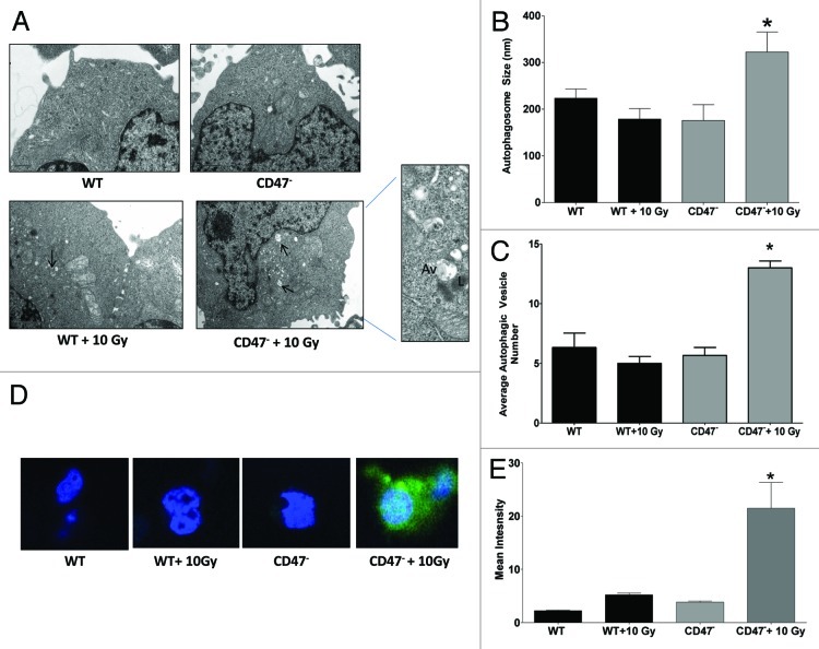
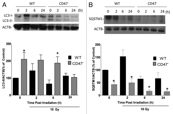
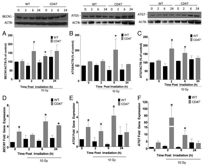
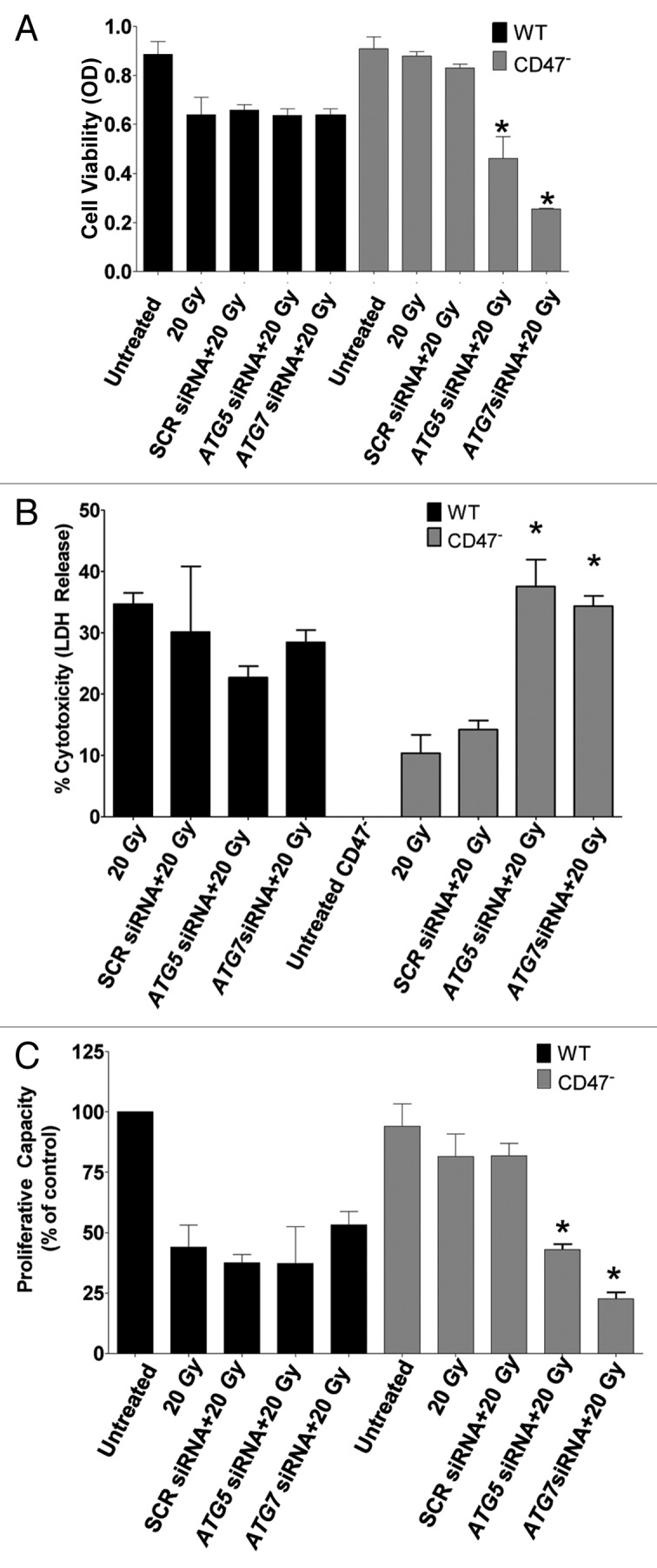
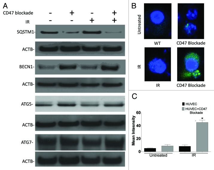
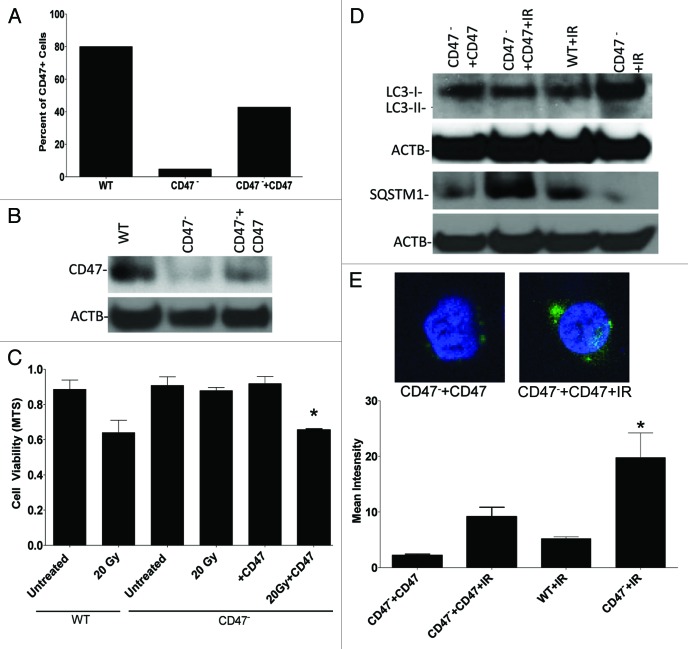
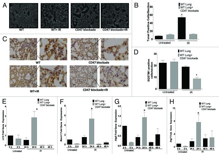
Accidental or therapeutic exposure to ionizing radiation has severe physiological consequences and can result in cell death. We previously demonstrated that deficiency or blockade of the ubiquitously expressed receptor CD47 results in remarkable cell and tissue protection against ischemic and radiation stress. Antagonists of CD47 or its ligand THBS1/thrombospondin 1 enhance cell survival and preserve their proliferative capacity. However the signaling pathways that mediate this cell-autonomous radioprotection are unclear. We now report a marked increase in autophagy in irradiated T-cells and endothelial cells lacking CD47. Irradiated T cells lacking CD47 exhibit significant increases in formation of autophagosomes comprising double-membrane vesicles visualized by electron microscopy and numbers of MAP1LC3A/B(+) puncta. Moreover, we observed significant increases in BECN1, ATG5, ATG7 and a reduction in SQSTM1/p62 expression relative to irradiated wild-type T cells. We observed similar increases in autophagy gene expression in mice resulting from blockade of CD47 in combination with total body radiation. Pharmacological or siRNA-mediated inhibition of autophagy selectively sensitized CD47-deficient cells to radiation, indicating that enhanced autophagy is necessary for the prosurvival response to CD47 blockade. Moreover, re-expression of CD47 in CD47-deficient T cells sensitized these cells to death by ionizing radiation and reversed the increase in autophagic flux associated with survival. This study indicates that CD47 deficiency confers cell survival through the activation of autophagic flux and identifies CD47 blockade as a pharmacological route to modulate autophagy for protecting tissue from radiation injury.

Keywords: ATG5; ATG7; BECN1; CD47; MAP1A/1BLC3; autophagosome; ionizing radiation; p62.


Figures

None
Figure 1. Radioprotection of CD47 Jurkat cells. (A) WT and CD47-deficient Jurkat cells were irradiated at the indicated doses, and cell viability was measured by MTS assay (n = 4; one-way ANOVA-Bonferroni post-test; *p < 0.05). (B) Cells were irradiated at 20 Gy, and cell cytotoxicity was measured by LDH release (n = 3; Student’s t-test, *p < 0.05). (C) Cells were irradiated at 20 Gy, and cell density was measured by crystal violet staining 10 d after radiation treatment (n = 3; one-way ANOVA-Bonferroni post-test *p < 0.05)
None
Figure 2. Deficiency of CD47 regulates autophagosome formation after IR. (A) Electron microscopy of Jurkat WT and CD47 6 h post IR. (B) Autophagic vacuole (Av) size was measured from photographs taken from sections and measured using ImageJ software (n = 3 for each condition; Student’s t-test, *p < 0.05). (C) (Av) number was measured in sections of cells differentiating from lysosomal (L) structures (n = 3 for each condition, one-way ANOVA-Bonferroni post-test *p < 0.05). (D) Representative confocal microscopy images and quantification (E) of Jurkat WT and CD47 transfected with GFP-LC3 6 h post-IR.
None
Figure 3. Deficiency of CD47 increases the autophagic flux. WT and CD47-deficient Jurkat T cells were cultured in complete medium and exposed to a 10 Gy dose of ionizing radiation. Cells were harvested at 0, 2, 6 and 24 h after IR. Protein expression was assessed by western blot hybridization using specific antibodies to LC3 (A) and SQSTM1 (B). Data were quantified by densitometry and normalizing the signal to that of ACTB/β-actin in the respective cells (n = 3; one-way ANOVA-Bonferroni post-test, *p < 0.05).
None
Figure 4. Deficiency of CD47 regulates BECN1, ATG5 and ATG7 gene expression. WT and CD47-deficient Jurkat T cells were cultured in complete medium and exposed to a 10 Gy dose of ionizing radiation. Cells were harvested at 0, 2, 6 and 24 h after IR. Protein and gene expression for BECN1 (A and D), ATG5 (B and E) and ATG7 (C and F) were measured by western blot hybridization and real time PCR, respectively (n = 3, Student’s t-test *p < 0.05).
None
Figure 5. Pharmacological inhibition of autophagy sensitizes CD47-deficient Jurkat T cells to IR. WT and CD47-deficient Jurkat T cells were plated in complete medium in 96-well plates. One hour prior to IR, cells were treated with 25 µM hydroxychloroquine (A) or 5 mM 3-MA (B). Cell viability was measured using MTS reagent after 72 h (n = 3 in triplicate, one-way ANOVA-Bonferroni post-test *p < 0.0001)
None
Figure 6. Silencing ATG5 and ATG7 sensitizes CD47-deficient Jurkat T cells to IR. WT and CD47 Jurkat T cells were plated in complete media and transfected with siRNAs against ATG5 and ATG7 or nonspecific scrambled control (SCR). Twenty-four hours after transfection cells were irradiated at 20 Gy. Cell viability was measured by MTS (A), cell cytotoxicity by LDH release (B), and proliferative capacity by crystal violet (n = 3 in triplicate; one-way ANOVA-Bonferroni post-test, *p < 0.001).
None
Figure 7. Blockade of CD47 increases autophagy in endothelial cells. HUVEC were plated in complete medium, and one group was treated with 10 µM CD47 antisense morpholino. After 48 h, cells were exposed to a 10 Gy dose of IR, and monolayers were harvested 6 h after IR exposure. (A) Representative images of protein expression of SQSTM1, BECN1, ATG7 and ATG5 in HUVEC measured by western blot hybridization. (B) Confocal microscopy images and quantification (C) of HUVEC treated in the same manner and transfected with GFP-LC3.
None
Figure 8. Re-expression of CD47 in CD47 Jurkat cells reverses radioprotection and reduces the autophagic flux. (A) Flow cytometry analysis using FITC-conjugated CD47 antibody B6H12 demonstrates re-expression of CD47 in CD47 Jurkat cells. (B) Western blot hybridization analysis of CD47 expression in Jurkat, CD47, and CD47 transfected with a plasmid expressing CD47. (C) Re-expression of CD47 in CD47 cells sensitizes this cell line to IR as measured by MTS assay (n = 3 one way ANOVA, *p < 0.04). (D) WT, CD47 and CD47 + CD47 and were cultured in complete medium and harvested before and after exposure to a 10 Gy dose of IR. Protein expression was assessed by western blot hybridization using LC3 and SQSTM1 antibodies. (E) Representative confocal images and quantitative analysis show LC3 puncta formation of cells transfected with GFP-LC3.
None
Figure 9. Suppression of CD47 upregulates autophagy and inhibits apoptotic lung tissue cell death in vivo after exposure to total body IR. WT mice were treated with saline or 10 µM CD47 morpholino. After 48 h of treatment, a group of each was exposed to TBI and sacrificed 24 h or 48 h after IR exposure. (A) TUNEL staining of representative sections of lung tissue from mice treated with saline and CD47 morpholino with or without exposure to IR fluorescent nuclei are indicative of apoptotic cells. (B) Quantitative analysis of TUNEL positive nuclei in lung tissue from control and morpholino-treated mice. (C) Immunohistochemical analysis and quantification (D) of SQSTM1 expression in lung tissue of control and irradiated mice treated with CD47 morpholino. Measurement of mRNA levels of LC3 (E) Becn1 (F), Atg5 (G), and Atg7 (H) in lung tissue of mice exposed to total body irradiation after 24 h otherwise noted at 48 h (n = 3, *p < 0.05).

Similar articles

Cited by

References

    1. Maxhimer JB, Soto-Pantoja DR, Ridnour LA, Shih HB, Degraff WG, Tsokos M, et al. Radioprotection in normal tissue and delayed tumor growth by blockade of CD47 signaling. Sci Transl Med. 2009;1:ra7. doi: 10.1126/scitranslmed.3000139. - DOI - PMC - PubMed
    1. Isenberg JS, Maxhimer JB, Hyodo F, Pendrak ML, Ridnour LA, DeGraff WG, et al. Thrombospondin-1 and CD47 limit cell and tissue survival of radiation injury. Am J Pathol. 2008;173:1100–12. doi: 10.2353/ajpath.2008.080237. - DOI - PMC - PubMed
    1. Roberts DD, Miller TW, Rogers NM, Yao M, Isenberg JS. The matricellular protein thrombospondin-1 globally regulates cardiovascular function and responses to stress via CD47. Matrix Biol. 2012;31:162–9. doi: 10.1016/j.matbio.2012.01.005. - DOI - PMC - PubMed
    1. Isenberg JS, Martin-Manso G, Maxhimer JB, Roberts DD. Regulation of nitric oxide signalling by thrombospondin 1: implications for anti-angiogenic therapies. Nat Rev Cancer. 2009;9:182–94. doi: 10.1038/nrc2561. - DOI - PMC - PubMed
    1. Isenberg JS, Qin Y, Maxhimer JB, Sipes JM, Despres D, Schnermann J, et al. Thrombospondin-1 and CD47 regulate blood pressure and cardiac responses to vasoactive stress. Matrix Biol. 2009;28:110–9. doi: 10.1016/j.matbio.2009.01.002. - DOI - PMC - PubMed

Publication types

MeSH terms