. 2012 Nov;26(11):4685-95.
doi: 10.1096/fj.12-212282. Epub 2012 Aug 8.

Snf1-related kinase inhibits colon cancer cell proliferation through calcyclin-binding protein-dependent reduction of β-catenin

Affiliations

Snf1-related kinase inhibits colon cancer cell proliferation through calcyclin-binding protein-dependent reduction of β-catenin

Amy K Rines et al. FASEB J. 2012 Nov.

Abstract

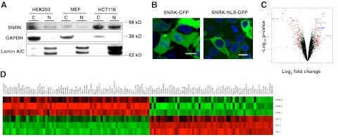
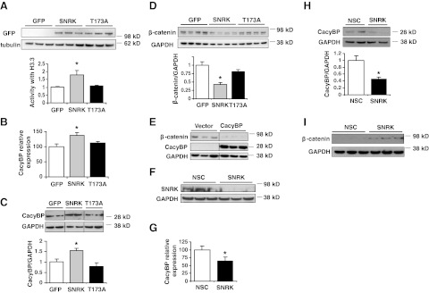
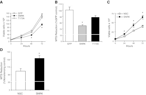
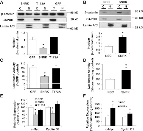
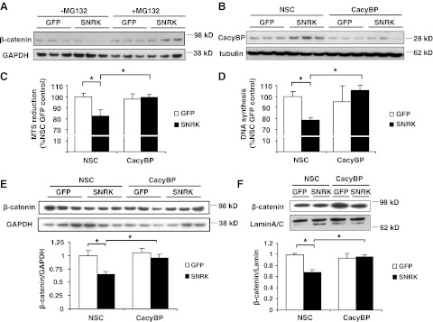
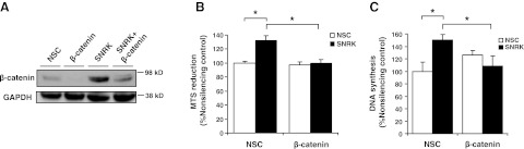
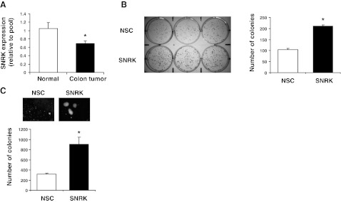
Sucrose nonfermenting 1 (Snf1)-related kinase (SNRK) is a serine/threonine kinase with sequence similarity to AMP-activated protein kinase (AMPK); however, its function is not well characterized. We conducted a gene array to determine which genes are regulated by SNRK. The array demonstrated that SNRK overexpression increased the levels of genes involved in cell proliferation, including calcyclin-binding protein (CacyBP), a member of the ubiquitin ligase complex that targets nonphosphorylated β-catenin for degradation. We confirmed that SNRK increased CacyBP mRNA and protein, and decreased β-catenin protein in HCT116 and RKO colon cancer cells. Furthermore, SNRK inhibited colon cancer cell proliferation, and CacyBP down-regulation reversed the SNRK-mediated decrease in proliferation and β-catenin. SNRK overexpression also decreased β-catenin nuclear localization and target gene transcription, and β-catenin down-regulation reversed the effects of SNRK knockdown on proliferation. SNRK transcript levels were reduced in human colon tumors compared to normal tissue by 35.82%, and stable knockdown of SNRK increased colon cancer cell tumorigenicity. Our results demonstrate that SNRK is down-regulated in colon cancer and inhibits colon cancer cell proliferation through CacyBP up-regulation and β-catenin degradation, resulting in reduced proliferation signaling. These findings reveal a novel function for SNRK in the regulation of colon cancer cell proliferation and β-catenin signaling.


Figures

Figure 1.
Figure 1.
SNRK is present in the nucleus and cytoplasm and alters the expression of genes associated with DNA synthesis and proliferation. A) SNRK protein levels in cytoplasmic (C) and nuclear (N) fractions from HEK 293, MEF, and HCT116 cells. GAPDH was used as a cytoplasmic loading control, and Lamin A/C was used as a nuclear loading control. B) Confocal microscopy images of HEK 293 cells transfected with wild-type SNRK (SNRK-GFP) and SNRK with a mutation in its NLS (SNRK-NLS-GFP). Scale bars = 10 μm. C) Volcano plot of the gene array. The volcano plot indicates the size of the biological effect (fold change) vs. the reproducibility of the result (statistical P value). Each gene is represented as a dot. Red dots represent the genes with P < 0.01, false discovery rate < 0.05, and fold change > 1.5. See Table 1 for gene ontology analysis based on biological processes. D) Heat map of the gene array. Each row is one sample; each column is one probe. Red represents higher expression than average, green represents lower expression than average; n = 3.
Figure 2.
Figure 2.
SNRK increases CacyBP levels and decreases β-catenin protein levels in colon cancer cells. A) Expression and activity of SNRK-GFP and SNRK-T173A-GFP in HCT116 cells; n = 3. B) Levels of CacyBP mRNA in HCT116 cells with overexpression of SNRK or controls; n = 9. C) Levels of CacyBP protein in HCT116 cells with overexpression of SNRK or controls. Top panel: representative Western blots (lanes have been rearranged from the same blot for clarity). Bottom panel: summary of densitometry; n ≥ 3. D) Levels of β-catenin protein in HCT116 cells with overexpression of SNRK or controls. Top panel: representative Western blots. Bottom panel: summary of densitometry; n = 7. E) Levels of β-catenin protein in HCT116 cells with overexpression of CacyBP or vector control (lanes shown are rearranged from the same blot). F) Western blot of SNRK protein in RKO cells treated with SNRK or nonsilencing control (NSC) shRNA. G) Levels of CacyBP mRNA with SNRK down-regulation in RKO cells; n = 10. H) Levels of CacyBP protein with SNRK down-regulation in RKO cells. Top panel: representative Western blots. Bottom panel: summary of densitometry; n = 3. I) Levels of β-catenin protein with SNRK down-regulation in RKO cells. *P < 0.05 vs. GFP and T173A or vs. NSC.
Figure 3.
Figure 3.
SNRK decreases colon cancer cell proliferation. A) Proliferation in HCT116 cells transfected with SNRK-GFP, SNRK-T173A-GFP, or GFP, as determined by viable cell counting with trypan blue exclusion at 24, 48, and 72 h; n ≥ 3. B) Proliferation in HCT116 cells after 72 h, as measured by MTS reduction; n ≥ 3. C) Proliferation in RKO cells treated with nonsilencing control (NSC) or SNRK shRNA, as measured by viable cell number at 24, 48, and 72 h; n = 3. D) Proliferation in RKO cells with down-regulation of SNRK at 72 h, as measured by MTS reduction; n = 3. *P < 0.05 vs. GFP and T173A or vs. NSC.
Figure 4.
Figure 4.
SNRK regulates β-catenin nuclear accumulation and β-catenin target expression in colon cancer cells. A, B) Top panels: Western blots of β-catenin in cytoplasmic (C) and nuclear (N) fractions in transfected HCT116 cells (A) and RKO cells treated with nonsilencing control (NSC) or SNRK shRNA (B). Bottom panels: summary of densitometry for nuclear β-catenin; n = 3. C) TOPflash luciferase reporter assay in transfected HCT116 cells; n ≥ 4. D) TOPflash luciferase reporter assay in RKO cells with SNRK down-regulation; n = 3. E) c-Myc and cyclin D1 mRNA expression in transfected HCT116 cells; n ≥ 10. F) c-myc and cyclin D1 mRNA expression in RKO cells with down-regulated SNRK; n ≥ 10. *P < 0.05 vs. GFP and T173A or vs. NSC.
Figure 5.
Figure 5.
CacyBP down-regulation reverses the decrease in proliferation caused by SNRK. A) Western blots of β-catenin protein in HCT116 cells transfected with GFP or SNRK and with or without 24 h of 5 μM MG132 treatment. B) Western blots of CacyBP protein in HCT116 cells transfected with GFP or SNRK and treated with CacyBP or nonsilencing control (NSC) siRNA. C, D) Proliferation measured by MTS reduction (C) and DNA synthesis measured by BrdU incorporation (D) in HCT116 cells transfected with SNRK or GFP and treated with CacyBP or NSC siRNA; n = 6. E, F) Top panels: Western blots of total β-catenin (E) and nuclear β-catenin (F) in HCT116 cells transfected with GFP or SNRK and treated with CacyBP or NSC siRNA. Bottom panels: summary of densitometry; n = 3. Legends indicate plasmids used; axis labels indicate siRNA used. CacyBP, CacyBP siRNA. *P < 0.05.
Figure 6.
Figure 6.
β-Catenin is necessary for the increase in proliferation caused by SNRK knockdown. A) Western blots of β-catenin protein in RKO cells treated with NSC or SNRK shRNA and NSC or β-catenin siRNA. NSC, treatment with NSC shRNA and siRNA; β-catenin, treatment with NSC shRNA and β-catenin siRNA; SNRK, treatment with SNRK shRNA and NSC siRNA; SNRK+β-catenin, treatment with SNRK shRNA and β-catenin siRNA. B) Proliferation as measured by MTS reduction in RKO cells treated with NSC or SNRK shRNA and NSC or β-catenin siRNA. Legend indicates shRNA used; axis labels indicate siRNA used; n ≥ 3. C) DNA synthesis as measured by BrdU incorporation in RKO cells treated with NSC or SNRK shRNA and NSC or β-catenin siRNA. Legend indicates shRNA used; axis labels indicate siRNA used; n ≥ 7. *P < 0.05.
Figure 7.
Figure 7.
SNRK is reduced in human colon cancer tissue, and stable SNRK knockdown increases the tumorigenicity of colon cancer cells in vitro. A) Quantitative real-time PCR using primers for human SNRK was conducted on samples from matched normal and tumor human colon tissue samples using an Origene TissueScan cDNA array; n = 22–23. B) Colony formation assay in RKO cells with stable knockdown of SNRK; n = 6. NSC, stable nonsilencing control shRNA treatment. C) Soft agar colony assay in RKO cells with stable knockdown of SNRK; n = 3. *P < 0.05.

Similar articles

Cited by

References

    1. Celenza J. L., Carlson M. (1984) Cloning and genetic mapping of SNF1, a gene required for expression of glucose-repressible genes in Saccharomyces cerevisiae. Mol. Cell. Biol. 4, 49–53 - PMC - PubMed
    1. Hardie D. G. (2007) AMP-activated/SNF1 protein kinases: conserved guardians of cellular energy. Nat. Rev. Mol. Cell. Biol. 8, 774–785 - PubMed
    1. Fogarty S., Hardie D. G. (2010) Development of protein kinase activators: AMPK as a target in metabolic disorders and cancer. Biochim. Biophys. Acta 1804, 581–591 - PubMed
    1. Wong A. K., Howie J., Petrie J. R., Lang C. C. (2009) AMP-activated protein kinase pathway: a potential therapeutic target in cardiometabolic disease. Clin. Sci. (Lond.) 116, 607–620 - PMC - PubMed
    1. Bright N. J., Thornton C., Carling D. (2009) The regulation and function of mammalian AMPK-related kinases. Acta Physiol. (Oxf.) 196, 15–26 - PubMed

Publication types

MeSH terms