. 2012 Dec;82(11):1231-5.
doi: 10.1038/ki.2012.277. Epub 2012 Aug 8.

A reproducible mouse model of chronic allograft nephropathy with vasculopathy

Affiliations

A reproducible mouse model of chronic allograft nephropathy with vasculopathy

Abolfazl Zarjou et al. Kidney Int. 2012 Dec.

Abstract

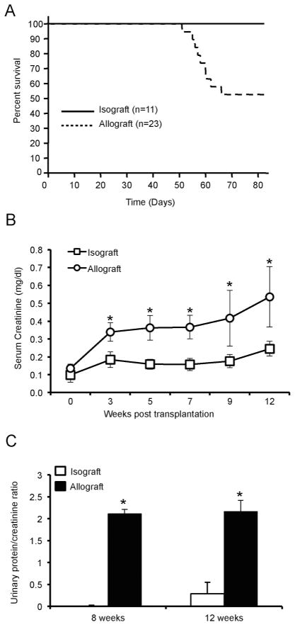
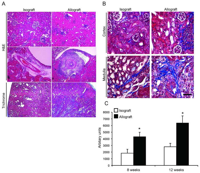
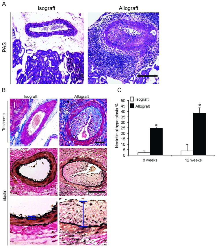
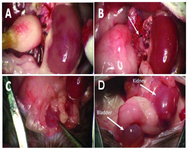
Although short-term outcomes in kidney transplantation have improved dramatically, long-term survival remains a major challenge. A key component of long-term, chronic allograft injury in solid organ transplants is arteriosclerosis characterized by vascular neointimal hyperplasia and inflammation. Establishing a model of this disorder would provide a unique tool not only to identify mechanisms of disease but also to test potential therapeutics for late graft injury. To this end, we utilized a mouse orthotopic renal transplant model in which C57BL/6J (H-2b) recipients were given either a kidney allograft from a completely mismatched Balb/cJ mouse (H-2d) or an isograft from a littermate. A unilateral nephrectomy was performed at the time of transplant followed by a contralateral nephrectomy on post-transplant day 7. Recipients were treated with daily cyclosporine subcutaneously for 14 days and then studied 8 and 12 weeks post transplantation. Renal function was significantly worse in allograft compared with isograft recipients. Moreover, the allografts had significantly more advanced tubulointerstitial fibrosis and profound vascular disease characterized by perivascular leukocytic infiltration and neointimal hyperplasia affecting the intrarenal blood vessels. Thus, we describe a feasible and reproducible murine model of intrarenal transplant arteriosclerosis that is useful to study allograft vasculopathy.


Figures

Figure 1
Figure 1. Survival and renal function in allograft and isograft recipients
(A) Survival rate was monitored after transplantation for 12 weeks. (B) Serum was separated from blood that was collected via retro-orbital puncture and serum creatinine was determined by LC-MS/MS at the indicated time points. (n=7/group, *P < 0.05). (C) Urine was collected at the time of sacrifice and urinary albumin and creatinine were determined. Data are represented as urinary albumin/creatinine ratio. (n=7/group, *P < 0.01).
Figure 2
Figure 2. Increased renal interstitial fibrosis in the allograft recipient group
(A) Histological analysis of renal isografts (left panels) and allografts (right panels). Isografts demonstrate preserved architecture (upper panels), normal intrarenal arteries (middle panels) and less fibrosis (lower panels). (B) Higher magnification of Masson Trichrome staining demonstrates the collagen deposition in the cortex (upper panels) and medulla (lower panels). Ten random fields (five cortical and five medullary) were selected from each graft recipient for digital quantification of collagen deposition. (n=7/group, *P < 0.05). Horizontal bar = 100 μm.
Figure 3
Figure 3. Perivascular leukocytic infiltrate and neointimal hyperplasia in allografts
(A) PAS-staining of renal allografts depicts significant perivascular mononuclear cell infiltrates and extensive intrarenal arterial neointimal hyperplasia. (B) Masson Trichrome staining was performed on kidney representative image showing severe neointimal hyperplasia and luminal obliteration that is exclusively found in the allografts (upper panels). Elastin stain was performed (middle panels) to demonstrate the extent of neointimal hyperplasia. Higher-magnification images of the neointimal layer (vertical bar) in the respective groups (lower panels). (D) Quantification of neointimal hyperplasia as described in the methods. *P < 0.05. Horizontal bar = 100 μm.
Figure 4
Figure 4. Mouse orthotopic kidney transplantation
(A) Transplanted kidney a few seconds following cross-clamp removal. Note the patchy red areas indicating restoration of normal blood flow. (B) The same kidney after full reperfusion. The upper suture line on the abdominal aorta can also be seen (arrow). (C) The forceps in the lower right corner indicates the suture line where the donor and recipient bladders are joined dome to dome. (D) Gross morphology of a kidney allograft and bladder at eight weeks post-transplantation. Note the presence of urine in the bladder.

Similar articles

Cited by

References

    1. Nankivell BJ, Kuypers DR. Diagnosis and prevention of chronic kidney allograft loss. Lancet. 2011;378:1428–1437. - PubMed
    1. Li C, Yang CW. The pathogenesis and treatment of chronic allograft nephropathy. Nat Rev Nephrol. 2009;5:513–519. - PubMed
    1. Djamali A, Premasathian N, Pirsch JD. Outcomes in kidney transplantation. Semin Nephrol. 2003;23:306–316. - PubMed
    1. Jevnikar AM, Mannon RB. Late kidney allograft loss: what we know about it, and what we can do about it. Clin J Am Soc Nephrol. 2008;3 (Suppl 2):S56–67. - PMC - PubMed
    1. Rahmani M, Cruz RP, Granville DJ, et al. Allograft vasculopathy versus atherosclerosis. Circ Res. 2006;99:801–815. - PubMed

Publication types