. 2012 Sep;139(18):3432-41.
doi: 10.1242/dev.080762. Epub 2012 Aug 8.

Atonal and EGFR signalling orchestrate rok- and Drak-dependent adherens junction remodelling during ommatidia morphogenesis

Affiliations

Atonal and EGFR signalling orchestrate rok- and Drak-dependent adherens junction remodelling during ommatidia morphogenesis

Francesca Robertson et al. Development. 2012 Sep.

Abstract

Morphogenesis of epithelial tissues relies on the interplay between cell division, differentiation and regulated changes in cell shape, intercalation and sorting. These processes are often studied individually in relatively simple epithelia that lack the complexity found during organogenesis when these processes might all coexist simultaneously. To address this issue, we are making use of the developing fly retinal neuroepithelium. Retinal morphogenesis relies on a coordinated sequence of interdependent morphogenetic events that includes apical cell constriction, localized alignment of groups of cells and ommatidia morphogenesis coupled to neurogenesis. Here, we use live imaging to document the sequence of adherens junction (AJ) remodelling events required to generate the fly ommatidium. In this context, we demonstrate that the kinases Rok and Drak function redundantly during Myosin II-dependent cell constriction, subsequent multicellular alignment and AJ remodelling. In addition, we show that early multicellular patterning characterized by cell alignment is promoted by the conserved transcription factor Atonal (Ato). Further ommatidium patterning requires the epidermal growth factor receptor (EGFR) signalling pathway, which transcriptionally governs rok- and Drak-dependent AJ remodelling while also promoting neurogenesis. In conclusion, our work reveals an important role for Drak in regulating AJ remodelling during retinal morphogenesis. It also sheds new light on the interplay between Ato, EGFR-dependent transcription and AJ remodelling in a system in which neurogenesis is coupled with cell shape changes and regulated steps of cell intercalation.


Figures
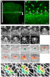
Fig. 1.
Fig. 1.
Multicellular patterning in the developing Drosophila eye. (A) WT Drosophila eye imaginal disc. E-Cad::GFP (green). The MF is indicated by a dashed line. An arrow indicates the direction of MF movement. Scale bar: 20 μm. (B) Magnification of the boxed region in A. At the posterior margin of the MF, cells form patterned structures referred to as arcs and lines (indicated by the arrowheads and asterisks, respectively). These multicellular structures are then patterned into 5-cell pre-clusters (arrow). Scale bar: 4 μm. (C) Reconstruction of the sequence of events involved in ommatidium assembly (a-i). a, line; b-e, line-to-arc transition. Type1-arcs mature towards a Type2-arc configuration (e). g is a 7-cell rosette, which resolves into a typical 6-cell rosette (n=7 out of 7 ommatidia examined in independent retinas) (h) and then into a 5-cell pre-cluster (i). Scale bars: 3 μm. A schematic of these stages has been illustrated in Ca′-i′. Lines of cells (pink, a′) form the outer cells of the arcs, which reside adjacent to the core cells (green, c′). The R8 (red, e′) is adjacent to the R2/R5 precursors (dark pink, e′), and adjacent to those are the R3/R4 (orange, e′). These cells (R8, R2, R5, R3 and R4) form the 5-cell pre-cluster (i′). (D-F) Stills from a time-lapse movie (supplementary material Movies 1, 3) of whole-mount pupae ubiquitously expressing E-Cad::GFP depicting a line of five cells (D), a 6-cell rosette configuration (E) and a 5-cell pre-cluster (F). Red is R8, the presumptive R2/R5 are yellow, the presumptive R3/R4 are green and the Mystery cell 1 is in turquoise (Wolff and Ready, 1976).
Fig. 2.
Fig. 2.
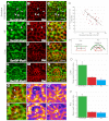
E-Cad, Baz and MyoII are planar polarized during ommatidia patterning. (A-C) Anterior compartment of a WT Drosophila eye disc stained for SerP19-MyoII (green) and Baz (red). Strong foci of Ser19P-MyoII at the ZA correspond with a reduction in Baz levels at the ZA (arrows). Arrowheads point to medial meshwork of MyoII. Scale bar: 4 μm. (D-F) MF of a WT eye disc stained for SerP19-MyoII (green) and Baz (red). Strong foci of Ser19P-MyoII correspond with a reduction in Baz levels at the ZA (arrowheads). Scale bar: 1 μm. (G) Quantification of the pixel intensity of Ser19P-MyoII and that of Baz at the ZA of MF cells. Each data point reflects a single pixel, measured randomly at the level of ZA across MF. (H) A schematic of a line and an arc. MyoII (green) is planar polarized in the posterior AJs of the lines and arcs, whereas E-Cad and Baz (red) are enriched in orthogonal, mediolateral AJs. (I-N) Ser19P-MyoII (green) and Baz (red) staining reveal a typical line (I-K) and a typical arc (L-N). Scale bars: 2 μm. (O,P) Quantification of the level of E-cad (red), SerP19MyoII (green) and Baz (blue) in posterior AJs versus mediolateral AJs, in lines (O) or arcs (P). For each protein, the average pixel intensity of all posterior AJs per ommatidium was compared with the average pixel intensity of all mediolateral AJs per ommatidium. Error bars indicate the s.d. between ommatidia. (Q-Q″) Representative transition phase from a type2-arc towards a 6-cell rosette (number of transitions monitored=9). SerP19-MyoII (green), Baz (red). MyoII is enriched at the AJs that are undergoing suppression (boxed area). White arrowheads point to AJs that are associated with SerP19-MyoII and that show a relative depletion of Baz (red). Images in Q′and Q″ were created using the Fire filter from FIJI. (R-R″) Representative 6-cell rosette in the process of becoming a 5-cell pre-cluster (number of transitions monitored=7). SerP19-MyoII (green), Baz (red). SerP19-MyoII is strongly associated with the central vertex of the rosette (white arrowhead). Images in R′ and R″ were created using the Fire filter from FIJI.
Fig. 3.
Fig. 3.
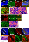
rok and Drak are required for MyoII-dependent multicellular patterning in Drosophila. (A-D) Clones expressing the mYFP-MyoIIDN transgene (blue) also stained for Arm (green) and Baz (red). B′ and C′ show a high magnification of the area expressing the mYFP-MyoIIDN transgene in A-D. The fire filter from FIJI is used to reveal the regions of high concentration versus low concentration of Baz (B′) and Arm (C′) at the cell’s ZA. (E-H2″′) rok2 clones are indicated by the absence of GFP (in blue). Green, Arm; red, Baz. White arrowheads point to abnormally configured ommatidial clusters (1 and 2) that are magnified in H1-H1″′ and H2-H2″′. H1′ and H2′ show the Arm channel and H1″ and H2″ show the Baz channel. The supernumerous AJs that should have been suppressed in order to accommodate the transition between type2-arc and 6-cell rosette are annotated with a red line in H1″′ and H2″′. (I-K) rok2/+, DrakDel whole mutant eyes stained for Arm (red) and SerP19MyoII (green). rok2/+ DrakDel mutant eyes display local instances of impaired constriction (indicated by asterisks). (L-O) rok2, DrakDel clones lack GFP (blue). Green, Arm; red, Baz. (P-S) Sagittal section of clones mutant for rok2, DrakDel (lacking blue). Green, DaPKC; red, Dlg. (T-W) rok2, DrakDel clone lacking GFP (blue). Green, Arm; red, Baz. Arrowheads indicate a developing ommatidium. (X) Cluster observed in the absence of rok and Drak function. Scale bars: 5 μm. Dashed lines outline the mutant tissue.
Fig. 4.
Fig. 4.
baz is required for AJ remodelling during ommatidia morphogenesis in Drosophila. (A-D) baz4 clone lacking GFP (blue). Green, Ser19P-MyoII; red, Arm. The arrowhead points to a line and the arrow points to an arc. Asterisks indicate type2 arcs having failed to generate 6-cell rosettes. Dashed lines outline the mutant tissue. (E-G) Higher magnification of the line and arc shown in A-D. A dotted line highlights the supra-cellular cable of MyoII at the posterior AJs. Ser19P-MyoII (F) and Arm (G). White arrows point to ectopic Ser19P-MyoII invading the mediolateral AJs. (H-K) Whole eyes mutant for bazGO484, lacking lacZ (indicated by β-galactosidase, blue). Green, Arm; red, Baz. Mutant clusters are denoted by the arrowheads 1-3. AJs separating R2 and R5 from the R8 take on a ‘Y’ conformation (arrowhead 1). More mature arcs elongate into a ‘string of paired cells’ (arrowhead 2). This defect leads to the loss of the common vertex normally shared between the R2, R8, and R5 precursors (arrowhead 3). A white asterisk marks a line emerging from the MF. (L,M) Higher magnification of ommatidia clusters 1 and 2, respectively. A dashed line highlights the ‘Y’ conformation found between the R8 and R2/5 neurons (L′,M′) Schematic of clusters 1 and 2, respectively. AJs that have failed to be suppressed during the transition from a type2-arc towards the 6-cell rosette configuration are depicted in red. Scale bars: 5 μm.
Fig. 5.
Fig. 5.
Ato is required to set up the posterior and mediolateral AJs in the wake of the MF in Drosophila. (A-D′) ato1 clone lacking GFP (blue). Green, Ser19P-MyoII; red, Baz. The dashed lines indicate the position of the MF. Dotted lines delineate the mutant tissue. (B′-D′) Higher magnification of the ato1 mutant clone shown in A-D. D′ shows Ser19P-MyoII (green) and Baz (red); B′ shows Baz channel; C′ shows Ser19P-MyoII channel. Arrowheads point to examples of AJs that are rich in Ser19P-MyoII and depleted for Baz. (E-G) ato1 mutant cells (arrowhead) lack GFP (red). Green, E-Cad. The WT MF is indicated by the arrow. (H,I) Higher magnification of the ato1 mutant clone shown in E-G. (J-M) Clones ectopically expressing UAS-ato in the anterior compartment of the eye disc lack GFP (blue). A dashed line marks the ato+/ato-interface. (N) Close up and schematic of the interface between the Ato+ and Ato– cells as seen in J-M and marked by the white arrowhead. From top to bottom: top panel is the GFP channel where the ato+/ato-interface is highlighted by the dashed line. The next panel shows Ser19P-MyoII, followed by Baz. The last panel is a drawing of the corresponding cells with WT cells (ato–) in blue, the MyoII cable in red and Ato+ cells in black. (O-Q) Clones ectopically expressing a UAS-ato transgene in the eye disc lack GFP (red). Green, E-Cad. Scale bars: 5 μm.
Fig. 6.
Fig. 6.
Ligand-dependent EGFR signalling orchestrates AJ remodelling during ommatidia patterning in Drosophila. (A-D) Whole eyes mutant for pntΔ88. Green, Ser19P-MyoII; red, Baz; blue, Arm. ‘Proto-arcs’ are denoted by the arrows. Ser19P-MyoII, Baz and Arm retain their WT polarization in lines (arrowheads) and ‘proto-arcs’ (arrows) in the MF. Two representative R8 cells are denoted by asterisks. (E-H) Higher magnification of the line and proto-arc depicted in A-D. Ser19P-MyoII (E), Baz (F), Arm (G), merge (H). A dashed line delineates the supra-cellular cable of Ser19P-MyoII. (I-L) rho,ru clones lack GFP (blue). Green, Ser19P-MyoII; red, Baz. The posterior boundary of the MF is denoted by a dashed line. Lines are denoted by the arrowhead. (M-P) WT MF stained for dp-ERK (MAPK; green), Ser19P-MyoII (red) and Arm (grey). Arm is in blue in P. White arrowheads point to lines. A dashed line delineates the posterior boundary of the MF (lines and arcs). Scale bars: 5 μm.
Fig. 7.
Fig. 7.
Coordinated function of Hh, Ato and EGFR signalling during ommatidia patterning. Genetic network involved in promoting neuroepithelial patterning in the developing fly retina. Hh and Dpp signalling transcriptionally promote apical cell constriction via Ci (Corrigall et al., 2007; Escudero et al., 2007; Schlichting and Dahmann, 2008). rok and Drak function redundantly during cell constriction by phosphorylating Myosin Regulatory Light Chain (MRLC; Sqh – FlyBase). Transcription of atonal is activated downstream of Hh/Dpp and this factor in turn upregulates E-Cad transcription at the posterior boundary of the MF (Brown et al., 2006). As a consequence of both cell constriction and E-Cad upregulation, cells in the MF present higher levels of E-Cad compared with their neighbours in the posterior compartment. We hypothesize that this leads to differential adhesion, a process that causes the accumulation of an acto-myosin cable at the interface (indicated by a dashed line) between high and low levels of ZA-associated E-Cad (Steinberg, 2007). In the posterior compartment (green) EGFR functions downstream of atonal during multicellular patterning in the eye (Brown et al., 2006) and transcriptionally promotes (via Pointed, Pnt) discrete steps of AJ remodelling that require both rok and Drak function.

Similar articles

Cited by

References

    1. Abramoff M. D., Magelhaes P. J., Ram S. J. (2004). Image processing with Image J. Biophotonics International 11, 36–42
    1. Baonza A., Casci T., Freeman M. (2001). A primary role for the epidermal growth factor receptor in ommatidial spacing in the Drosophila eye. Curr. Biol. 11, 396–404 - PubMed
    1. Bertet C., Sulak L., Lecuit T. (2004). Myosin-dependent junction remodelling controls planar cell intercalation and axis elongation. Nature 429, 667–671 - PubMed
    1. Blankenship J. T., Backovic S. T., Sanny J. S., Weitz O., Zallen J. A. (2006). Multicellular rosette formation links planar cell polarity to tissue morphogenesis. Dev. Cell 11, 459–470 - PubMed
    1. Brodu V., Casanova J. (2006). The RhoGAP crossveinless-c links trachealess and EGFR signaling to cell shape remodeling in Drosophila tracheal invagination. Genes Dev. 20, 1817–1828 - PMC - PubMed

Publication types

MeSH terms