. 2012 Oct;93(Pt 10):2171-2182.
doi: 10.1099/vir.0.043224-0. Epub 2012 Aug 8.

Identification and complete genome characterization of a novel picornavirus in turkey (Meleagris gallopavo)

Affiliations

Identification and complete genome characterization of a novel picornavirus in turkey (Meleagris gallopavo)

Ákos Boros et al. J Gen Virol. 2012 Oct.

Abstract

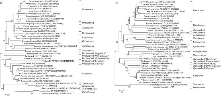
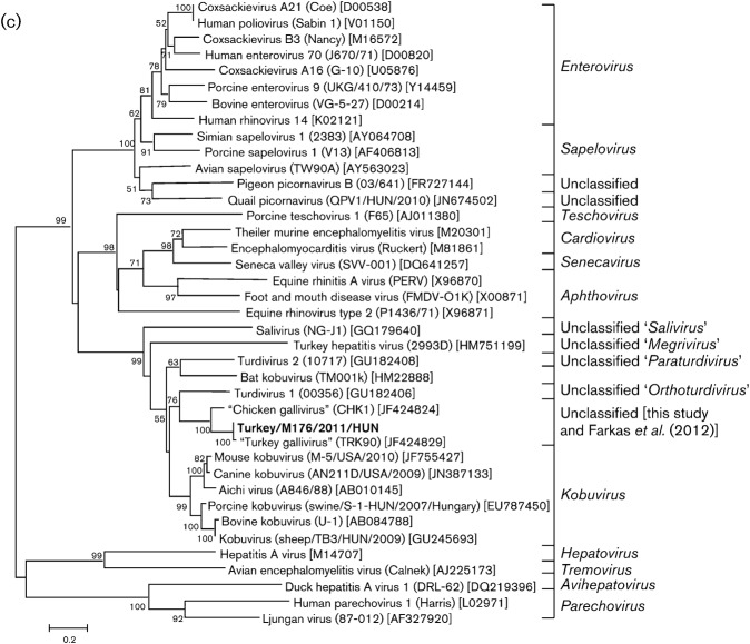
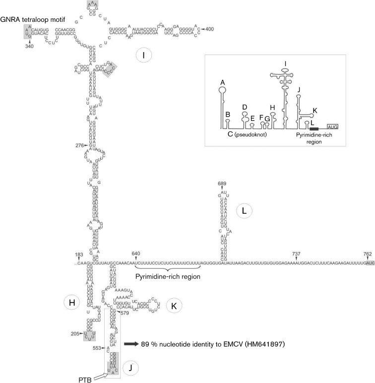
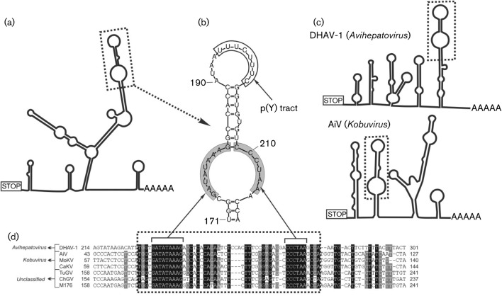
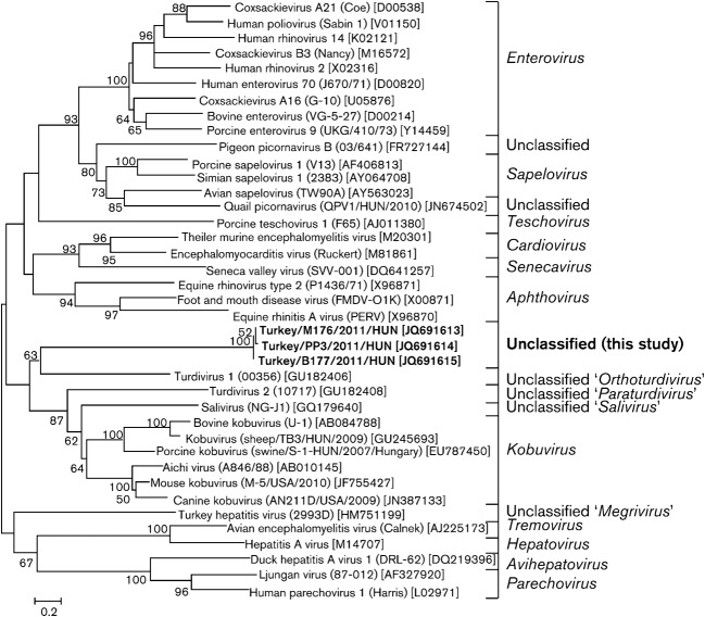
Members of the family Picornaviridae are important pathogens of humans and animals, although compared with the thousands of known bird species (>10 000), only a few (n = 11) picornaviruses have been identified from avian sources. This study reports the metagenomic detection and complete genome characterization of a novel turkey picornavirus from faecal samples collected from eight turkey farms in Hungary. Using RT-PCR, both healthy (two of three) and affected (seven of eight) commercial turkeys with enteric and/or stunting syndrome were shown to be shedding viruses in seven (88 %) of the eight farms. The viral genome sequence (turkey/M176/2011/HUN; GenBank accession no. JQ691613) shows a high degree of amino acid sequence identity (96 %) to the partial P3 genome region of a picornavirus reported recently in turkey and chickens from the USA and probably belongs to the same species. In the P1 and P2 regions, turkey/M176/2011/HUN is related most closely to, but distinct from, the kobuviruses and turdivirus 1. Complete genome analysis revealed the presence of characteristic picornaviral amino acid motifs, a potential type II-like 5' UTR internal ribosome entry site (first identified among avian-origin picornaviruses) and a conserved, 48 nt long 'barbell-like' structure found at the 3' UTR of turkey/M176/2011/HUN and members of the picornavirus genera Avihepatovirus and Kobuvirus. The general presence of turkey picornavirus - a novel picornavirus species - in faecal samples from healthy and affected turkeys in Hungary and in the USA suggests the worldwide occurrence and endemic circulation of this virus in turkey farms. Further studies are needed to investigate the aetiological role and pathogenic potential of this picornavirus in food animals.


Figures

Fig. 1.
Fig. 1.
Predicted genome organization of turkey picornavirus turkey/M176/2011/HUN (GenBank accession no. JQ691613). P1 represents viral structural proteins and P2 and P3 represent non-structural proteins. Nucleotide (upper numbers) and amino acid (lower numbers) lengths are indicated in each gene box. Conserved picornaviral amino acid motifs and predicted potential N-terminal cleavage sites along the coding region are indicated below and above the bar. Positions of the amino acid motifs are indicated by the position of the first amino acid of the motif. Above the gene box, sequence contigs acquired from pyrosequencing are indicated by grey bars.
Fig. 1.
Fig. 1.
Predicted genome organization of turkey picornavirus turkey/M176/2011/HUN (GenBank accession no. JQ691613). P1 represents viral structural proteins and P2 and P3 represent non-structural proteins. Nucleotide (upper numbers) and amino acid (lower numbers) lengths are indicated in each gene box. Conserved picornaviral amino acid motifs and predicted potential N-terminal cleavage sites along the coding region are indicated below and above the bar. Positions of the amino acid motifs are indicated by the position of the first amino acid of the motif. Above the gene box, sequence contigs acquired from pyrosequencing are indicated by grey bars.
Fig. 2.
Fig. 2.
Predicted RNA secondary structure of the IRES of the turkey picornavirus (turkey/M176/2011/HUN; GenBank accession no. JQ691613). The complete structure of the 5′ UTR, including domains A–L and the type II IRES, has been annotated as proposed previously (inset). The central 5′ IRES domains of turkey picornavirus are labelled from H to L to maintain the continuity of the current nomenclature. The positions of conserved type II IRES motifs and the predicted polyprotein AUG start codon are indicated by shaded boxes. Nucleotide sequence similarity in the apical region of domain J between EMCV (genus Cardiovirus) and turkey picornavirus is boxed and indicated by a black arrow). Analogous to EMCV, possible specific binding between domain J and PTB is indicated by a white arrow.
Fig. 3.
Fig. 3.
Conserved motif analysis of the 3′ UTR genome region. (a, c) The predicted secondary 3′ UTR structures of turkey/M176/2011/HUN (a) and of representative members of the genera Avihepatovirus and Kobuvirus (c). Dotted boxes indicate the conserved ‘barbell-like’ structure. (b) Detailed structure of the ‘barbell-like’ formation of the study sequence 3′ UTR. Grey boxes indicate identical nucleotides of different picornavirus species. (d) Sequence alignment of partial 3′ UTRs including the ‘barbell-like’ structure (dotted box) of duck hepatitis A virus 1 (DHAV-1; GenBank accession no. DQ249299), Aichi virus (AiV; FJ890523), mouse kobuvirus (MoKV; JF755427), canine kobuvirus (CaKV; JN088541), turkey gallivirus (TuGV; JF424829), chicken gallivirus (ChGV; JF424824) and the study sequence M176 (turkey/M176/2011/HUN; JQ691613). Note that the numbering of the 3′ UTR alignment begins from the STOP codon.
Fig. 4.
Fig. 4.
Phylogenetic relationship between turkey picornavirus (turkey/M176/2011/HUN; GenBank accession no. JQ691613; indicated in bold), representative members of the 12 picornavirus genera and unassigned picornaviruses, based on amino acid sequences of the different picornavirus coding regions: P1 (a), P2 (b) and P3 (c).
Fig. 5.
Fig. 5.
Unrooted phylogenetic tree based on the deduced VP1 capsid protein sequences of the study sequences (in bold) and representative members of the family Picornaviridae.

References

    1. Alexandersen S., Knowles N. J., Dekker A., Belsham G. J., Zhang Z., Koenen F. (2012). Picornaviruses. In Diseases of Swine, 10th edn, pp. 587–620 Edited by Zimmerman J. J., Karriker L. A., Ramirez A., Schwartz K. J., Stevenson G. W. Chichester, UK: Wiley
    1. Bailey D., Karakasiliotis I., Vashist S., Chung L. M., Rees J., McFadden N., Benson A., Yarovinsky F., Simmonds P., Goodfellow I. (2010). Functional analysis of RNA structures present at the 3′ extremity of the murine norovirus genome: the variable polypyrimidine tract plays a role in viral virulence. J Virol 84, 2859–2870 10.1128/JVI.02053-09 - DOI - PMC - PubMed
    1. Bazan J. F., Fletterick R. J. (1988). Viral cysteine proteases are homologous to the trypsin-like family of serine proteases: structural and functional implications. Proc Natl Acad Sci U S A 85, 7872–7876 10.1073/pnas.85.21.7872 - DOI - PMC - PubMed
    1. Blom N., Hansen J., Brunak S., Blaas D. (1996). Cleavage site analysis in picornaviral polyproteins: discovering cellular targets by neural networks. Protein Sci 5, 2203–2216 10.1002/pro.5560051107 - DOI - PMC - PubMed
    1. Boros Á., Pankovics P., Simmonds P., Reuter G. (2011). Novel positive-sense, single-stranded RNA (+ssRNA) virus with di-cistronic genome from intestinal content of freshwater carp (Cyprinus carpio). PLoS ONE 6, e29145 10.1371/journal.pone.0029145 - DOI - PMC - PubMed

Publication types

Substances

Associated data