

Dendritic morphology of pyramidal neurons in the chimpanzee neocortex: regional specializations and comparison to humans
- PMID: 22875862
- PMCID: PMC3767963
- DOI: 10.1093/cercor/bhs239
Dendritic morphology of pyramidal neurons in the chimpanzee neocortex: regional specializations and comparison to humans
Abstract
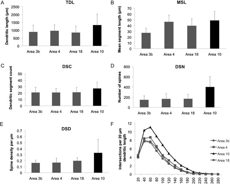
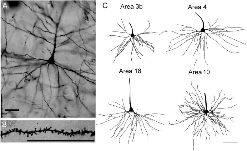
The primate cerebral cortex is characterized by regional variation in the structure of pyramidal neurons, with more complex dendritic arbors and greater spine density observed in prefrontal compared with sensory and motor cortices. Although there are several investigations in humans and other primates, virtually nothing is known about regional variation in the morphology of pyramidal neurons in the cerebral cortex of great apes, humans' closest living relatives. The current study uses the rapid Golgi stain to quantify the dendritic structure of layer III pyramidal neurons in 4 areas of the chimpanzee cerebral cortex: Primary somatosensory (area 3b), primary motor (area 4), prestriate visual (area 18), and prefrontal (area 10) cortex. Consistent with previous studies in humans and macaque monkeys, pyramidal neurons in the prefrontal cortex of chimpanzees exhibit greater dendritic complexity than those in other cortical regions, suggesting that prefrontal cortical evolution in primates is characterized by increased potential for integrative connectivity. Compared with chimpanzees, the pyramidal neurons of humans had significantly longer and more branched dendritic arbors in all cortical regions.
Keywords: Golgi; area 10; dendrites; evolution; primate cerebral cortex.
Figures


References
-
- Amici F, Aureli F, Call J. Fission-fusion dynamics, behavioral flexibility, and inhibitory control in primates. Curr Biol. 2008;18:1415–1419. - PubMed
-
- Anderson B, Rutledge V. Age and hemisphere effects on dendritic structure. Brain. 1996;119:1983–1990. - PubMed
-
- Anderson K, Yamamoto E, Kaplan J, Hannan M, Jacobs B. Neurolucida Lucivid versus Neurolucida camera: A quantitative and qualitative comparison of three-dimensional neuronal reconstructions. J Neurosci Methods. 2010;186:209–214. - PubMed
-
- Beran MJ, Savage-Rambaugh ES, Pate JL, Rumbaugh DM. Delay of gratification in chimpanzees. Devel Psychobiol. 1999;34:119–127. - PubMed
Publication types
MeSH terms
Grants and funding
LinkOut - more resources
Full Text Sources
Miscellaneous
