. 2012 Oct 4;87(4):81.
doi: 10.1095/biolreprod.112.102293. Print 2012 Oct.

DBA-lectin reactivity defines mouse uterine natural killer cell subsets with biased gene expression

Affiliations

DBA-lectin reactivity defines mouse uterine natural killer cell subsets with biased gene expression

Zhilin Chen et al. Biol Reprod. .

Abstract

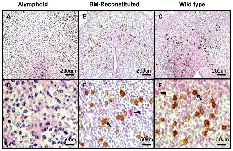
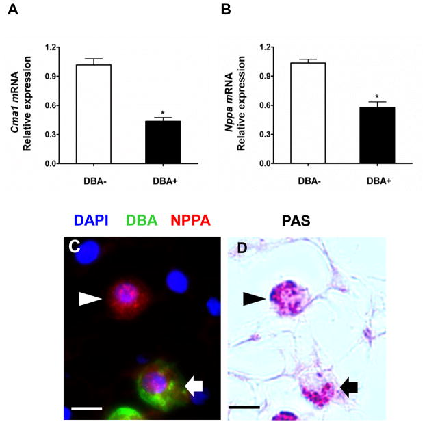
Endometrial decidualization, a process essential for blastocyst implantation in species with hemochorial placentation, is accompanied by an enormous but transient influx of natural killer (NK) cells. Mouse uterine NK (uNK) cell subsets have been defined by diameter and cytoplasmic granule number, reflecting stage of maturity, and by histochemical reactivity with Periodic Acid Schiff (PAS) reagent with or without co-reactivity with Dolichos biflorus agglutinin (DBA) lectin. We asked whether DBA- and DBA+ mouse uNK cells were equivalent using quantitative RT-PCR analyses of flow-separated, midpregnancy (Gestation Day [gd] 10) cells and immunohistochemistry. CD3E (CD3)-IL2RB (CD122)+DBA cells were identified as the dominant Ifng transcript source. Skewed IFNG production by uNK cell subsets was confirmed by analysis of uNK cells from eYFP-tagged IFNG-reporter mice. In contrast, CD3E-IL2RB+DBA+ uNK cells expressed genes compatible with significantly greater potential for IL22 synthesis, angiogenesis, and participation in regulation mediated by the renin-angiotensin system (RAS). CD3E-IL2RB+DBA+ cells were further divided into VEGFA+ and VEGFA- subsets. CD3E-IL2RB+DBA+ uNK cells but not CD3E-IL2RB+DBA- uNK cells arose from circulating, bone marrow-derived progenitor cells by gd6. These findings indicate the heterogeneous nature of mouse uNK cells and suggest that studies using only DBA+ uNK cells will give biased data that does not fully represent the uNK cell population.


Conflict of interest statement

The authors have no conflicting financial interests.

Figures

Figure 1
Figure 1. DBA+ but not DBA− uNK cells arise from trafficked, BM-derived progenitors
Gd6.5 DB dually stained with PAS and DBA lectin. Mesometrial side of the uterus is to top. (A, D) Alymphoid Rag2−/−Il2rg−/− DB lacks PAS+DBA− and PAS+DBA+ uNK cells. (B, E) uNK cells in alymphoid Rag2−/−Il2rg−/− reconstituted with BALB/c BM 3 wk previous to mating were PAS+DBA+; PAS+DBA− cells were very rare (1/230 cells-0.0044% total uNK cells). (C, F) About 50% of uNK cells in gd6.5 BALB/c+/+ implant sites are PAS+DBA−, 50% are PAS+DBA+. Arrowheads-PAS+DBA−; Arrows-PAS+DBA+; Size bars 200 μm in (A–C); 50 μm in (D–F).
Figure 2
Figure 2. Expression of Ifng by gd9.5-10.5 uNK cells
(A) Relative Ifng mRNA expression by sorted gd10.5 CD3E-IL2RB+DBA− and CD3E-IL2RB+CD122+DBA+ cells using qRT-PCR normalized to Hprt. Data are means±SEM from cells prepared in three independent sorting experiments. ** P<0.01. (B–C) show the same microscope field with (B) a 3 color merged image shows DAPI, FITC-DBA+ uNK cells and Alexa Fluor® 594-IFNG, (C) shows post-fluorescence staining with PAS to identify all uNK cells. PAS+DBA− uNK cells show intense IFNG staining. PAS+ DBA+ uNK cells are weakly reactive. (D) CD3E-122+ cells from DB and MLAp of gd9 Yeti implantation sites were analysed for ITGA2 (DX5) and KLRB1C (NK1.1). The less frequent ITGA2+ KLRB1C+ cell population, defined previously as DBA− [14], was the strongly YFP+ cell population. Arrowheads-PAS+DBA-cells; Arrows-PAS+DBA+ cells; Size bar (B–C) 20μm.
Figure 3
Figure 3. Expression of IL22 and PGF by gd10.5 uNK cells
Relative mRNA expression of Il22 (A) and Pgf (D) by sorted CD3E-IL2RB+DBA− and CD3E-IL2RB+DBA+ decidual cells using qRT-PCR normalized to Hprt. Data are means±SEM from cells prepared in three independent sorting experiments. * P<0.05, ** P<0.01. (B–C) show the same microscope field with (B) a 3 color merged image showing DAPI, FITC-DBA+ uNK cells and Alexa Fluor® 594-IL22. (C) shows post-fluorescence staining of these cells with PAS to identify all uNK cells. (E–F) show the same images of a different microscope field with (E) a 3 color merged image showing DAPI, FITC-DBA+ uNK cells and Alexa Fluor® 594-PGF. (F) shows post-fluorescence staining with PAS to identify all uNK cells. Arrowheads-PAS+DBA-cells; Arrows-PAS+DBA+ cells; Size bar represents 20μm.
Figure 4
Figure 4. Expression of VEGFA by uNK cells
(A) Relative Vegfa mRNA expression by CD3E-IL2RB+DBA− and CD3E-IL2RB+DBA+ gd10.5 decidual cells using by qRT-PCR normalized to Hprt. Data are means±SEM from cells prepared in three independent sorting experiments. * P<0.05. (B–C) show the same microscope field with (B) a 3 color merged image showing DAPI, FITC-DBA+ uNK cells and Alexa Fluor® 594-VEGFA. (C) shows post-fluorescence staining with PAS to identify all uNK cells. Arrowheads-PAS+DBA-cells; Arrows-PAS+DBA+ cells. (D) summarizes DBA lectin and VEFGA reactive DAPI+ mesometrial cells in BALB/c implant sites at gd7, 9 and14. White bar DBA+VEGFA−; Grey bar DBA+VEGFA+; Black bar DBA-VEGFA+ cells (gd7, 22 fields from 2 pregnancies; gd9, 43 fields from 3 pregnancies and gd14, 61 fields from 2 pregnancies scored at 200X magnification). * P<0.05, *** P<0.001. (E) Dually stained DB showing in (Ei) PRF1 (perforin, red) that is associated with DBA+ granules (green). Many but not all cells expressed PRF1. (Eii) shows uNK cells containing DBA+ cytoplasmic granules and non granule-associated VEGFA. (Eiii) co-localizes PRF1granules and plasma membrane-associated VEGFA (pseudocolored violet) co-localized to single DBA+ uNK cells. Size bars are 20 μm.
Figure 5
Figure 5. Expression of Cma1 and NPPA by gd10.5 uNK cells
Relative mRNA expression by DBA− and DBA+ gd10 CD3E-IL2RB+ decidual cells of (A) Cma1 and (B) Nppa using qRT-PCR normalized to Hprt expression. Data are mean±SEM from cells prepared in three independent sorting experiments. * P<0.05. (C–D) show the same microscope field with (C) a 3 color merged image showing DAPI, FITC-DBA+ uNK cells and Alexa Fluor® 594-NPPA. (D) shows post-fluorescence staining with PAS to identify all uNK cells. PAS+DBA- uNK cells show more NPPA reactivity than PAS+DBA-cells but both subsets stain. Arrowheads-PAS+DBA-cells; Arrows-PAS+DBA+ cells; Size bar (C–D) 20μm
Figure 6
Figure 6. Expression of REN1 and ACE by gd10.5 uNK cells
Relative mRNA expression of Ren1 (A) and Ace (D) by sorted CD3E-IL2RB+DBA− and CD3E-IL2RB+DBA+ decidual cells by qRT-PCR normalized to Hprt. Data are means±SEM from cells prepared in three independent sorting experiments. * P<0.05, ** P<0.01. (B–C) show the same microscope field with (B) a 3 color merged image showing DAPI, FITC-DBA+ uNK cells and Alexa Fluor® 594–REN1. (C) shows post-fluorescence staining with PAS to identify all uNK cells. REN1 reactivity was largely localized over uNK cells with random weaker stromal staining. (E–F) show the same microscope field from a different section showing (E) a 3 color merged image showing DAPI, FITC-DBA+ uNK cells and Alexa Fluor® 594-ACE. (F) post-fluorescence staining with PAS to identify all uNK cells. ACE expression was almost exclusively over uNK cells. Arrowheads-PAS+DBA-cells; Arrows-PAS+DBA+ cells; Size bars represent 20μm.

Similar articles

Cited by

References

    1. Cherrier M, Ohnmacht C, Cording S, Eberl G. Development and function of intestinal innate lymphoid cells. Curr Opin Immunol. 2012;24:227–283. - PubMed
    1. Croy BA, van den Heuvel MJ, Borzychowski AM, Tayade C. Uterine natural killer cells: a specialized differentiation regulated by ovarian hormones. Immunol Rev. 2006;214:161–185. - PubMed
    1. Peel S. Granulated metrial gland cells. Adv Anat Embryol Cell Biol. 1989;115:1–112. - PubMed
    1. Paffaro VA, Jr, Bizinotto MC, Joazeiro PP, Yamada AT. Subset classification of mouse uterine natural killer cells by DBA lectin reactivity. Placenta. 2003;24:479–488. - PubMed
    1. Parr EL, Young LH, Parr MB, Young JD. Granulated metrial gland cells of pregnant mouse uterus are natural killer-like cells that contain perforin and serine esterases. J Immunol. 1990;145:2365–2372. - PubMed

Publication types

MeSH terms