. 2012 Aug 8;32(32):10833-40.
doi: 10.1523/JNEUROSCI.5628-11.2012.

Chronic-pain-associated astrocytic reaction in the spinal cord dorsal horn of human immunodeficiency virus-infected patients

Affiliations

Chronic-pain-associated astrocytic reaction in the spinal cord dorsal horn of human immunodeficiency virus-infected patients

Yuqiang Shi et al. J Neurosci. .

Abstract

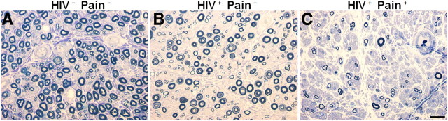
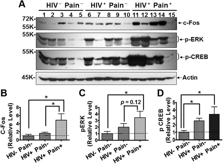
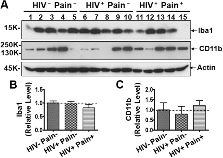
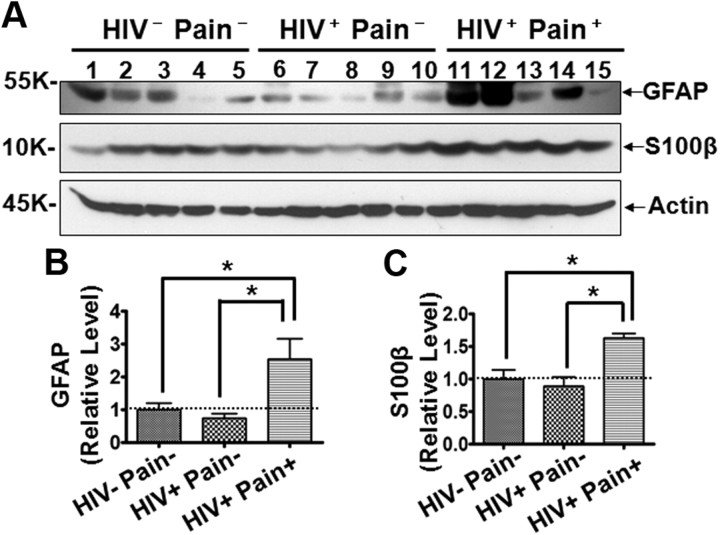
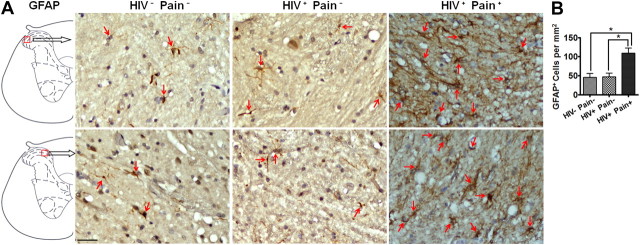
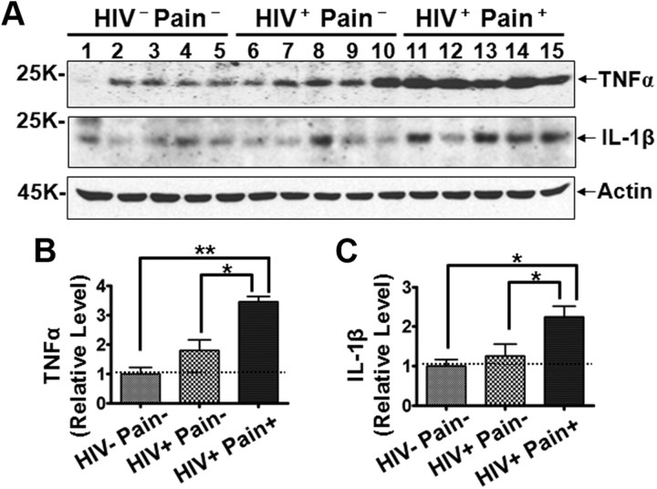
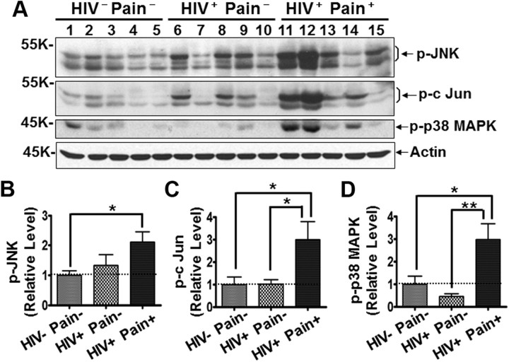
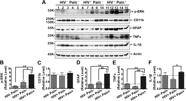
Studies with animal models have suggested that reaction of glia, including microglia and astrocytes, critically contributes to the development and maintenance of chronic pain. However, the involvement of glial reaction in human chronic pain is unclear. We performed analyses to compare the glial reaction profiles in the spinal dorsal horn (SDH) from three cohorts of sex- and age-matched human postmortem tissues: (1) HIV-negative patients, (2) HIV-positive patients without chronic pain, and (3) HIV patients with chronic pain. Our results indicate that the expression levels of CD11b and Iba1, commonly used for labeling microglial cells, did not differ in the three patient groups. However, GFAP and S100β, often used for labeling astrocytes, were specifically upregulated in the SDH of the "pain-positive" HIV patients but not in the "pain-negative" HIV patients. In addition, proinflammatory cytokines, TNFα and IL-1β, were specifically increased in the SDH of pain-positive HIV patients. Furthermore, proteins in the MAPK signaling pathway, including pERK, pCREB and c-Fos, were also upregulated in the SDH of pain-positive HIV patients. Our findings suggest that reaction of astrocytes in the SDH may play a role during the maintenance phase of HIV-associated chronic pain.


Figures

Figure 1.
Figure 1.
Examples of myelinated nerve fiber loss in distal sural nerves. Semithin transverse sections of three nerves obtain at autopsy were fixed in 4% glutaraldehyde, embedded in plastic, and stained with toluidine blue. Round myelinated nerve fibers are stained blue. Decreasing endoneurial myelinated nerve fiber density in the nerves is illustrated. The nerve in A is from an HIV-negative control (subject 3) and contains a density of myelinated nerve fibers within the normal range. The nerve in B is from an HIV-infected patient (subject 9) who did not have HIV-associated distal sensory peripheral neuropathy clinically and did not report having pain symptoms. The density of myelinated nerve fibers shows possible slight nerve fiber loss that is diagnostically borderline and within the normal limits. The nerve in C is an abnormal specimen from a subject with HIV-associated distal sensory peripheral neuropathy who reported having neuropathic pain symptoms (subject 15). The nerve contains a markedly lower density of myelinated nerve fibers and severe nerve fiber loss, consistent with marked chronic axonal neuropathy (compare C with A or B). The normal range of sural nerve fiber dropout is highly variable in human populations; consequently, one cannot distinguish a patient with pain versus one without pain neuropathologically. Scale bar: 20 μm.
Figure 2.
Figure 2.
Levels of neuronal activity-regulated proteins in the SDH. A, Increased c-Fos, p-ERK, and p-CREB revealed by immunoblotting. Shown is the same membrane blotted with different antibodies after stripping; same for blots in other figures, unless indicated otherwise. B–D, Quantitative summary of c-Fos (B), p-ERK (C), and p-CREB (D). Error bars indicate SEM. *p < 0.05.
Figure 3.
Figure 3.
Expression of Iba1 and CD11b in the SDH. A, Immunoblots of Iba1 and CD11b. B, C, Quantitative summary of Iba1 (B) and CD11b (C). Error bars indicate SEM.
Figure 4.
Figure 4.
Levels of GFAP and S100β proteins in the SDH. A, Immunoblots of GFAP and S100β. B, C, Quantitative summary of GFAP (B) and S100β (C). Error bars indicate SEM. *p < 0.05.
Figure 5.
Figure 5.
Increase of astrocytes in the SDH of pain-positive HIV patients revealed by immunohistochemistry. A, Representative images of GFAP staining from two different patients for each group. B, The density of GFAP-positive (brown) cells in the SDH. To facilitate cell counting, the sections were counterstained to label the nuclei (blue, arrows). Staining of the sections from the HIV Pain, HIV+ Pain, and HIV+ Pain+ patients was performed in parallel to keep the same experimental conditions among different groups. The dark staining of the HIV+ Pain+ sections was due to high GFAP expression. Scale bar, 50 μm. Error bars indicate SEM. *p < 0.05.
Figure 6.
Figure 6.
Expression levels of TNFα and IL-1β in the SDH. A, Immunoblots of TNFα and IL-1β. B, C, Quantitative summary of TNFα (B) and IL-1β (C). Error bars indicate SEM. *p < 0.05; **p < 0.01.
Figure 7.
Figure 7.
MAPK signaling in the SDH. A, Immunoblots of phosphorylated JNK (p-JNK), phosphorylated c-Jun (p-c-Jun), and phosphorylated p38 (p-p38 MAPK). B–D, Quantitative summary of p-JNK (B), p-c-Jun (C), and p-p38 MAPK (D). Error bars indicate SEM. *p < 0.05; **p < 0.01.
Figure 8.
Figure 8.
Characterization of molecular markers in the cervical SDH. A, Immunoblots of phosphorylated ERK (p-ERK), CD11b, GFAP, TNFα, and IL-1β. B–F, Quantitative summary of p-ERK (B), CD11b (C), GFAP (D), TNFα (E), and IL-1β (F). Error bars indicate SEM. *p < 0.05; **p < 0.01.

References

    1. Cao H, Gao YJ, Ren WH, Li TT, Duan KZ, Cui YH, Cao XH, Zhao ZQ, Ji RR, Zhang YQ. Activation of extracellular signal-regulated kinase in the anterior cingulate cortex contributes to the induction and expression of affective pain. J Neurosci. 2009;29:3307–3321. - PMC - PubMed
    1. Che Y, Yu YM, Han PL, Lee JK. Delayed induction of p38 MAPKs in reactive astrocytes in the brain of mice after KA-induced seizure. Brain Res Mol Brain Res. 2001;94:157–165. - PubMed
    1. Chung IY, Benveniste EN. Tumor necrosis factor-α production by astrocytes. Induction by lipopolysaccharide, IFN-γ, and IL-1β. J Immunol. 1990;144:2999–3007. - PubMed
    1. Clark AK, Gentry C, Bradbury EJ, McMahon SB, Malcangio M. Role of spinal microglia in rat models of peripheral nerve injury and inflammation. Eur J Pain. 2007a;11:223–230. - PubMed
    1. Clark AK, Yip PK, Grist J, Gentry C, Staniland AA, Marchand F, Dehvari M, Wotherspoon G, Winter J, Ullah J, Bevan S, Malcangio M. Inhibition of spinal microglial cathepsin S for the reversal of neuropathic pain. Proc Natl Acad Sci U S A. 2007b;104:10655–10660. - PMC - PubMed

Publication types

MeSH terms