. 2012 Aug 8;32(32):11082-94.
doi: 10.1523/JNEUROSCI.0064-12.2012.

Anti-NMDA receptor encephalitis antibody binding is dependent on amino acid identity of a small region within the GluN1 amino terminal domain

Affiliations

Anti-NMDA receptor encephalitis antibody binding is dependent on amino acid identity of a small region within the GluN1 amino terminal domain

Amy J Gleichman et al. J Neurosci. .

Abstract

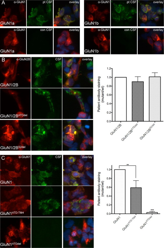
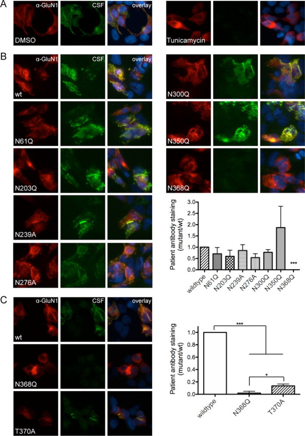
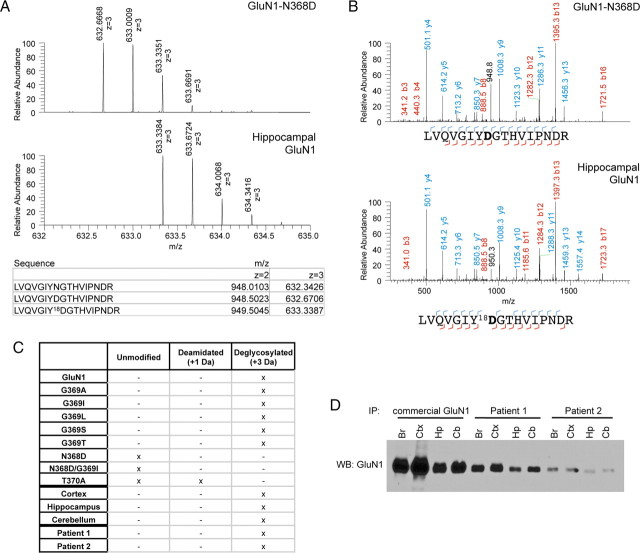
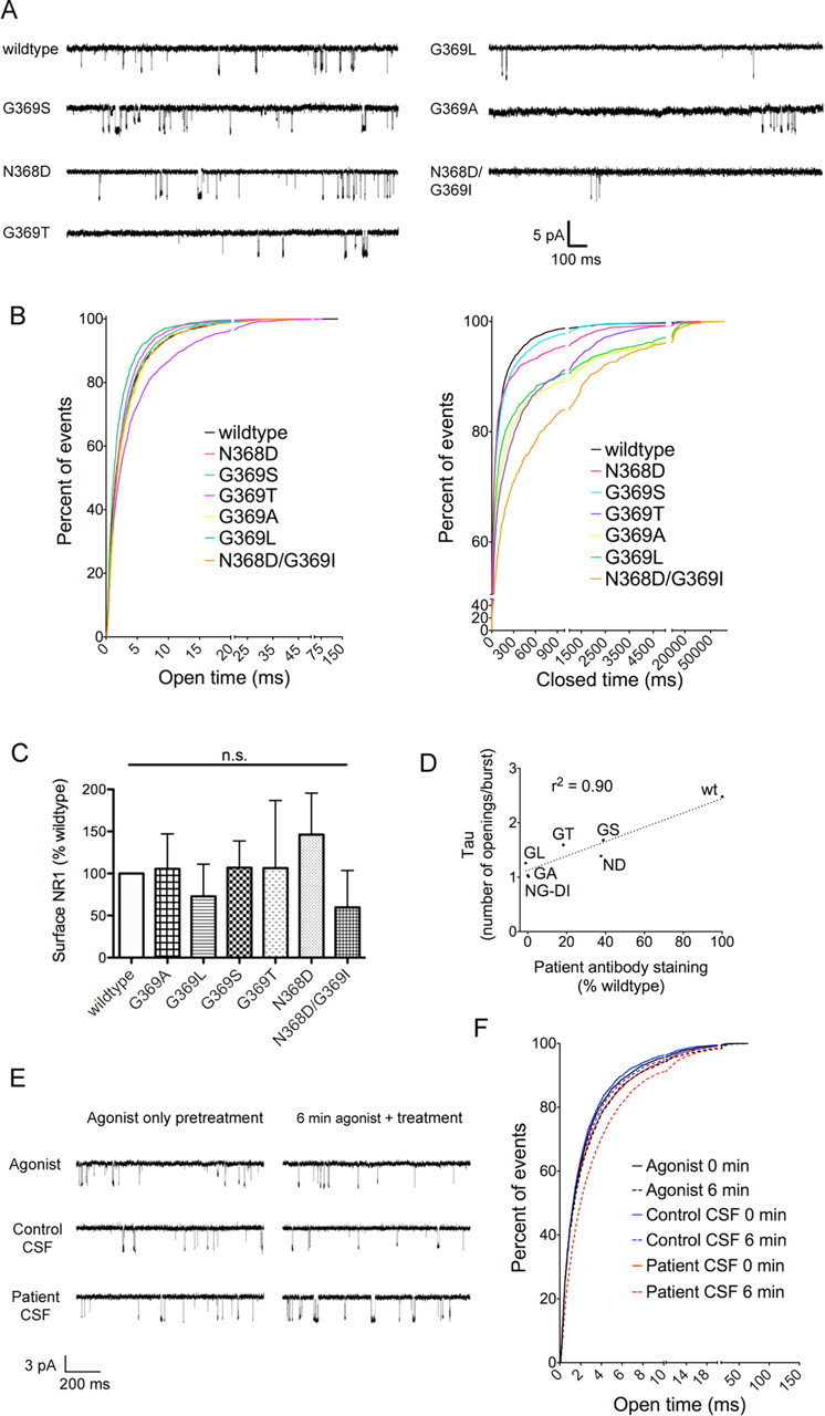
Anti-NMDA receptor (NMDAR) encephalitis is a newly identified autoimmune disorder that targets NMDARs, causing severe neurological symptoms including hallucinations, psychosis, and seizures, and may result in death (Dalmau et al., 2008). However, the exact epitope to which these antibodies bind is unknown. A clearly defined antigenic region could provide more precise testing, allow for comparison of immunogenicity between patients to explore potential clinically relevant variations, elucidate the functional effects of antibodies, and make patients' antibodies a more effective tool with which to study NMDAR function. Here, we use human CSF to explore the antigenic region of the NMDAR. We created a series of mutants within the amino terminal domain of GluN1 that change patient antibody binding in transfected cells in stereotyped ways. These mutants demonstrate that the N368/G369 region of GluN1 is crucial for the creation of immunoreactivity. Mass spectrometry experiments show that N368 is glycosylated in transfected cells and rat brain regions; however, this glycosylation is not directly required for epitope formation. Mutations of residues N368/G369 change the closed time of the receptor in single channel recordings; more frequent channel openings correlates with the degree of antibody staining, and acute antibody exposure prolongs open time of the receptor. The staining pattern of mutant receptors is similar across subgroups of patients, indicating consistent immunogenicity, although we have identified one region that has a variable role in epitope formation. These findings provide tools for detailed comparison of antibodies across patients and suggest an interaction between antibody binding and channel function.


Figures

Figure 1.
Figure 1.
Patients' antibodies recognize the ATD of GluN1 and are not dependent on splice variant or GluN2 domains. A, Patients' antibodies (pt CSF) recognize both splice variants that lack (GluN1a) and include (GluN1b) exon 5 (GluN1a = 0.482 ± 0.088 AU; GluN1b = 0.586 ± 0.073 AU (where AU is arbitrary units); n = 5 patients, p > 0.05, t test with Welch's correction). CSF from individuals without anti-NMDAR encephalitis (con CSF) does not stain GluN1-transfected HEK cells. B, Antibody staining is not affected by the absence of domains within GluN2B (n = 7 patients, p > 0.05, one-way ANOVA plus Tukey's post hoc testing). C, The ATD of GluN1 is both necessary and sufficient for staining by patients' antibodies, although the lack of other extracellular and transmembrane domains does decrease antibody staining (n = 7–10 patients, **p < 0.01, ***p < 0.001, one-way ANOVA plus Tukey's post hoc testing).
Figure 2.
Figure 2.
GluN1–N368 affects epitope formation, independent of glycosylation state. A, Blocking N-linked glycosylation with tunicamycin (2 μg/ml) blocks antibody staining (DMSO = 0.605 ± 0.117 AU, tunicamycin = −0.016 ± 0.020 AU (where AU is arbitrary units); n = 10 patients, p < 0.001, t test with Welch's correction). B, Mutation of six of the seven N-linked glycosylation sites within the ATD of GluN1 does not affect staining by patients' antibodies, while mutation of N368 to Q blocks antibody staining (n = 6–9 patients; wild-type vs N368Q, p < 0.001; wild-type vs all others, p > 0.05; Kruskal–Wallis test). C, Blocking N-linked glycosylation of N368 by mutating T370 to A, however, decreases but does not abolish antibody staining (n = 11 patients; *p < 0.05, one-way ANOVA plus Tukey's post hoc testing, ***p < 0.001).
Figure 3.
Figure 3.
Patient antibody staining has the same structural components as deamidation. A, Mutating residue G369 decreases staining by patients' antibodies to different extents (n = 8–9 patients, ***p < 0.001, one-way ANOVA plus Tukey's post hoc testing). B, Mutation of G369 to residues that increasingly slow deamidation in model peptides correlate with decreased patient antibody staining (r2 = 0.87, nonlinear correlation, one phase decay; where wt is wild type). C, Mutation of N368 to D maintains antibody staining at a decreased level while dual mutation of N368D/G369I blocks staining (n = 8–9 patients, ***p < 0.001, one-way ANOVA plus Tukey's post hoc testing).
Figure 4.
Figure 4.
GluN1–N368 is fully glycosylated in heterologous systems and rat brain regions. A, Peptide ion isotope abundance profiles. GluN1–N368D has a one Da increase in peptide mass due to the presence of D instead of N (top), while deglycosylating hippocampal GluN1 with PNGase F in 18O water leads to a further two Da increase in peptide size due to transfer of 18O during deglycosylation (bottom) and conversion of N to D. B, Example MS-MS spectra of GluN1–368-containing peptides from N368D (top) or deglycosylated hippocampal GluN1 (bottom). All hippocampal fragments that include deglycosylated N368 show a 2 Da increase in mass due to 18O incorporation. C, Observed unmodified, deamidated (+1 Da), or deglycosylated (+3 Da) GluN1–368 peptides from N368/G369 mutants and rat brain regions, immunoprecipitated with commercial GluN1 antibody, and rat hippocampus, immunoprecipitated with CSF from two patients. “Unmodified” refers to peptides of the expected mass; N368D and N368D/G369I show no evidence of post-translational modification but do show a 1 Da increase due to the presence of Asp residues. D, Western blot (WB) of GluN1 immunoprecipitations (IP) using commercial antibody or CSF from two different patients. Br, Whole brain; Ctx, cortex; Hp, hippocampus; Cb, cerebellum; all rat.
Figure 5.
Figure 5.
GluN1–N368/G369 modulates patient antibody staining with no contribution from GluN2/3 subunits. A, Example overlay pictures of wild-type GluN1, G369S, and G369L subunits coexpressed with GluN2 subunits, GluN3A, or vector. Red, Commercial GluN1 antibody; green, patient CSF. B, Quantification of coexpression studies. GluN1 mutant accounts for most of the total variance (n = 3 patients; GluN1 = 69.2%, p < 0.0001, two-way ANOVA), while GluN2 subunit effects are nonsignificant (2.52%, p = 0.14). GluN3A also does not impact patient antibody staining (n = 3 patients; GluN1 = 86.4%, p < 0.0001; GluN3A = 0.2%, p = 0.52, two-way ANOVA).
Figure 6.
Figure 6.
GluN1–N368/G369 mutations affect receptor closed duration but not surface expression, and antibody application prolongs open time. A, Example single-channel outside-out traces from GluN1 mutants, coexpressed with GluN2B in HEK293 cells. All traces were recorded with coapplication of 100 nm glutamate and 1 μm glycine. B, Cumulative frequency histograms of open duration and closed duration of single-channel traces (n = 4–5 patches per mutant). C, Cell-surface biotinylations show no difference in surface expression of GluN1, normalized to transfection efficiency (lysate GFP levels) and a surface loading control (transferrin receptor) (n = 3, p > 0.05, one-way ANOVA). D, The number of events in a burst correlates with patient antibody staining of N368/G369 mutants, as measured by the tau of the exponential fit of event number histogram (r2 = 0.90, linear regression; n (single channel burst analysis) = 4–5 patches, n (staining) = 8–11 patients). E, Example single-channel outside-out traces from GluN1/GluN2B-transfected HEK293 cells exposed to a minimum of 2 min 100 nm glutamate/1 μm glycine (agonist only pretreatment) followed by a minimum of 7 min agonist only, agonist with control CSF (1:100), or agonist with patient CSF (1:100) (6 min agonist plus treatment). F, Cumulative frequency histograms of open duration (n = 7–8 patches per condition). 0 min = open duration during 1 min of recordings in agonist immediately preceding treatment; 6 min = open duration during 1 min of recordings in agonist only, agonist + control CSF (1:100, 2 individuals), or agonist + patient CSF (1:100, 3 individuals), measured after 6–7 min of treatment.
Figure 7.
Figure 7.
Adult male and pediatric patients show similar reactivity to GluN1 mutants as the original adult female patient population. A, Example staining patterns of a patient from the originally identified patient population (female with ovarian teratoma) and a more recently identified patient (pediatric male). B, Quantification of staining patterns in patients that match the original cohort (n = 9–11 patients) and those that have been identified more recently (males and pediatric patients without tumors, n = 7 patients). The patient population does not contribute to the variability within the samples (0.01%, p = 0.82, two-way ANOVA); most variability is accounted for by the GluN1 mutant (76.71%, p < 0.0001). Most of the significant differences between GluN1 mutants are identical between the two cohorts: wild-type versus all mutants, p < 0.001 to p < 0.0001; N368Q and G369I versus G369S and N368D, p < 0.05 to p < 0.0001. However, the differences between T370A and N368D are significant in the male/pediatric cohort (p < 0.05) but not the original (p > 0.05). *p < 0.05, **p < 0.01, ***p < 0.001.
Figure 8.
Figure 8.
Additional deletions of GluN1-ATD subdomains support an epitope located near the hinge of the top and bottom lobes and reveal a degree of antibody heterogeneity. AC, Binding of patients' antibodies is blocked by deletion of an α-helix spanning residues 144–156, which is near N368/G369. Deletion of the top lobe (top del) of the ATD, however, has variable effects. It can preserve antibody staining at levels close to that of wild-type (A), destroy antibody staining (B), or increase antibody staining (C). D, Deletion of residues 144–156 uniformly destroys antibody binding (n = 4 patients, ****p < 0.0001 vs wild-type, Student's t test), while deletion of the top lobe has different effects on different patients (n = 15 patients, p > 0.05 vs wild-type, Student's t test). E, Individual staining intensities of ATD top lobe deletion versus wild-type. F, Model of ATD with proposed antibody binding site and effect of G369 mutations. Open-cleft conformation of the ATD both allows antibody binding and leads to channel opening, and the nearby α-helix spanning residues 144–156 also seems to play a role. Antibody binding to this open conformation then stabilizes the conformation and prolongs open time. The proximity of the top lobe of the ATD makes it likely that a small shift in epitope location could result in the variable staining patterns of the ATD top lobe deletion mutants. G, Substitution of larger residues at position 369 block antibody binding and promote a closed-cleft conformation that keeps the channel closed.

References

    1. Akazawa C, Shigemoto R, Bessho Y, Nakanishi S, Mizuno N. Differential expression of five N-methyl-d-aspartate receptor subunit mRNAs in the cerebellum of developing and adult rats. J Comp Neurol. 1994;347:150–160. - PubMed
    1. Chapman MR, Vause HE. Anti-NMDA receptor encephalitis: diagnosis, psychiatric presentation, and treatment. Am J Psychiatry. 2011;168:245–251. - PubMed
    1. Chazot PL, Cik M, Stephenson FA. An investigation into the role of N-glycosylation in the functional expression of a recombinant heteromeric NMDA receptor. Mol Membr Biol. 1995;12:331–337. - PubMed
    1. Colquhoun D, Sakmann B. Fast events in single-channel currents activated by acetylcholine and its analogues at the frog muscle end-plate. J Physiol. 1985;369:501–557. - PMC - PubMed
    1. Coyle JT. Glutamate and schizophrenia: beyond the dopamine hypothesis. Cell Mol Neurobiol. 2006;26:365–384. - PMC - PubMed

Publication types

MeSH terms