. 2012 Oct;23(19):3863-72.
doi: 10.1091/mbc.E12-02-0142. Epub 2012 Aug 8.

Physical association of the WC-1 photoreceptor and the histone acetyltransferase NGF-1 is required for blue light signal transduction in Neurospora crassa

Affiliations

Physical association of the WC-1 photoreceptor and the histone acetyltransferase NGF-1 is required for blue light signal transduction in Neurospora crassa

Andrea Brenna et al. Mol Biol Cell. 2012 Oct.

Abstract

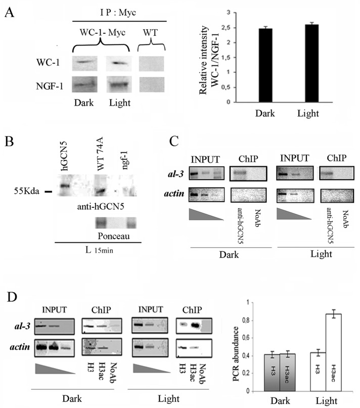
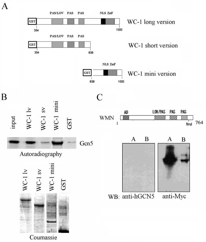
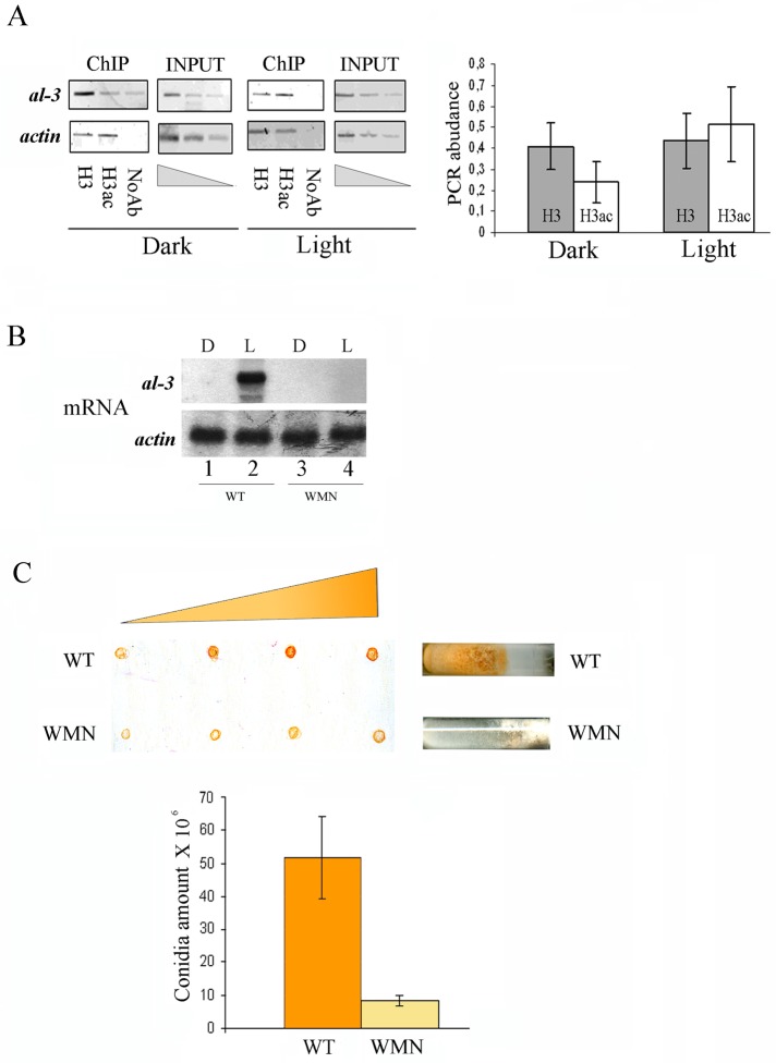
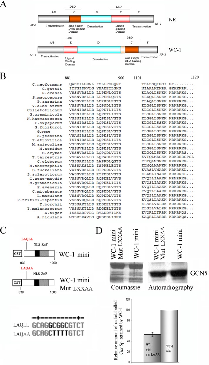
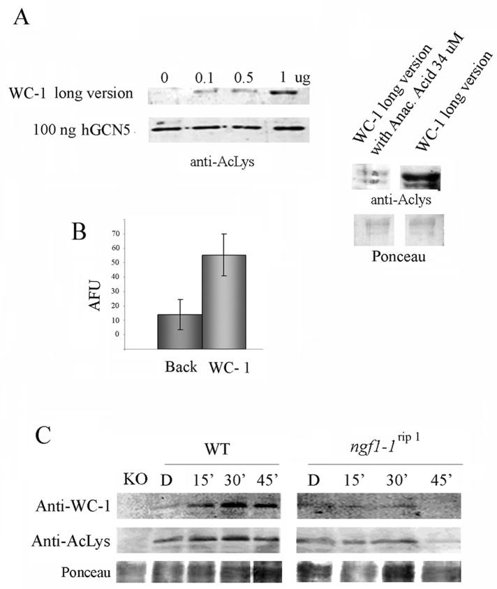
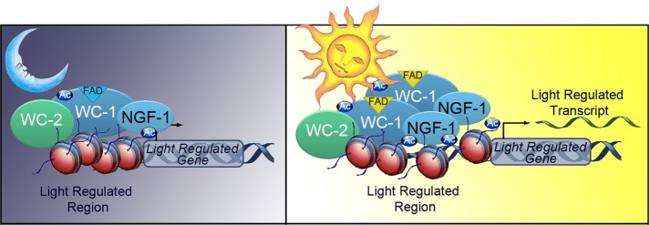
In Neurospora crassa and other filamentous fungi, light-dependent-specific phenomena are regulated by transcription factors WC-1 and WC-2. In addition to its transcriptional activity, WC-1 is able to directly sense light stimuli through a LOV sensor domain. Its location in the nucleus and heterodimerization with WC-2, together with the presence of a zinc-finger DNA-binding domain and an environmental sensor domain, all resemble the functional evolutionary architecture adopted by vertebrate nuclear receptors (NRs). Here we describe a scenario in which WC-1 represents a functional orthologue of NRs and acts through association with the chromatin-modifying coactivator NGF-1, which encodes a homologue of the yeast Gcn5p acetyltransferase. To support this view, we show a direct association between WC-1 and NGF-1 that depends on a WC-1 region containing a conserved functional LXXLL motif, a signature previously described as being an exclusive feature of NR/coactivator interaction. Our data suggest that a WC-1/NGF-1 complex is preassembled in the dark on light-inducible promoters and that, after exposure to light stimulation, NGF-1-associated HAT activity leads to histone H3 acetylation and transcriptional activation. Finally, we provide evidence for a NGF-1-independent acetylated form of WC-1. Overall our data indicate that Neurospora and higher eukaryotes share a common mechanism for the signal transduction of environmental stimuli.


Figures

FIGURE 1:
FIGURE 1:
NGF-1 physically interacts with WC-1 in dark and light conditions. (A) Myc WC-1 was immunoprecipitated (IP) using a commercial anti-Myc antibody from 400 μg of total protein extracts from Neurospora mycelia grown for 3 d in the dark before harvesting (Dark) or exposed to saturating white light for 5 min and then returned to the dark for 20 min before harvesting (Light 25 min). Precipitated WC-1 and coprecipitated NGF-1 were detected with anti-Myc and anti-hGCN5, respectively. As control of the specificity of the signal obtained, the same experiment was performed on the WT strain without the Myc epitope (left). Quantification of the ratio between the relative optical density of WC-1 and NFG-1 bands from three independent experiments under Dark and Light conditions (right). The data are the mean ± SEM from three independent experiments. (B) Western blot with anti-hGCN5 against hGCN5 purified protein, Neurospora WT, and ngf-1 strains confirms the absence of cross-hybridization of the heterologous antibody. Ponceau staining was used to check the presence of similar protein amounts on the membrane. (C) ChIP assay on Neurospora mycelia grown for 3 d in the dark before harvesting (Dark) or exposed to saturating white light for 5 min and then returned to the dark for 20 min before harvesting (Light 25 min). DNA immunoprecipitated with anti-hGCN5 antibody was amplified for the al-3 light-responsive promoter region. A sequence for actin was used as negative control. INPUT, PCR on chromatin samples before immunoprecipitation. ChIP, PCR from anti-hGCN5 IP samples. Noab, PCR from samples without antibody. (D) ChIP analysis as described in C was performed with anti–histone H3 (H3) and anti–acetylated histone H3 (H3ac; left). Quantification of the PCR abundance for the al-3 promoter was calculated as described in Materials and Methods (right). The data are the mean ± SEM from three independent experiments.
FIGURE 2:
FIGURE 2:
WC-1 needs the C-terminal region between 838 and 1000 aa to bind Gcn5p in vitro and in vivo. (A) Schematic representation of the functional domains of the recombinant proteins used in the pull-down experiments: GST, glutathione S-transferase; LOV, light sensor domain; NLS, nuclear localization signal; PAS, protein–protein interaction domain; ZnF, zinc-finger DNA-binding domain. The numbers refer to the amino acid residues of WC-1 in relation to the initial methionine in the full-length protein. (B) Pull-down experiments using the same recombinant proteins as shown in A and [35S]Gcn5-radiolabeled protein. A representative autoradiography of the [35S]GCN5 protein retained by the GST fused proteins is shown (top). Input corresponds to 30% of total incorporation of [35S]methionine in the GCN5. Coomassie staining of the SDS–PAGE illustrates the amount of the recombinant WC-1 proteins used for the pull-down experiment shown in the top (bottom). (C) Scheme showing the NruI deletion in the Myc-tagged WC-1 protein (WMN; Cheng et al, 2003). Deleted WC-1 (aa 1–764) was immunoprecipitated with an anti-Myc antibody from 800 μg (lane A) and 400 μg (lane B) of total protein extract (bottom). IP samples were analyzed by Western blot with the indicated antibodies.
FIGURE 3:
FIGURE 3:
Impairment of light-mediated H3 acetylation in the WMN strain. (A) ChIP assay on Neurospora WMN mycelia grown for 3 d in the dark before harvesting (Dark) or exposed to saturating white light for 5 min and then returned to the dark for 20 min before harvesting (Light 25 min). DNA immunoprecipitated with the anti–histone H3 (H3) and anti–acetyl histone H3 (H3ac) antibodies was amplified for the al-3 light-responsive promoter region. A sequence for actin was used as negative control. INPUT, PCR on chromatin samples before immunoprecipitation. ChIP, PCR from anti–hGCN5 IP samples. Noab, PCR from samples without antibody. Quantification of the PCR abundance for the al-3 promoter was calculated as described in Materials and Methods (right). The data are the mean ± SEM from three independent experiments. (B) Northern blot analysis of al−3mRNA in Neurospora WT and WMN strains kept in the dark (Dark) or after photoinduction (Light 25 min). The membrane was rehybridized using a probe against the actin mRNA as loading control (bottom). (C) WT and WMN strains were grown in slant for 4 d in the dark, illuminated for 5 h, and then kept in the dark for 8 h at 4°C to accumulate carotenoids. Increasing amounts of the conidia resuspended in water (5, 10, 20, and 25 μl) were spotted on a nitrocellulose filter to observe the increment of the red color corresponding to carotenoid content (top). Conidia count in WT and WMN was measured using a Burker cell counter (bottom). The data are the mean ± SEM from three independent experiments.
FIGURE 4:
FIGURE 4:
Identification of a WC-1 LXXLL motif involved in the association with HAT. (A) Schematic representation of a generic nuclear receptor (NR) and WC-1 structural organization, evidencing architectural and functional similarities. (B) Multialignment of the regions containing the LXXLL motifs within Neurospora WC-1 and several WC-1–like proteins present in the National Center for Biotechnology Information databank (LLDLL and LAQLL, indicated by a dashed line). Consensus analysis reveals conservation only for the LXXLL motif present at position 1008–1012 in Neurospora WC-1. Organisms and protein accession numbers: Cryptococcus neoformans, XP_567995.1; Cryptococcus gattii, XP_003193431.1; Neurospora crassa, CAA63964; Sordaria macrospora, XP_003351224.1; Podospora anserina, CAD60767; Verticillium albo-atrum, XP_003001986.1; Colletotrichum higginsianum, CCF31796.1; Glomerella graminicola, EFQ30482.1; Nectria haematococca, XP_003040715.1; Fusarium oxysporum, ABY47609; Gibberella fujikuroi, CAO85915; Gibberella zae, XP_388117; Hypocrea jecorina, AAV80185; Trichoderma atroviride, AAU14171.1; Metarhizium anisopliae, EFY99524.1; Metarhizium acridum, EFY92414; Magnaporthe oryzae, XP_360995.2; Thielavia terrestris, XP_003654033; Chaetomium globosum XP_001219613; Myceliophthora thermophila, XP_003665493.1; Botryotinia fuckeliana, XP_001547999.1; Sclerotinia sclerotiorum, XP_001586924.1; Cercospora zeae-maydis, AEH41590.1; Mycosphaerella graminicola, EGP83257.1; Phaeosphaeria avenaria, ACS74812.1; Cochliobolus miyabeanus, BAF35570.1; Leptosphaeria maculans, CBX98033.1; Pyrenophora tritici-repentis, XP_001933567.1; Tuber borchii, CAE01390.1; M., Tuber melanosporum, CAZ83744.1; Aspergillus niger, XP_001395039.1; Aspergillus nidulans, XP_661040.1. (C) Schematic representation of the domains in the two WC-1 mini versions and WT and point mutation mutant (Mut LXXAA) used in the pull-down experiments. The LAQLL mutagenized version is changed in LAQAA. (D) Recombinant proteins (C) were used in pull-down assays to evaluate their interaction with a radiolabeled Sc-Gcn5p. A representative Coomassie-stained gel and its autoradiography are shown (top). Quantification of the relative amount of 35S-ScGcn5p retained by the two recombinant products. The signal corresponding to WC-1 mini/ScGcn5p interaction was set to 100% (bottom). The data are the mean ± SEM from three independent experiments.
FIGURE 5:
FIGURE 5:
WC-1 is an acetylated protein. (A) Progressive amounts of GST–WC-1 long version (Figure 2A) were tested in an in vitro acetylation assay in the presence of human GCN5, as described in Materials and Methods, using an anti–Pan acetylated-lysine antibody. Anacardic acid, a common HAT inhibitor, was used to prove the dependence of the acetylated signal on the acetyltransferase reaction. Ponceau staining was used to check the presence of similar WC-1 amounts on the membrane (right). (B) WC-1 acetylation was confirmed by a fluorescence in vitro acetylation assay. The data are the mean ± SEM from three independent experiments. AFU, arbitrary fluorescence units. (C) Western blot analysis with anti–WC-1 and anti–Pan acetylated- lysine antibodies of WT and ngf-1 RIP strains grown for 3 d in the dark before harvesting (Dark) or exposed to saturating white light for 5 min and then returned to the dark for 10, 25, and 55 min before harvesting (Light 15, 30, 60 min). A wc-1–null strain (KO) was used as a control of the specificity of the anti–WC-1 antibody. Ponceau staining was used to check the presence of similar WC-1 amounts on the membrane.
FIGURE 6:
FIGURE 6:
Diagram showing a possible working model. WC-1 and NGF-1 are preassembled in the dark on the promoter of light-inducible genes, but the histone tails are not accessible for NGF-1–mediated acetylation. (B) On photoinduction, the White Collar complex undergoes a conformational change that allows NGF-1 to acetylate histone H3 and promote transcriptional activation.

Similar articles

Cited by

References

    1. Aoyagi S, Archer TK. Dynamics of coactivator recruitment and chromatin modifications during nuclear receptor mediated transcription. Mol Cell Endocrinol. 2008;280:1–5. - PMC - PubMed
    1. Baima S, Macino G, Morelli G. Photoregulation of the albino-3 gene in Neurospora crassa. J Photochem Photobiol. 1991;11:107–115. - PubMed
    1. Bain DL, Heneghan AF, Connaghan-Jones KD, Miura MT. Nuclear receptor structure: implications for function. Annu Rev Physiol. 2007;69:201–220. - PubMed
    1. Ballario P, Talora C, Galli D, Linden H, Macino G. Roles in dimerization and blue light photoresponse of the PAS and LOV domains of Neurospora crassa white collar proteins. Mol Microbiol. 1998;29:719–729. - PubMed
    1. Ballario P, Vittorioso P, Magrelli A, Talora C, Cabibbo A, Macino G. White collar-1, a central regulator of blue light responses in Neurospora, is a zinc finger protein. EMBO J. 1996;15:1650–1657. - PMC - PubMed

MeSH terms

LinkOut - more resources