. 2012 Oct;23(19):3873-81.
doi: 10.1091/mbc.E12-04-0276. Epub 2012 Aug 8.

Semaphorin-7a reverses the ERF-induced inhibition of EMT in Ras-dependent mouse mammary epithelial cells

Affiliations

Semaphorin-7a reverses the ERF-induced inhibition of EMT in Ras-dependent mouse mammary epithelial cells

Maryline Allegra et al. Mol Biol Cell. 2012 Oct.

Abstract

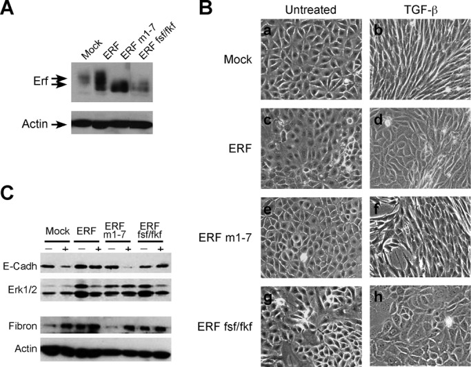
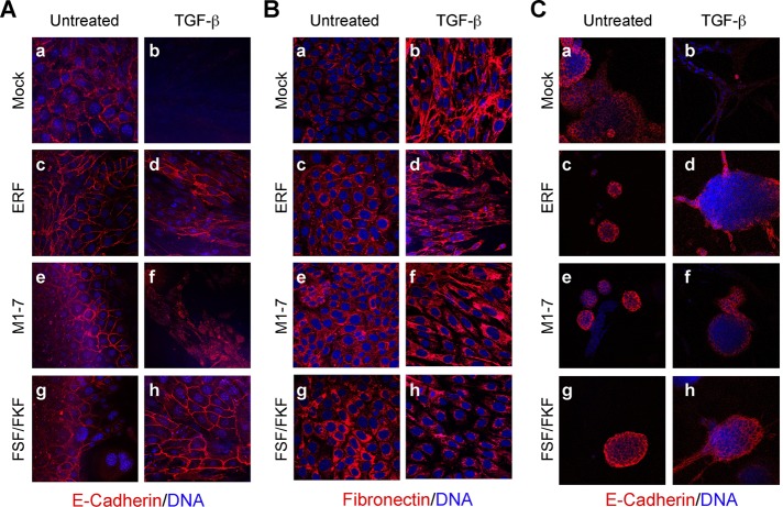
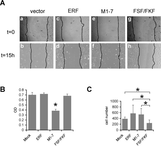
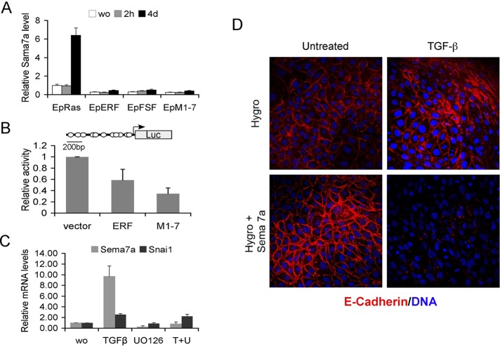
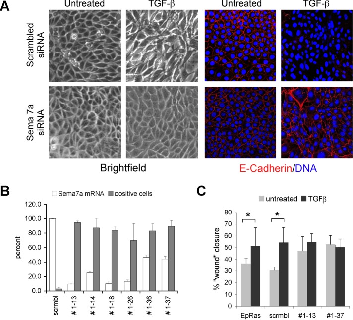
Epithelial-to-mesenchymal transition (EMT) is a key process in cancer progression and metastasis, requiring cooperation of the epidermal growth factor/Ras with the transforming growth factor-β (TGF-β) signaling pathway in a multistep process. The molecular mechanisms by which Ras signaling contributes to EMT, however, remain elusive to a large extent. We therefore examined the transcriptional repressor Ets2-repressor factor (ERF)-a bona fide Ras-extracellular signal-regulated kinase/mitogen-activated protein kinase effector-for its ability to interfere with TGF-β-induced EMT in mammary epithelial cells (EpH4) expressing oncogenic Ras (EpRas). ERF-overexpressing EpRas cells failed to undergo TGF-β-induced EMT, formed three-dimensional tubular structures in collagen gels, and retained expression of epithelial markers. Transcriptome analysis indicated that TGF-β signaling through Smads was mostly unaffected, and ERF suppressed the TGF-β-induced EMT via Semaphorin-7a repression. Forced expression of Semaphorin-7a in ERF-overexpressing EpRas cells reestablished their ability to undergo EMT. In contrast, inhibition of Semaphorin-7a in the parental EpRas cells inhibited their ability to undergo TGF-β-induced EMT. Our data suggest that oncogenic Ras may play an additional role in EMT via the ERF, regulating Semaphorin-7a and providing a new interconnection between the Ras- and the TGF-β-signaling pathways.


Figures

FIGURE 1:
FIGURE 1:
Characterization of the EpRas cell clones overexpressing wt or mutated ERF. (A) Selected clones of EpRas cells transfected with empty vector, wt ERF, the M1-7 ERF mutation, or the FSF/FKF ERF mutation were examined for ERF expression by immunoblotting using S17S Erf-specific antibody (top) and an actin-specific antibody (bottom) as a loading control. The lanes shown are not consecutive on the gel. All clones expressed 3- to 20-fold higher amounts of ERF compared with the vector control. (B) Phase contrast images of the mentioned clones were grown in the absence (a, c, e, g) or presence (b, d, f, h) of TGF-β for 5 d. Vector-transfected cells acquired a spindle-like morphology after exposure to TGF-β (b), whereas ERF-expressing cells maintained to a large extent their epithelial morphology (d, f, h). (C) The levels of E-cadherin (top) and fibronectin (second from bottom) in cells cultured for 5 din the presence or absence of TGF-β were examined by immunoblotting. Erk2/1 (second from top) and actin (bottom) levels were used respectively as loading controls. Vector-transfected and ERFm1-7–expressing cells show decreased E-cadherin and increased fibronectin levels after treatment with TGF-β. In contrast, E-cadherin and fibronectin levels in wt ERF– and ERF-FSF/FKF–expressing cells remained constant.
FIGURE 2:
FIGURE 2:
ERF-expressing EpRas cells resist TGF-β–induced EMT. (A, B) Vector-transfected EpRas cells (a, b) and clones expressing wt ERF (c, d), ERFm1-7 (e, f), or ERF-FSF/FKF (g, h) were grown for 6 d on porous support in the absence (a, c, e, g) or presence (b, d, f, h) of 5 ng/ml TGF-β. The cells were stained for E-cadherin (A) or fibronectin (B) with the respective specific antibody (red) and TOPRO-3 for DNA (blue) and analyzed by confocal microscopy. Vector-transfected cells lost E-cadherin expression (A, b) and secreted fibronectin (B, b) as a result of TGF-β treatment. In contrast, wt ERF– (d) and ERF-FSF/FKF–expressing cells (h) maintained basolateral E-cadherin expression and mostly cytoplasmic fibronectin. ERFm1-7–expressing cells have cytoplasmic E-cadherin (A, f) and high levels of cytoplasmic fibronectin (B, f). (C) The same cells were cultured in three-dimensional collagen gels in serum-free media for 5 d in the absence (a, c, e, g) or presence (b, d, f, h) of 5 ng/ml TGF-β and stained for E-cadherin (red) and DNA (blue). Vector-transfected cells failed to express E-cadherin after treatment with TGF-β (b). In contrast, all ERF-expressing cells maintained basolateral expression of E-cadherin (d, f, h).
FIGURE 3:
FIGURE 3:
ERF mutations decreased EpRas cell motility. (A) Phase contrast images of vector-transfected (a, b) and ERF-expressing cells (c–h) subjected to “wound-healing” assay in the presence of TGF-β. With the exception of ERFm1-7–expressing cells (f), gap closure for all cell lines was comparable. (B) Cell proliferation rate as assessed colorimetrically with MTT. All cell lines had comparable proliferation rates, except the ERFm1-7–expressing cells, which exhibited statistically significant reduced proliferation compared with all other lines (asterisk). (C) Transwell motility assay of EpRas cells treated with TGF-β for 24 h and induced to migrate using serum as a chemoattractant. A minimum of three random fields from each Transwell were scored for cell presence. The average of four independent experiments is shown. The two-tailed homoscedastic p-values of standard Student's test for wtERF and ERFm1-7 compared with vector-transfected cells were 0.113 and 0.103, respectively. For ERF-FSF/FKF–expressing cells the p-values against Mock-, wtERF-, and ERFm1-7–expressing cells were 0.021, 0.018, and 0.011, respectively. Statistically significant differences are indicated by an asterisk.
FIGURE 4:
FIGURE 4:
Semaphorin-7a inhibition mediates the Erf EMT phenotype. (A) Semiquantitative PCR analysis of the Sema7a mRNA levels among the different EpRas lines in the absence or presence of 5 ng/ml TGF-β for 2 h or 4 d. Sema7a levels for each sample were normalized to CPH and compared with the RNA of the parental EpRas cells growing in normal media. Sema7a mRNA levels are lower in all ERF clones and fail to elevate after 4 d of TGF-β treatment. (B) Luciferase assays from Ref1 cells cotransfected with the pGL3-Sema7a reporter, empty vector, or an ERF-expressing plasmid and pRSV-LacZ as transfection efficiency control. Luciferase activity was normalized to β-galactosidase activity, and they were all compared with vector-transfected cells. Three independent experiments in duplicate were evaluated, indicating a transcriptional repression by ERF. The cartoon shows the structure of the plasmid used with the putative ets-binding sites marked by circles. (C) Semiquantitative PCR analysis of the Sema7a and Snai1 mRNA levels in the parental EpRas cells in the presence or absence of TGF-β and the Mek1/2 inhibitor U0126. Cells were exposed to TGF-β for 5 d and to U0126 twice 16 and 2 h before harvest. The values were normalized to the values of the untreated parental cells. (D) Confocal images of Ep-ERFm1-7 cells expressing either the hygromycin resistance gene or hygromycin and Semaphorin-7a and growing on untreated glass coverslips. Cells were exposed to 5 ng/ml TGF-β for 4 d, fixed, and stained with an anti–E-cadherin antibody (red) and TOPRO-3, a DNA-intercalating dye (blue). Cells overexpressing Semaphorin-7a (bottom) undergo EMT as determined by the loss of E-cadherin. Similar results were obtained from EpRas-ERF sema7a–expressing cells.
FIGURE 5:
FIGURE 5:
Semaphorin-7a is required for EMT. (A) Phase contrast (left) and confocal images (right) of EpRas cells expressing either a scrambled siRNA or a Semaphorin-7a–specific siRNA and growing on untreated glass coverslips. Cells were exposed to 5 ng/ml TGF-β for 5 d, fixed, and stained with an anti–E-cadherin antibody (green) and TOPRO-3, a DNA-intercalating dye (blue). Loss of Semaphorin-7a (bottom) inhibits EMT as determined by the E-cadherin presence. (B) Graphic representation of Sema7a mRNA levels quantified by real-time quantitative PCR (white bars) and the fraction of E-cadherin–positive cells (gray bars) after 5 d of exposure to TGF-β. The E-cadherin–positive cells were quantified by averaging the independent fields of view for each cell line, stained as in A. (C) Wound-healing assay of parental EpRas cells and clones expressing either a scramble or Sema7a siRNA in the presence or absence of TGF-β. Wounds were photographed at 0, 6, and 16 h, and closure was measured at identical points. The 16-h time point is shown. Statistically significant differences (t test, p < 0.05) are indicated by an asterisk.

References

    1. Aitola M, Carlsson P, Mahlapuu M, Enerback S, Pelto-Huikko M. Forkhead transcription factor FoxF2 is expressed in mesodermal tissues involved in epithelio-mesenchymal interactions. Dev Dyn. 2000;218:136–149. - PubMed
    1. Athanasiou M, LeGallic L, Watson DK, Blair DG, Mavrothalassitis G. Suppression of the Ewing's sarcoma phenotype by FLI1/ERF repressor hybrids. Cancer Gene Ther. 2000;7:1188–1195. - PubMed
    1. Bielenberg DR, Klagsbrun M. Targeting endothelial and tumor cells with semaphorins. Cancer Metastasis Rev. 2007;26:421–431. - PubMed
    1. Bolos V, Peinado H, Perez-Moreno MA, Fraga MF, Esteller M, Cano A. The transcription factor Slug represses E-cadherin expression and induces epithelial to mesenchymal transitions: a comparison with Snail and E47 repressors. J Cell Sci. 2003;116:499–511. - PubMed
    1. Boyer B, Vallâes AM, Edme N. Induction and regulation of epithelial-mesenchymal transitions. Biochem Pharmacol. 2000;60:1091–1099. - PubMed

Publication types

MeSH terms

LinkOut - more resources