. 2012:18:1907-17.
Epub 2012 Jul 14.

A novel cytarabine crystalline lipid prodrug: hexadecyloxypropyl cytarabine 3',5'-cyclic monophosphate for proliferative vitreoretinopathy

Affiliations

A novel cytarabine crystalline lipid prodrug: hexadecyloxypropyl cytarabine 3',5'-cyclic monophosphate for proliferative vitreoretinopathy

Jae Suk Kim et al. Mol Vis. 2012.

Abstract

Purpose: The objectives of this study were to synthesize and characterize two types of cytarabine (Ara-C) lipid produgs and evaluate the prodrugs for sustained intraocular delivery after administration by intravitreal injection.

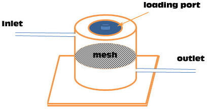
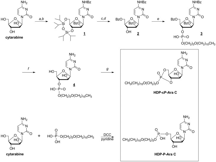
Methods: Hexadecyloxypropyl cytarabine 5'-monophosphate (HDP-P-Ara-C) and hexadecyloxypropyl cytarabine 3',5'-cyclic monophosphate (HDP-cP-Ara-C) were synthesized starting from cytarabine (1-β-D-arabinofuranosylcytosine). Their vitreal clearance profile was simulated using a custom dissolution chamber, in vitro cytotoxicity was evaluated using cell proliferation assays, and in vivo ocular properties in rat and rabbit eyes were assessed using biomicroscopy, indirect ophthalmoscopy, tonometry, electroretinography, and histology.

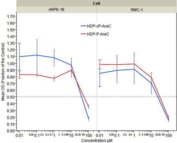
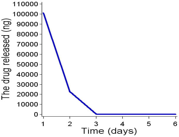
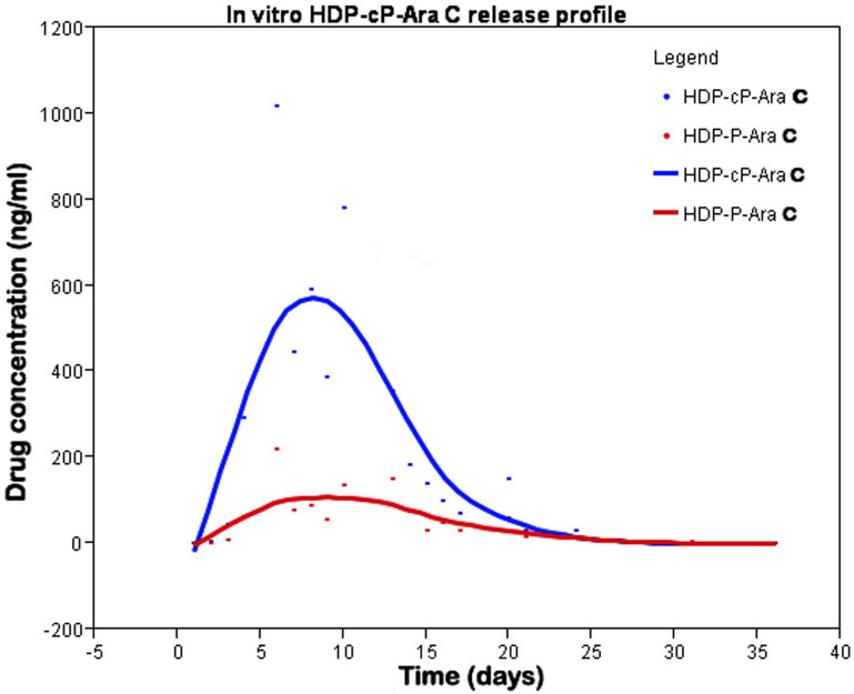
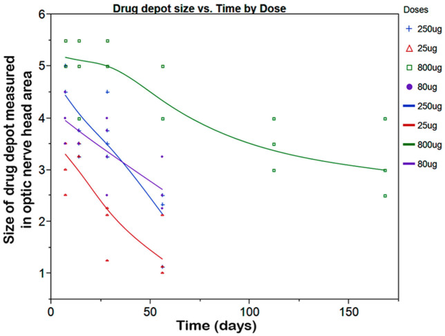
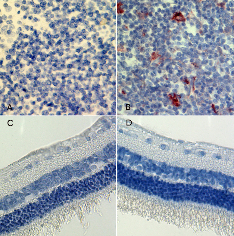
Results: HDP-P-Ara-C was cleared from the dissolution chamber (flow rate 2 µL/min) within 7 days. In contrast, HDP-cP-Ara-C, a much more insoluble prodrug, was still detectable 36 days after the dissolution process was started. HDP-P-Ara-C had a 50% cytotoxicity concentration of 52±2.6 μM in human retinal pigment epithelium (ARPE-19) and 32±2.2 µM in a rat Müller cell line, rMC-1. The 50% cytotoxicity concentration values for HDP-cP-Ara-C in ARPE-19 and rMC-1 cells were 50 µM and 25 µM, respectively. HDP-P-Ara-C was not detectable 2 weeks after the highest intravitreal dose (228 µg/rat eye) was injected, and no ocular toxicity was found. With HDP-cP-Ara-C, the drug depot was visible for 26 weeks following a single intravitreal injection (800 µg/rabbit eye). For both compounds, the electroretinogram, intraocular pressure, and other toxicity studies were negative except for the highest dose of HDP-cP-Ara-C (800 µg/eye), which had focal toxicity from the direct touch of the retina and decreased dark adapted a-waves and decreased flicker electroretinogram amplitudes (generalized estimating equations, p=0.039 and 0.01).

Conclusions: The cyclic monophosphate prodrug, HDP-cP-Ara-C, was found to have physiochemical properties better suited for sustained delivery of cytarabine to posterior segments of the eye. These properties included limited aqueous solubility, in vitro antiproliferative activity, and good tolerability after injection into rabbit eyes.


Figures

Figure 1
Figure 1
The dissolution chamber is made of poly methyl methacrylate. Inlet tubing connects from a syringe pump to the upper part of the chamber, and the outlet tubing leaves from the lower part of the chamber to the sample collector. In the center of the dissolution chamber, a titanium mesh circle is installed to prevent the large drug particles reaching the lower chamber to possibly clog the outgoing tubing. The mesh contains 5-µM pores.
Figure 2
Figure 2
Synthesis of hexadecyloxypropyl cytarabine 5’-monophosphate and hexadecyloxypropyl cytarabine 3’,5’-cyclic monophosphate. The following reagents were used: a) 1,3-dichloro-1,1,3,3-tetraisopropyldisiloxane, pyridine; b) benzoyl chloride, pyridine; c) tetrabutylammonium fluoride, HOAc, CH3CN; d) benzoyl chloride, pyridine, e) hexadecyloxypropyl phosphate, N,N-dicyclohexylcarbodiimide, pyridine; f) ammonia/methanol; g) 2,4,6-triisopropylbenzenesulfonyl chloride, pyridine.
Figure 3
Figure 3
Dose response curve showing the effect of hexadecyloxypropyl cytarabine 3’,5’-cyclic monophosphate (HDP-cP-Ara-C) and hexadecyloxypropyl cytarabine 5’-monophosphate (HDP-P-Ara-C)on human retinal pigment epithelium (ARPE-19) cells and rat Müller (rMC-1) cells proliferation. The y-axis represents the fraction of mean optical density (OD) values of testing wells for the compounds by the mean OD values of the cells for medium control using WST-1 or XTT cell proliferation assay.
Figure 4
Figure 4
In vitro hexadecyloxypropyl cytarabine 5’-monophosphate (HDP-P-Ara-C) release profile. HDP-P-Ara-C completely released from the dissolution chamber within 6 days.
Figure 5
Figure 5
In vitro hexadecyloxypropyl cytarabine 3’,5’-cyclic monophosphate (HDP-cP-Ara-C) release profile. HDP-cP-Ara-C had a peak release around day 10, and the compound was still detectable at the end of the experiment (day 36). HDP-cP-Ara-C and hexadecyloxypropyl cytarabine 5’-monophosphate (HDP-P-Ara-C) were detected in the dissolution chamber eluate.
Figure 6
Figure 6
Color fundus photograph of an experimental eye showing hexadecyloxypropyl cytarabine 3’,5’-cyclic monophosphate (HDP-cP-Ara-C) drug depot at 16 weeks (A) and at 28 weeks (B) after the intravitreal injection of 800 μg of crystals. Panel B shows about 40% to 50% decrease of the drug depot over 12 weeks.
Figure 7
Figure 7
This graph illustrates the change in hexadecyloxypropyl cytarabine 3’,5’-cyclic monophosphate (HDP-cP-Ara-C) intravitreal drug depot size over time. The drug depot size was measured in the unit of optic nerve disc area by an indirect ophthalmoscope at each checking point. The drug depots from all the doses were visible 2 months after the initial intravitreal injection. The drug depot of the highest dose was still visible 7 months before the animals were euthanized.
Figure 8
Figure 8
Light microscopic graphs of the retina and spleen. A and B are from the spleen tissue. The A serves as a negative control, and the B serves as a positive control for the terminal deoxynucleotidyl transferase dUTP nick end labeling (TUNEL) assay for the retina. D is from study eye that received an intravitreal injection of 250 μg of hexadecyloxypropyl cytarabine 3’,5’-cyclic monophosphate (HDP-cP-Ara-C), and frame C is from the fellow control eye of the same rabbit. Both retinas show normal structures and negative apoptosis staining (82.5X).

References

    1. The classification of retinal detachment with proliferative vitreoretinopathy. Ophthalmology. 1983;90:121–5. - PubMed
    1. Charteris DG. Proliferative vitreoretinopathy: pathobiology, surgical management, and adjunctive treatment. Br J Ophthalmol. 1995;79:953–60. - PMC - PubMed
    1. Pastor JC. Proliferative vitreoretinopathy: an overview. Surv Ophthalmol. 1998;43:3–18. - PubMed
    1. Asaria RH, Kon CH, Bunce C, Charteris DG, Wong D, Khaw PT, Aylward GW. Adjuvant 5-fluorouracil and heparin prevents proliferative vitreoretinopathy: Results from a randomized, double-blind, controlled clinical trial. Ophthalmology. 2001;108:1179–83. - PubMed
    1. Charteris DG, Aylward GW, Wong D, Groenewald C, Asaria RH, Bunce C. A randomized controlled trial of combined 5-fluorouracil and low-molecular-weight heparin in management of established proliferative vitreoretinopathy. Ophthalmology. 2004;111:2240–5. - PubMed

Publication types

MeSH terms

Substances

LinkOut - more resources