. 2012:18:2012-21.
Epub 2012 Jul 20.

TGFBI gene mutations in a Korean population with corneal dystrophy

Affiliations

TGFBI gene mutations in a Korean population with corneal dystrophy

Kyong Jin Cho et al. Mol Vis. 2012.

Abstract

Purpose: To investigate the clinical and genetic features of Korean patients with corneal dystrophies associated with mutations in the human transforming growth factor-β-induced (TGFBI) gene.

Methods: In this study, 387 subjects (71 families and 89 individuals - 268 patients having TGFBI corneal dystrophies and 119 normal relatives) were assessed. All subjects underwent a complete ophthalmologic evaluation, including biomicroscopic inspection and dilated fundus examination. As a control, 100 individuals without corneal disease were selected from the general population. The polymerase chain reaction (PCR) and direct sequencing were used to screen for mutations in TGFBI.

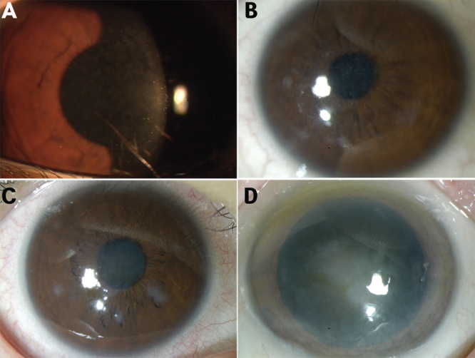
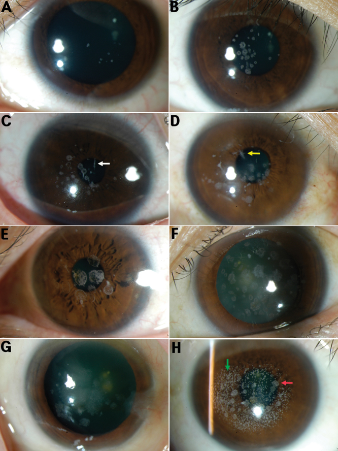
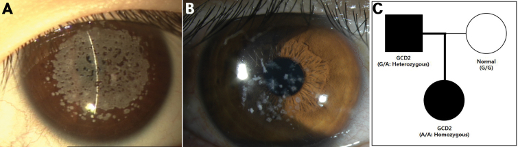
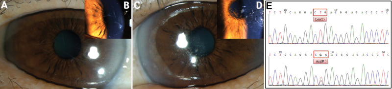
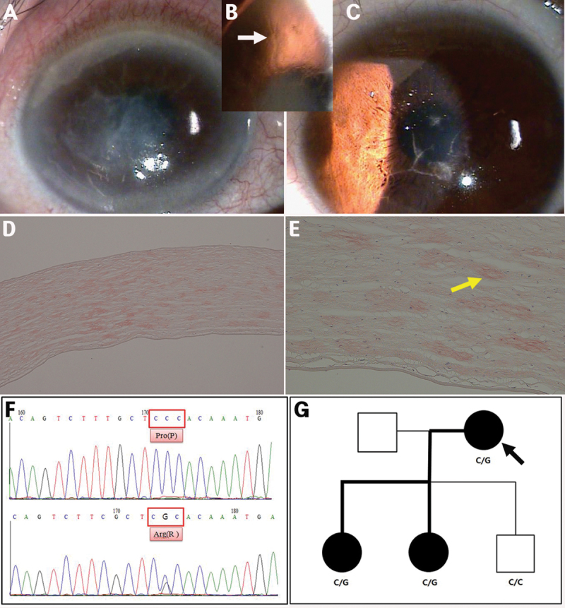
Results: All subjects recruited exhibited a range of corneal dystrophies, including Thiel-Behnke corneal dystrophy (TBCD, R555Q; 6 families and 4 individuals), granular corneal dystrophy type 2 (GCD2, R124H; 61 families and 80 individuals), lattice corneal dystrophy (LCD; 4 families and 5 individuals; 7 with type 1 [R124C], and 2 with a variant [L527R, P542R]). The disease showed an autosomal dominant inheritance pattern in all families.

Conclusions: R124H in GCD2 was the most common mutation. GCD1 and Reis-Bucklers corneal dystrophy were not found. In the GCD2 patients there were a large number of laser refractive surgery-induced corneal opacities. A spontaneous R124H mutation was confirmed in an already mutated allele that resulted in a change from a heterozygous into a homozygous form. Also, a novel mutation, P527R, was identified in LCD.


Figures

Figure 1
Figure 1
Phenotypes of Thiel-Behnke corneal dystrophy (all carried the R555Q mutation of the heterozygous form). A: An 11-month-old female, irregularly shaped scattered opacities with non-involvement of the peripheral cornea in the Bowman layer. B: A 26-year-old female, subepithelial reticular (honeycomb) opacities with non-involvement of the peripheral cornea. C: A 43-year-old female, opacities united, resulting in denser and larger opacities. D: A 69-year-old female, opacities, progressing to the deep stromal layers and corneal periphery.
Figure 2
Figure 2
Phenotypes of granular corneal dystrophy, type 2 (all carried the R124H mutation). A: A 16-year-old male, subtle superficial stromal tiny whitish dots. B: A 24-year-old female, rings or stellate-shaped snowflake stromal opacities appeared between the superficial stroma and the mid stroma. C: A 34-year-old male, lattice lines in the deeper cornea (white arrow). D: A 47-year-old female, star-shaped stromal opacities (yellow arrow). E: A 55-year-old male. F: A 65-year-old female, more superficial, translucent, flattened breadcrumb opacities. G: A 77-year-old female, opacities coalesced in the anterior stroma. H: A 39-year-old female with a history of LASIK surgery, diffuse, confluent, white, and small opacities (green arrow) coexisted with discrete and granular opacities (red arrow).
Figure 3
Figure 3
Family with granular corneal dystrophy, Type 2, in which a spontaneous R124H mutation was added to an already mutated allele, resulting in a change from a heterozygous to a homozygous form. A: A 6-year-old female (proband, homozygous form), dense and confluent opacities with peripheral cornea non-involvement in the stromal layer. B: A 32-year-old male (father of proband, heterozygous form), star- and disc- shaped opacities. C: Pedigree, homozygous R124H mutation (CGC→CAC), heterozygous R124H mutation, and no mutations in the proband, father or mother.
Figure 4
Figure 4
Phenotype of variant lattice corneal dystrophy (L527R mutation) with asymmetric corneal opacity in eye laterality, in a 67-year-old female. A, B: Several bifurcating, thick lattice lines in the superficial stroma of the right cornea. C, D: Discrete and nodular opacities were noted in the deep stroma of the central cornea. E: DNA sequence analysis of the TGFBI gene (upper) and a heterozygous point mutation, CTG→CGG (c.1580 T>G: Leu527Arg in exon 12) in codon 527 (bottom).
Figure 5
Figure 5
Family with variant lattice corneal dystrophy (novel mutation R542P). A, B: An 87-year-old female (proband), relatively thick lattice lines (white arrow) that extended from limbus to limbus in the superficial and deep stromal layers. C: A 55-year-old male (daughter of proband), lattice lines discrete in the superficial stroma of the central cornea. D, E: Histopathology (proband, 40× and 100×, respectively), Congo red-positive deposits in almost an entire corneal stroma layer. F: DNA sequence analysis of the TGFBI gene (upper) and a heterozygous point mutation, CCC→CGC (c.1625 C>G: Pro542Arg in exon 12) in codon 542 (bottom). G: Pedigree, heterozygous P542R mutation in the proband and her two daughters.

References

    1. Weiss JS, Moller HU, Lisch W, Kinoshita S, Aldave AJ, Belin MW, Kivela T, Busin M, Munier FL, Seitz B, Sutphin J, Bredrup C, Mannis MJ, Rapuano CJ, Van Rij G, Kim EK, Klintworth GK. The IC3D classification of the corneal dystrophies. Cornea. 2008;27(Suppl 2):S1–83. - PMC - PubMed
    1. Pieramici SF, Afshari NA. Genetics of corneal dystrophies: the evolving landscape. Curr Opin Ophthalmol. 2006;17:361–6. - PubMed
    1. Aldave AJ, Sonmez B. Elucidating the molecular genetic basis of the corneal dystrophies: are we there yet? Arch Ophthalmol. 2007;125:177–86. - PubMed
    1. Munier FL, Korvatska E, Djemai A, Le Paslier D, Zografos L, Pescia G, Schorderet DF. Kerato-epithelin mutations in four 5q31-linked corneal dystrophies. Nat Genet. 1997;15:247–51. - PubMed
    1. Escribano J, Hernando N, Ghosh S, Crabb J, Coca-Prados M. cDNA from human ocular ciliary epithelium homologous to beta ig-h3 is preferentially expressed as an extracellular protein in the corneal epithelium. J Cell Physiol. 1994;160:511–21. - PubMed

Publication types

MeSH terms

Substances

LinkOut - more resources