

Amniotic membrane traps and induces apoptosis of inflammatory cells in ocular surface chemical burn
- PMID: 22876141
- PMCID: PMC3413422
Amniotic membrane traps and induces apoptosis of inflammatory cells in ocular surface chemical burn
Abstract
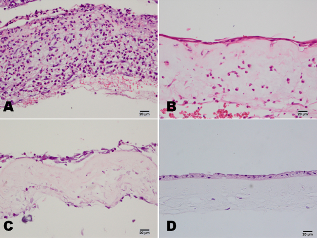
Purpose: Severe chemical burns can cause necrosis of ocular surface tissues following the infiltration of inflammatory cells. It has been shown that amniotic membrane transplantation (AMT) is an effective treatment for severe chemical burns, but the phenotypes of cells that infiltrate the amniotic membrane and the clinical significance of these cellular infiltrations have not previously been reported. The present work studies the inflammation cell traps and apoptosis inducing roles of the amniotic membrane after AMT in patients with acute chemical burns.
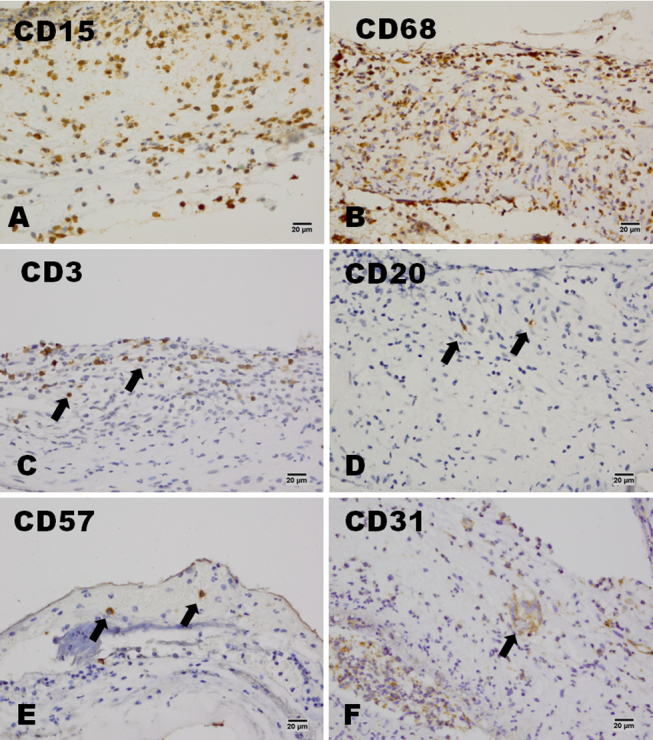
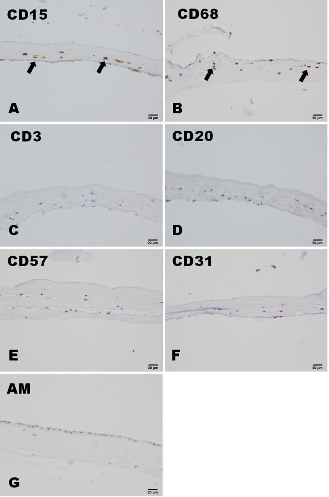
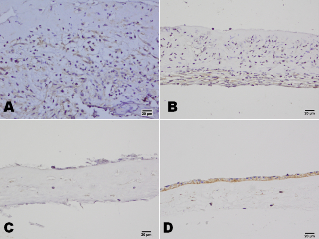
Methods: A total of 30 patients with acute alkaline burns were classified as having either moderate or severe burns. In all participants, AMT was performed within one week of his/her injury. After 7-9 days, the transplanted amniotic membranes were removed. Histopathological and immunohistochemical techniques were used for the examination and detection of infiltrating cells, and tests for the expression of CD (cluster of differentiation)15, CD68, CD3, CD20, CD57, CD31, CD147, and CD95 (Fas) were performed. A TUNEL (TdT-mediated dUTP nick end labeling) assay was used to confirm apoptosis of the infiltrating cells. Three patients with herpes simplex-induced keratitis who had undergone AMT to treat persistent epithelium defects were used as a control group. Amniotic membrane before transplantation was used as another control.
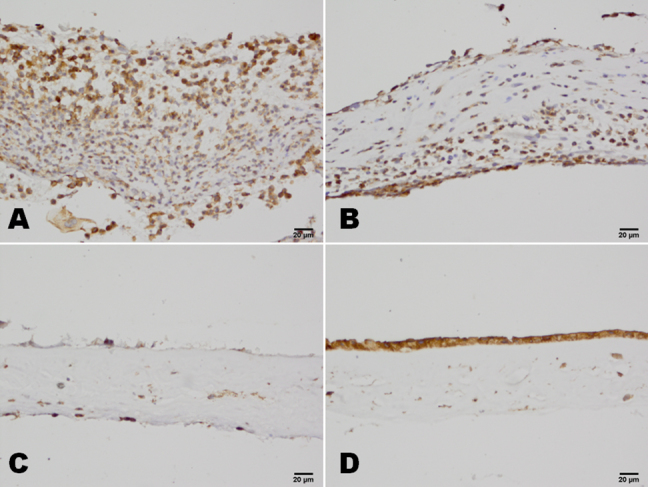
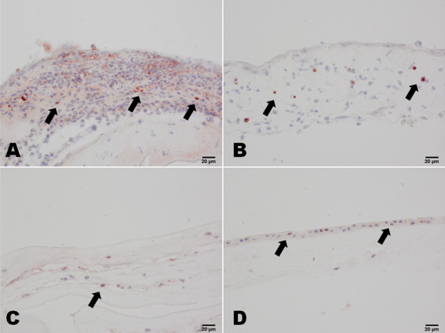
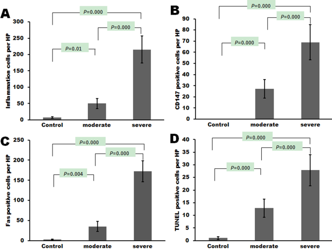
Results: After amniotic membrane transplantation, the number of infiltrating cells in patients with severe burns was significantly higher than in patients with moderate burns or in control patients (p<0.05). Among the severe burns patients, CD15 and CD68 were widely expressed in the infiltrating cells, and CD3, CD20, and CD57 were only found in a small number of cells. Occasionally, CD31-positive cells were found in the amniotic membranes. More cells that were CD147, Fas, and TUNEL positive were found in patients with severe burns than in patients with moderate burns or in control patients.
Conclusions: Neutrophils and macrophages were the main cells that had infiltrated into the amniotic membrane during the acute phase of healing from a chemical burns. AMT can trap different inflammatory cells and induce apoptosis of inflammatory cells in acute ocular chemical burns.
Figures
References
-
- Tamhane A, Vajpayee RB, Biswas NR, Pandey RM, Sharma N, Titiyal JS, Tandon R. Evaluation of amniotic membrane transplantation as an adjunct to medical therapy as compared with medical therapy alone in acute ocular burns. Ophthalmology. 2005;112:1963–9. - PubMed
-
- Fish R, Davidson RS. Management of ocular thermal and chemical injuries, including amniotic membrane therapy. Curr Opin Ophthalmol. 2010;21:317–21. - PubMed
-
- Resch MD, Schlötzer-Schrehardt U, Hofmann-Rummelt C, Sauer R, Cursiefen C, Kruse FE, Beckmann MW, Seitz B. Adhesion structures of amniotic membranes integrated into human corneas. Invest Ophthalmol Vis Sci. 2006;47:1853–61. - PubMed
-
- Said DG, Nubile M, Alomar T, Hopkinson A, Gray T, Lowe J, Dua HS. Histologic features of transplanted amniotic membrane: implications for corneal wound healing. Ophthalmology. 2009;116:1287–95. - PubMed
-
- Sangwan VS, Basu S, Vemuganti GK, Sejpal K, Subramaniam SV, Bandyopadhyay S, Krishnaiah S, Gaddipati S, Tiwari S, Balasubramanian D. Clinical outcomes of xeno-free autologous cultivated limbal epithelial transplantation: a 10-year study. Br J Ophthalmol. 2011;95:1525–9. - PubMed
Publication types
MeSH terms
Substances
LinkOut - more resources
Full Text Sources
Research Materials
Miscellaneous