. 2012;8(8):e1002830.
doi: 10.1371/journal.ppat.1002830. Epub 2012 Aug 2.

Structural and biochemical basis for development of influenza virus inhibitors targeting the PA endonuclease

Affiliations

Structural and biochemical basis for development of influenza virus inhibitors targeting the PA endonuclease

Rebecca M DuBois et al. PLoS Pathog. 2012.

Abstract

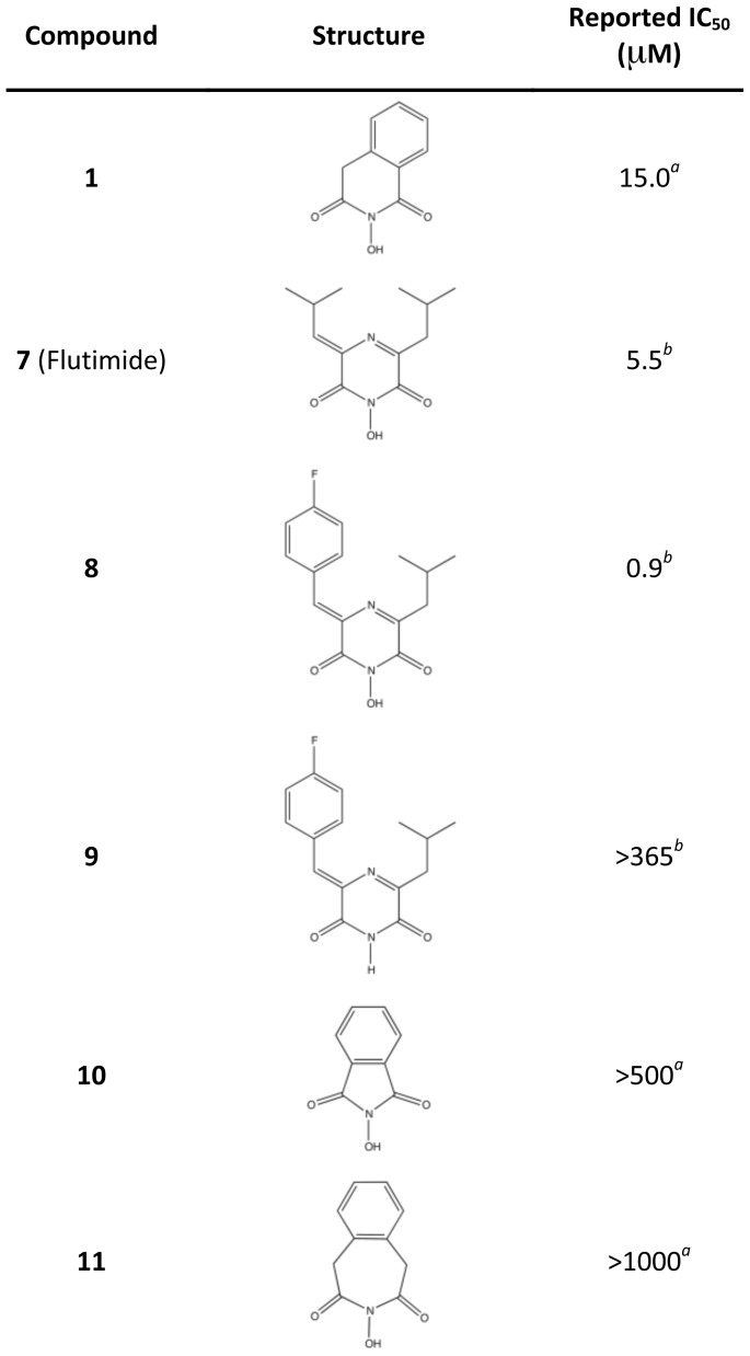
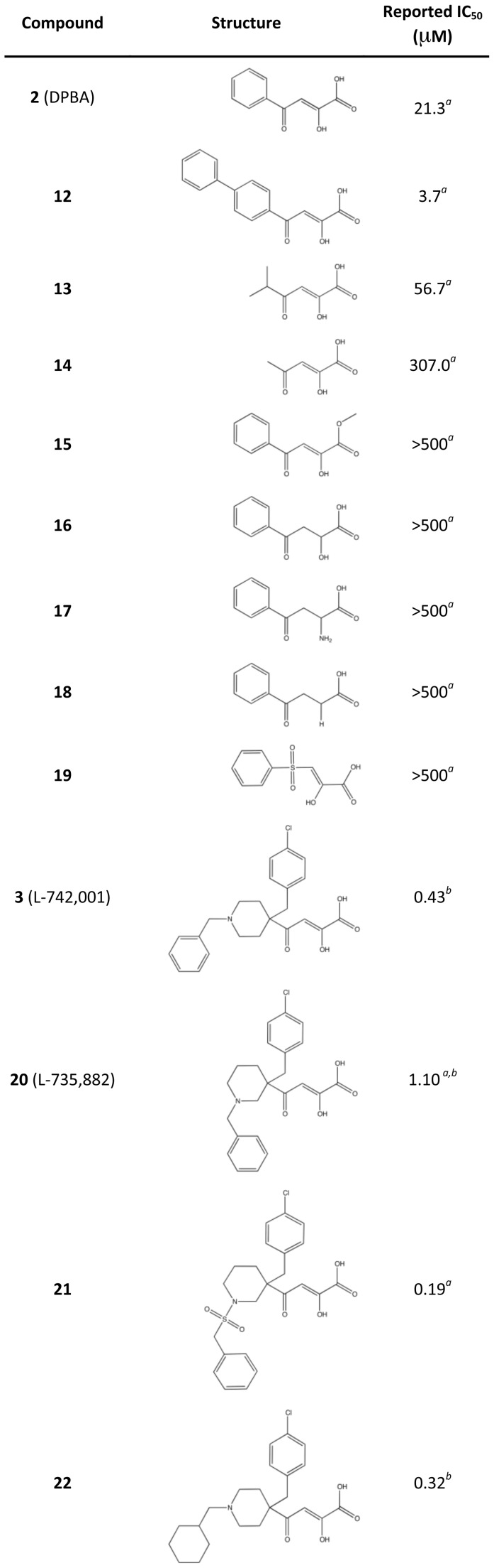
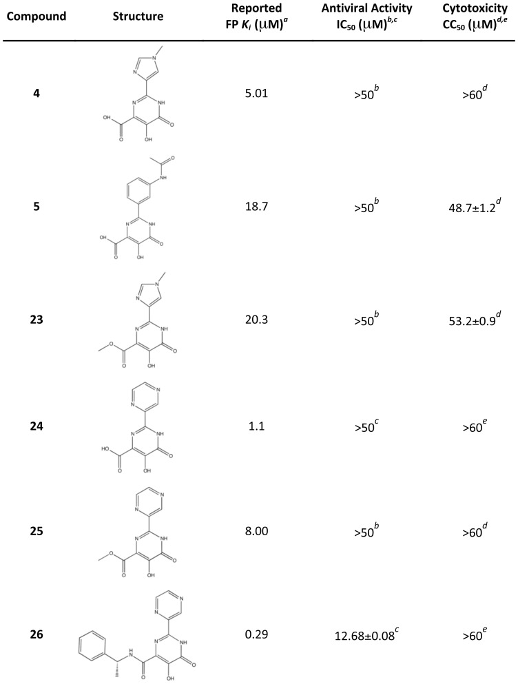
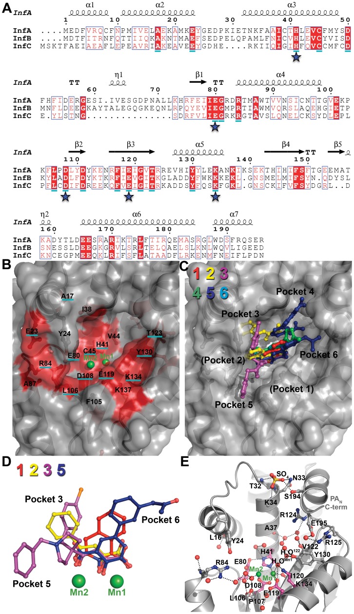
Emerging influenza viruses are a serious threat to human health because of their pandemic potential. A promising target for the development of novel anti-influenza therapeutics is the PA protein, whose endonuclease activity is essential for viral replication. Translation of viral mRNAs by the host ribosome requires mRNA capping for recognition and binding, and the necessary mRNA caps are cleaved or "snatched" from host pre-mRNAs by the PA endonuclease. The structure-based development of inhibitors that target PA endonuclease is now possible with the recent crystal structure of the PA catalytic domain. In this study, we sought to understand the molecular mechanism of inhibition by several compounds that are known or predicted to block endonuclease-dependent polymerase activity. Using an in vitro endonuclease activity assay, we show that these compounds block the enzymatic activity of the isolated PA endonuclease domain. Using X-ray crystallography, we show how these inhibitors coordinate the two-metal endonuclease active site and engage the active site residues. Two structures also reveal an induced-fit mode of inhibitor binding. The structures allow a molecular understanding of the structure-activity relationship of several known influenza inhibitors and the mechanism of drug resistance by a PA mutation. Taken together, our data reveal new strategies for structure-based design and optimization of PA endonuclease inhibitors.


Conflict of interest statement

The authors have declared that no competing interests exist.

Figures

Figure 1
Figure 1. Crystal structure and endonuclease activity of PAN ΔLoop.
(A) Schematic of PAN (magenta) and PAN ΔLoop (green), and location of the 22-residue loop (orange) replaced by a Gly-Gly-Ser linker in the PAN ΔLoop construct. (B) Two orthogonal views of the overlay of the crystal structures of PAN and PAN ΔLoop, colored as in A. Key active site residues are shown in stick representation, the paired manganese ions are shown as spheres, and the N- and C-termini are labeled. The coordinates for PAN are from PDB entry 2W69. The atomic coordinates and structure factors for PAN ΔLoop have been deposited in the Protein Data Bank as PDB entry 4E5E. (C) Coomassie-stained SDS-PAGE of PAN and PAN ΔLoop showing the amount of protein (µM) in a 10 µl endonuclease activity assay reaction. Molecular weight (MW) markers (kD) are shown on the left. (D) Endonuclease activity assay with PAN and PAN ΔLoop. Single-stranded DNA plasmid M13mp18 was incubated with increasing concentrations (µM) of PAN or PAN ΔLoop. Reactions products were resolved on a 1.0% agarose gel stained with ethidium bromide. Molecular weight (MW) ladder (kb) is shown on the left.
Figure 2
Figure 2. Chemical structures of compounds used in this study.
Oxygen atoms that coordinate manganese ions in the active site of PAN ΔLoop are colored red.
Figure 3
Figure 3. Inhibition of PAN endonuclease activity by known and predicted inhibitors.
Compounds 16 (AF, respectively) were incubated at increasing concentrations (µM) with 15 µM PAN and single-stranded DNA plasmid M13mp18. Reaction products were resolved on a 1.0% agarose gel and stained with ethidium bromide. Control lanes (‘C’) contained no PAN in the reaction mixture. Molecular weight (MW) ladder (kb) is shown on the left.
Figure 4
Figure 4. Crystal structures of PAN ΔLoop bound to compounds 1–6 (A–F, respectively).
PAN ΔLoop is shown as gray cartoon. Compounds are shown as ball-and-stick models and are colored yellow (carbon), blue (nitrogen), red (oxygen), and orange (chlorine). Manganese ions (Mn1 and Mn2) are shown as green spheres. The carbon atoms of key active site residues are colored magenta, and the carbon atoms of other residues interacting with the compound or discussed in the text are colored cyan. An ordered water molecule (H2O122) is shown as a red sphere. Black dotted lines represent molecular interactions less than 3.2 Å away. In the case of compounds 2 and 5, two molecules (yellow labels A and B) are bound in the PAN ΔLoop active site. Gray arrows (panels B and C) show the movement of helix-α3 residue Tyr24 compared with the structure of PAN ΔLoop-Apo. The atomic coordinates and structure factors for PAN ΔLoop bound to compounds 16 have been deposited in the Protein Data Bank as PDB entries 4E5F, 4E5G, 4E5H, 4E5I, 4E5J, and 4E5L, respectively.
Figure 5
Figure 5. Reported IC50 values of Flutimide, Flutimide-related, and N-hydroxyimide inhibitors determined in an in vitro transcription assay with influenza A polymerase.
aPublished results in an influenza virus in vitro transcription assay . bPublished results in an influenza virus in vitro transcription assay .
Figure 6
Figure 6. Reported IC50 values of 4-substituted 2,4-dioxobutanoic acid inhibitors determined in an in vitro transcription assay with influenza A polymerase.
aPublished results in an influenza virus in vitro transcription assay . bPublished results in an influenza virus in vitro transcription assay .
Figure 7
Figure 7. Reported PAN binding activities, antiviral activities, and cytotoxicities of compounds 4 and 5 and related compounds.
aPublished results in a competitive binding fluorescence polarization assay with PAN . b,cAntiviral activity as measured by inhibition of viral plaque formation in this studyb or previouslyc . d,eCompound cytotoxicity in MDCK cells after 72 hours as measured in this studyd or previouslye .
Figure 8
Figure 8. Conserved residues and ordered water molecules in the PAN active site cleft.
(A) Sequence alignment of PAN from influenza A, B, and C. Consensus sequences were determined from more than 13,000 sequences using the online database www.fludb.org. The secondary structure of PAN from influenza A is shown above the sequence alignment. Residues in a solid red background are identical between the influenza A, B, and C consensus sequences. Residues that are >99.9% conserved in all sequences analyzed are underlined in cyan. Stars indicate key active site residues. (B) Surface representation of the PAN ΔLoop active site cleft. Manganese ions (Mn1 and Mn2) are shown as green spheres. The highly conserved cleft is colored red. Residues that are identical between influenza A, B, and C consensus sequences are not underlined, and residues that are >99.9% conserved are underlined in cyan. (C) Surface representation of PAN ΔLoop active site cleft with overlays of compounds 1 (red), 2 (yellow), 3 (purple), 4 (green), 5 (blue), and 6 (cyan). The compounds occupy four (3–6) of the six binding pockets discussed in the text. The orientation and depicted surface are identical to those shown in panel B. (D) Superposition of bound compounds 1, 2, 3 and 5 (same coloring as panel C) after structural alignment of the entire PAN ΔLoop domain. For clarity, molecules B of 2 and 5 are not shown. Also not shown for clarity are compounds 4 and 6 because their metal-binding scaffolds overlay perfectly with 5. Manganese ions (Mn1 and Mn2) are shown as green spheres. (E) Ordered water molecules found in at least three of the four molecules in the crystallographic asymmetric unit are shown as red spheres. PAN ΔLoop is shown as gray cartoon. Manganese ions (Mn1 and Mn2) are shown as green spheres. Key active site residues are shown in magenta carbon atoms and other residues interacting with the compound or discussed in the text are shown in gray carbon atoms. A sulfate ion is shown as ball-and-stick and colored yellow (sulfur) and red (oxygen). This sulfate is bound at the same location as a predicted phosphate group of nucleic acid in a model of the PAN-substrate complex . Black dotted lines represent molecular interactions less than 3.2 Å away.

References

    1. Reid AH, Taubenberger JK, Fanning TG. The 1918 Spanish influenza: integrating history and biology. Microbes Infect. 2001;3:81–87. - PubMed
    1. Garten RJ, Davis CT, Russell CA, Shu B, Lindstrom S, et al. Antigenic and genetic characteristics of swine-origin 2009 A(H1N1) influenza viruses circulating in humans. Science. 2009;325:197–201. - PMC - PubMed
    1. WHO. Avian influenza in humans. 2012. Available: http://www.who.int/csr/disease/avian_influenza/
    1. Imai M, Watanabe T, Hatta M, Das SC, Ozawa M, et al. Experimental adaptation of an influenza H5 HA confers respiratory droplet transmission to a reassortant H5 HA/H1N1 virus in ferrets. Nature. 2012;486:420–8. - PMC - PubMed
    1. Herfst S, Schrauwen EJA, Linster M, Chutinimitkul S, de Wit E, et al. Airborne Transmission of Influenza A/H5N1 Virus Between Ferrets. Science. 2012;336:1534–1541. - PMC - PubMed

Publication types

MeSH terms