. 2012;8(8):e1002831.
doi: 10.1371/journal.ppat.1002831. Epub 2012 Aug 2.

Structural analysis of specific metal chelating inhibitor binding to the endonuclease domain of influenza pH1N1 (2009) polymerase

Affiliations

Structural analysis of specific metal chelating inhibitor binding to the endonuclease domain of influenza pH1N1 (2009) polymerase

Eva Kowalinski et al. PLoS Pathog. 2012.

Abstract

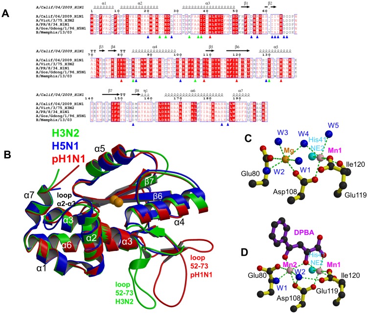
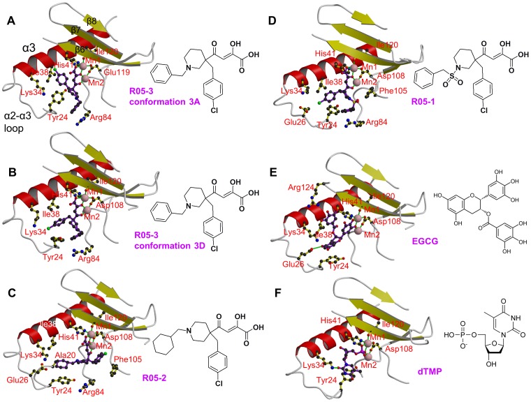
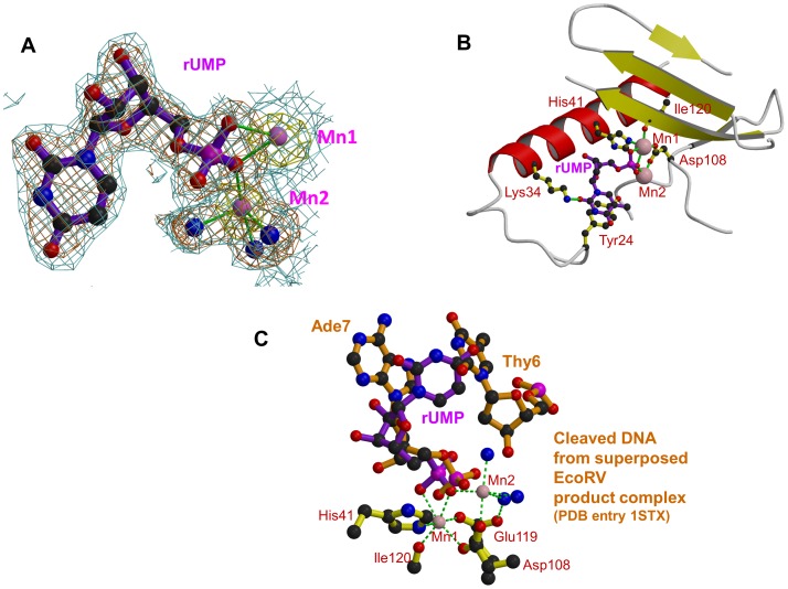
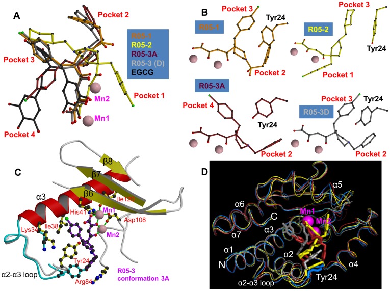
It is generally recognised that novel antiviral drugs, less prone to resistance, would be a desirable alternative to current drug options in order to be able to treat potentially serious influenza infections. The viral polymerase, which performs transcription and replication of the RNA genome, is an attractive target for antiviral drugs since potent polymerase inhibitors could directly stop viral replication at an early stage. Recent structural studies on functional domains of the heterotrimeric polymerase, which comprises subunits PA, PB1 and PB2, open the way to a structure based approach to optimise inhibitors of viral replication. In particular, the unique cap-snatching mechanism of viral transcription can be inhibited by targeting either the PB2 cap-binding or PA endonuclease domains. Here we describe high resolution X-ray co-crystal structures of the 2009 pandemic H1N1 (pH1N1) PA endonuclease domain with a series of specific inhibitors, including four diketo compounds and a green tea catechin, all of which chelate the two critical manganese ions in the active site of the enzyme. Comparison of the binding mode of the different compounds and that of a mononucleotide phosphate highlights, firstly, how different substituent groups on the basic metal binding scaffold can be orientated to bind in distinct sub-pockets within the active site cavity, and secondly, the plasticity of certain structural elements of the active site cavity, which result in induced fit binding. These results will be important in optimising the design of more potent inhibitors targeting the cap-snatching endonuclease activity of influenza virus polymerase.


Conflict of interest statement

Andrea Wolkerstofer and Oliver Szolar are employed by Savira pharmaceuticals (http://www.savira.at/). Stephen Cusack was involved in the foundation of Savira and is scientific advisor to the company.

Figures

Figure 1
Figure 1. The PA endonuclease carries a divalent cation binding site in its active center.
A: Sequence alignment of the PA-Nter endonuclease from four influenza A (including the three of known atomic structure) and one influenza B strain. The secondary structure of the pH1N1 domain is shown over the alignment. Red triangles indicate conserved cation binding (His41, Glu80, Asp108, Glu119) and catalytic (Lys134) residues. Blue triangles indicate naturally variable positions amongst influenza A strains. Green triangles indicate residues interacting with the inhibitors described in this paper. B: Superposition of PA endonuclease structure from H3N2 (green, PDB entry 2W69), H5N1 (blue, PDB entry 3EBJ) and pH1N1 (red, this work). The two bound divalent metal ions are represented by orange spheres. Flexible region 53–73 is at the bottom right and only ordered in certain chains from the H3N2 (B chain) and pH1N1 (e.g. D chain of dTMP complex) structures. For H5N1, region 53–73 is not visible. Major secondary structure elements are shown consistent with those in Figure 1A. C: Divalent ion co-ordination in the native endonuclease structure. Manganese and magnesium ions are respectively pink and orange spheres and co-ordinating water molecule blue spheres and the ion co-ordination is shown with green dotted lines. For clarity, only His41 NE2 is shown (cyan sphere). D: Divalent ion co-ordination in the DPBA bound structure. Manganese ions are pink spheres and co-ordinating water molecule blue spheres and the ion co-ordination is shown with green dotted lines. For clarity, only His41 NE2 is shown (cyan sphere).
Figure 2
Figure 2. Binding of diketo inhibitors, EGCG and dTMP in the active site of pH1N1 endonuclease.
Manganese ions are pink spheres and the ion co-ordination is shown with green lines. Side chains of key active site residues that interact with the compound or are close to it are shown. The orientation in each case is the same after superposition of the domain. Helix α3 (red), the α3-α3 loop and beta strands β6, β7 and β8 (yellow) are indicated in panel A. A: R05-3 in conformation 3A. B: R05-3 in conformation 3D. C: R05-2 (chain A). Ala20 is marked in addition (see discussion). D: R05-1. E: EGCG. F: dTMP.
Figure 3
Figure 3. Binding of dTMP/rUMP in the active site of pH1N1 endonuclease.
A: Electron density for rUMP in pH1N1 PA endonuclease. Manganese ions are pink spheres, co-ordinating water molecule blue spheres and the ion co-ordination is shown with green lines. Blue contour: final 2Fo-Fc electron density at 1.0σ. Brown contour: Fo-Fc unbiased difference map at 2.8σ. Yellow contour: anomalous density at 4.0σ. B: Binding site of rUMP in the active site following the same scheme as in Figure 2. C: rUMP bound in the active site of pH1N1 PA (purple) with superposed DNA from product complex of EcoRV (brown, pdb entry 1STX). Active site residues (yellow), manganese ions (pink) and water molecules (blue) are for the rUMP structure. The position of the two DNA bases either side of the cleavage site in the EcoRV product complex is shown.
Figure 4
Figure 4. Active site pockets of the PA endonuclease.
Each panel shows the pH1N1 domain in surface representation (green) in the same orientation with the manganese ions as pink spheres. Bound compounds are shown in surface and stick representation. Some residues underlying prominent surface features are indicated in white. A. Native unliganded structure. B. R05-3A (brick). C. EGCG (grey). D. R05-2 (yellow). In the native state, the active site cavity is large. The different compounds fill various sub-pockets of the cavity (indicated in red) and induced fit movements tend to close up the active site, but the cavity is never entirely filled.
Figure 5
Figure 5. Active site plasticity of the PA endonuclease.
A: Superposition of diketo inhibitors and EGCG bound in the PA active site after structural alignment of the entire endonuclease domain for each structure. R05-1 in orange, R05-2 in yellow, R05-3 conformation 3A in brick, R05-3 conformation 3D in light grey and EGCG in black. Manganese ions are pink spheres. The aromatic extensions from the metal binding scaffold are inserted into different sub-pockets of the active site. B: Configuration of the diketo inhibitors (coloured as in Figure 3A) with respect to residue Tyr24. All panels are in the same orientation. C: Diagram comparing the native (cyan) and R05-3 bound form in the conformation 3A (purple). Tyr24 side-chain moves to partially stack on the chlorobenzene and Arg84 is re-ordered to stack with the benzene ring of R05-3A. Manganese ions are pink spheres and the ion co-ordination is shown with green lines. D: Superposition of the Cα-trace of native (cyan) and R05-3A (brick), R05-2 (yellow) and EGCG (grey) pH1N1 structures. Manganese ions are pink spheres. Much of the structure, notably the metal binding catalytic center, is relatively rigid, but there is more flexibility in the α2-a3 loop (especially at Tyr24) and also in the α5 helix (which bears the catalytic lysine).

References

    1. Plotch SJ, Bouloy M, Ulmanen I, Krug RM. A unique cap(m7GpppXm)-dependent influenza virion endonuclease cleaves capped RNAs to generate the primers that initiate viral RNA transcription. Cell. 1981;23:847–858. - PubMed
    1. Poon LL, Pritlove DC, Fodor E, Brownlee GG. Direct evidence that the poly(A) tail of influenza A virus mRNA is synthesized by reiterative copying of a U track in the virion RNA template. J Virol. 1999;73:3473–3476. - PMC - PubMed
    1. Ruigrok RW, Crepin T, Hart DJ, Cusack S. Towards an atomic resolution understanding of the influenza virus replication machinery. Curr Opin Struct Biol. 2010;20:104–113. - PubMed
    1. Guilligay D, Tarendeau F, Resa-Infante P, Coloma R, Crepin T, et al. The structural basis for cap binding by influenza virus polymerase subunit PB2. Nat Struct Mol Biol. 2008;15:500–506. - PubMed
    1. Dias A, Bouvier D, Crepin T, McCarthy AA, Hart DJ, et al. The cap-snatching endonuclease of influenza virus polymerase resides in the PA subunit. Nature. 2009;458:914–918. - PubMed

Publication types

MeSH terms