. 2012;8(8):e1002866.
doi: 10.1371/journal.pgen.1002866. Epub 2012 Aug 2.

Multiple roles and interactions of Tbx4 and Tbx5 in development of the respiratory system

Affiliations

Multiple roles and interactions of Tbx4 and Tbx5 in development of the respiratory system

Ripla Arora et al. PLoS Genet. 2012.

Abstract

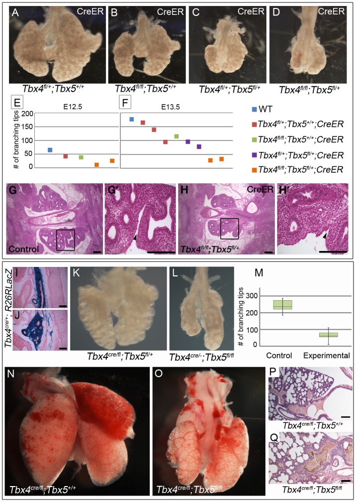
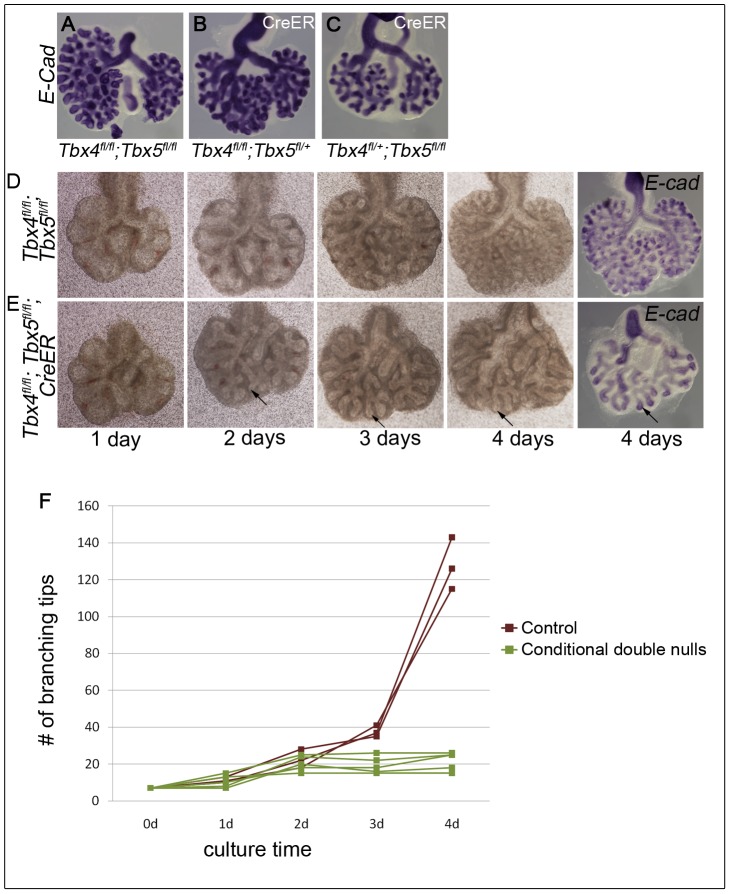
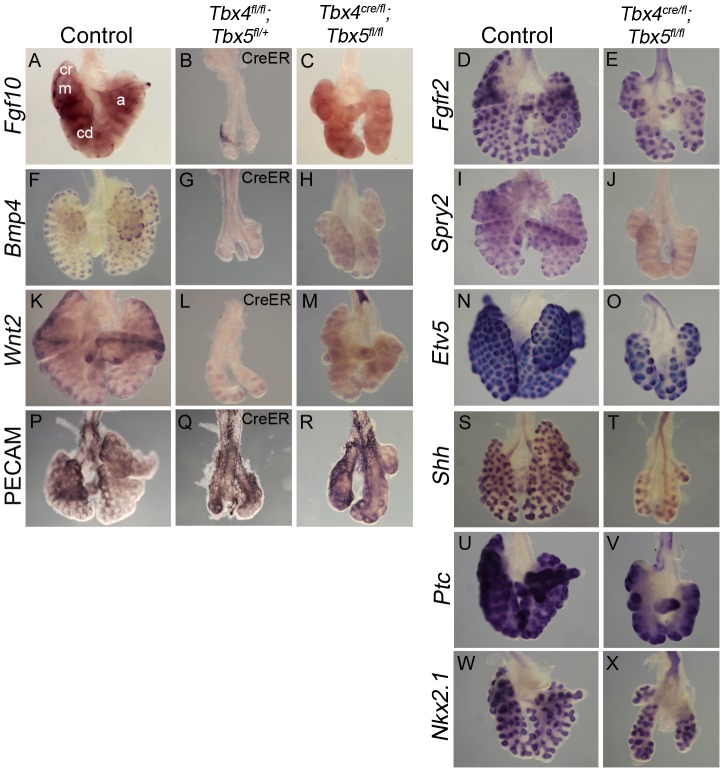
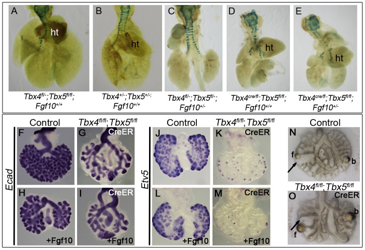
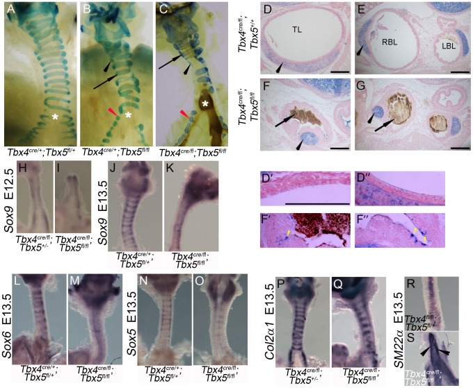
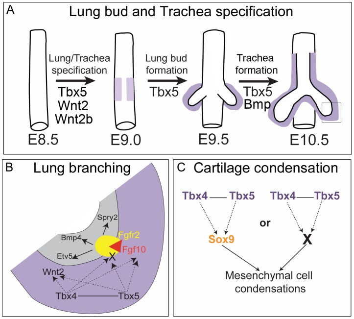
Normal development of the respiratory system is essential for survival and is regulated by multiple genes and signaling pathways. Both Tbx4 and Tbx5 are expressed throughout the mesenchyme of the developing lung and trachea; and, although multiple genes are known to be required in the epithelium, only Fgfs have been well studied in the mesenchyme. In this study, we investigated the roles of Tbx4 and Tbx5 in lung and trachea development using conditional mutant alleles and two different Cre recombinase transgenic lines. Loss of Tbx5 leads to a unilateral loss of lung bud specification and absence of tracheal specification in organ culture. Mutants deficient in Tbx4 and Tbx5 show severely reduced lung branching at mid-gestation. Concordant with this defect, the expression of mesenchymal markers Wnt2 and Fgf10, as well as Fgf10 target genes Bmp4 and Spry2, in the epithelium is downregulated. Lung branching undergoes arrest ex vivo when Tbx4 and Tbx5 are both completely lacking. Lung-specific Tbx4 heterozygous;Tbx5 conditional null mice die soon after birth due to respiratory distress. These pups have small lungs and show severe disruptions in tracheal/bronchial cartilage rings. Sox9, a master regulator of cartilage formation, is expressed in the trachea; but mesenchymal cells fail to condense and consequently do not develop cartilage normally at birth. Tbx4;Tbx5 double heterozygous mutants show decreased lung branching and fewer tracheal cartilage rings, suggesting a genetic interaction. Finally, we show that Tbx4 and Tbx5 interact with Fgf10 during the process of lung growth and branching but not during tracheal/bronchial cartilage development.


Conflict of interest statement

The authors have declared that no competing interests exist.

Figures

Figure 1
Figure 1. Expression of Tbx4 and Tbx5 in the developing lung and trachea.
(A–L) Tbx4 and Tbx5 expression analyzed using ISH on lungs. Tbx5 is first expressed at E9.0 (B,D) when the specification of lung primordia occurs, as seen by Nkx2.1 expression (A,C). Red arrows point to the posterior extent of the third pharyngeal pouch which marks the anterior of the expression domain of both Nkx2.1 and Tbx5. Right views (A,B), ventral views (C,D). Tbx4 is first expressed at E9.5 along with Tbx5 in the newly formed lung buds (E,F). Expression is seen in lung whole mounts at E11.5 (G,H), E13.5 (I,J) and in lung mesenchyme in cryosections at E15.5 (K,L). (M–S) Tbx4 and Tbx5 expression analyzed using ISH on tracheas. Tbx4 and Tbx5 are expressed throughout tracheal mesenchyme (m) at E13.5 (M,M′,N,N′) but not in the epithelium (e) or the mesothelium (arrowheads). Caudal view of cut tracheas after whole mount ISH (M–P). ISH on cryosections (M′–P′). At E13.5, Sox9 is expressed in the mesenchyme on the ventral side (O,O′) and SM22α is expressed on the dorsal side (P,P′) of the trachea. D-dorsal; V-ventral; R-right; L-left. At E15.5, Tbx4 and Tbx5 are expressed around the condensing cartilage mesenchyme and in the intercartilage mesenchyme (Q,R). Sox9 is expressed in the condensing cartilage rings (S). Asterisks indicate areas of cartilage condensation. Insets in (Q) and (S) show ISH on E15.5 sagittal cryosections with Tbx4 and Sox9 probe, respectively. Scale bars represent 50 µm.
Figure 2
Figure 2. Early loss of Tbx5 leads to aberrant lung bud and trachea specification.
(A–I) Foreguts were isolated between 8–16 somite stages and analyzed by ISH following culture. Nkx2.1 (A), Tbx4 (B) and Tbx5 (C) expression in lung buds (arrows) and tracheal primordia (yellow arrowhead) was confirmed in control cultures. Excision of Tbx5 using 4-OH tamoxifen in conditional null foreguts with CreER leads to the loss of Nkx2.1 expression in one of the lung buds at 3 (E) or 4 days (H). Conditional double nulls (F,I) carrying CreER show a phenotype similar to the conditional Tbx5 nulls, suggesting additional removal of Tbx4 does not exacerbate the phenotype. Nkx2.1 expression was absent in the foregut tube of conditional Tbx5 null and conditional double null foreguts after 3 or 4 days of culture (yellow arrowheads in E,F,H,I) compared to controls (D,G). Conditional Tbx5 nulls show reduced Wnt2 expression (K) and absence of Wnt2b expression (M) in the developing lung buds as compared to controls (J,L). Black arrowheads (J) point to Wnt2 expression in the heart in the controls. ht, heart; th, thyroid primordia.
Figure 3
Figure 3. Loss of Tbx4 and Tbx5 causes reduced lung branching and lethality at birth.
(A–H′) Tbx4fl and Tbx5fl alleles were excised using CreER by injecting tamoxifen at E8.75 and lungs were dissected at different time points. Loss of Tbx4 alone or both Tbx4 and Tbx5 leads to lung hypoplasia at E13.5 (A–D). The number of branching tips was quantitated following E-cadherin staining for different genotypes at E12.5 (E) and E13.5 (F). H & E on histological sections shows a decrease in lung size at E12.5 in the conditional Tbx4 null;Tbx5 heterozygous lungs (G,H,). (G′) and (H′) are magnified views of the boxed regions in (G) and (H), respectively. Black arrowheads indicate separation between the lobes of the right lung in the control (G′) and lack of separation in the mutants (H′). (I–Q) Tbx4cre was utilized for lung and trachea-specific excision of Tbx4fl and Tbx5fl alleles. Tbx4cre was expressed in most of the trachea mesenchyme (I) and lung mesenchymal cells (J) as seen by a R26RlacZ reporter expression at E13.5. Lung-specific Tbx4 heterozygous;Tbx5 null lungs show hypoplasia at E13.5 (K,L) and at birth (N,O). The number of branching tips in control lungs and the lung-specific Tbx4 heterozygous;Tbx5 null lungs, labeled as experimental in the box plot (M), are significantly different at E13.5. The green boxes contain 50% of the values; the median is indicated by a horizontal line in the box; bars represent the 5th and 95th percentiles. H & E staining on sections shows lung morphology for control (P) and mutants (Q) at birth. Scale bars represent 100 µm.
Figure 4
Figure 4. Loss of Tbx4 and Tbx5 leads to branching arrest ex vivo.
Lung buds were isolated at E11.5 and cultured in the presence of 4-OH tamoxifen. At the end of the culture airways were visualized using E-cadherin ISH. Conditional Tbx4 homozygous;Tbx5 heterozygous (B) and conditional Tbx4 heterozygous;Tbx5 null (C) lungs with CreER showed reduced branching after 4 days of culture compared to controls (A). Control, without CreER, (D) and conditional double null lung buds, with CreER, (E) were cultured for 4 days and photographed at 1 day, 2 days, 3 days and 4 days. Arrows in E show the progression of an elongating airway. A plot of the number of branching tips as a function of time for controls and the conditional double nulls (F), shows a branching arrest of the conditional double null lungs after 2 days of culture (Mann Whitney U test p = 0.036).
Figure 5
Figure 5. Loss of Tbx4 and Tbx5 affects the Fgf10 signaling pathway and Wnt2 expression.
(A–X) Marker analysis of control and Tbx4- and Tbx5-deficient lungs using whole mount ISH (A–O, S–X) and IHC (P–R): Fewer foci of Fgf10 expression were seen in the Tbx4 and Tbx5-deficient lungs (B,C) compared to control (A). cr, cranial; m, medial; cd, caudal; a, accesory lobes. Fgf10 target genes Bmp4 (F,G,H) and Spry2 (I,J) and canonical Wnt2 (K,L,M) were downregulated in Tbx4 and Tbx5-deficient lungs compared to controls. Fgfr2 (D,E), Etv5 (N,O), PECAM (P,Q,R), Shh (S,T), Ptc (U,V) and Nkx2.1 (W,X) were expressed similarly in controls and Tbx4 and Tbx5-deficient lungs.
Figure 6
Figure 6. Tbx4 and Tbx5 interact with Fgf10, but FGF10 fails to rescue Tbx4- and Tbx5-deficient lungs.
(A–E) Lungs of different genotypes at E18.5. Tbx4;Tbx5 double heterozygous lungs (B) are smaller than control lungs (A). Tbx4;Tbx5;Fgf10 triple heterozygous lungs (C) are smaller than the double heterozygous lungs (B) but comparable in size to the lung-specific Tbx4 heterozyous;Tbx5 null lungs (D). Removing an additional copy of Fgf10 from the lung-specific Tbx4 heterozyous;Tbx5 null lungs does not further affect lung size (E). ht, heart. (F–I) E-cad expression in epithelium of the control lungs and conditional double null lungs in the absence (F,G) and presence (H,I) of exogeneous Fgf10 showing a lack of rescue of branching morphogenesis. (J–M) Etv5 expression in control lungs and conditional double null lungs in the absence (J,K) and presence (L,M) of exogeneous Fgf10. Etv5 expression remains unchanged after addition of Fgf10. (N,O) Both control and conditional double null lungs respond to FGF10 coated beads (f) but not to BSA coated beads (b) as seen by swelling of tips (arrows) in proximity to the FGF10 bead.
Figure 7
Figure 7. Loss of Tbx4 and Tbx5 disrupts tracheal/bronchial cartilage and smooth muscle formation.
(A–G) Alcian blue staining was used to visualize tracheal/bronchial cartilage. Ten-eleven cartilage rings were seen in control tracheas (E18.5, A); normal (arrow) and incomplete rings were seen in tracheas and bronchi (black and red arrowheads respectively) of lung-specific Tbx5 null (E18.5, B) and lung-specific Tbx4 heterozygous;Tbx5 nulls (postnatal day (P) 0, C). Asterisks in (A–C) indicate the point of bronchial separation from the trachea. Alcian blue on P0 sections showed that the control trachea (D) and main stem bronchial lumens (E) were expanded and the chondrocytes were present as C shaped rings (arrowheads). The lung-specific Tbx4 heterozygous;Tbx5 null tracheal lumen (F) and main stem bronchial lumen (G) were collapsed and filled with a mucus like substance (arrows) and alcian blue positive foci were seen (black arrowheads). Higher magnification views of control (D′,D″) and lung-specific Tbx4 heterozygous;Tbx5 null (F′,F″) tracheas. Yellow arrowheads indicate alcian blue positive mucus-producing cells in the lung-specific Tbx4 heterozygous;Tbx5 null tracheas. TL, Tracheal lumen; RBL, Right bronchial lumen; LBL, Left bronchial lumen. (H–S) Genes important for chondrogenesis and smooth muscle development. Sox9 expression at E12.5 showed a comparable ventral expression pattern in control (H) and lung-specific Tbx4 heterozygous;Tbx5 null tracheas (I). At E13.5, Sox9 positive cells begin to condense and appear in a ring like pattern in controls (J) but not in the mutant tracheas (K). Sox6 (L,M) and Sox5 (N,O) expression was downregulated in the lung-specific Tbx4 heterozygous;Tbx5 null tracheas at E13.5. Col2α1 was expressed in its characteristic ring like pattern (P) similar to Sox9 in the control tracheas but there were fewer rings with apparently normal expression in the lung-specific Tbx4 heterozygous;Tbx5 null tracheas (Q). SM22α expression was analyzed on the dorsal trachea at E13.5 in the control (R) and in the lung-specific Tbx4 heterozygous;Tbx5 null tracheas (S). Arrowheads in (S) point to ectopic expression in an uncharacteristic pattern in the mutants. Scale bars represent 100 µm.
Figure 8
Figure 8. Lack of genetic interactions between Tbx4, Tbx5, and Fgf10 in trachea formation.
Alcian blue staining was used to analyze tracheal/bronchial cartilage development in tracheas from different genotypes at E18.5. Control tracheas (A), Tbx4;Fgf10 double heterozygous tracheas (B) and Tbx5;Fgf10 double heterozygous tracheas (C) have 10–11 C-shaped, ventral tracheal cartilage rings and the trachea forms the two main stem bronchi which have lateral C-shaped cartilage rings. Fgf10 homozygous mutants (D), Tbx4;Tbx5 double heterozygous mutants (E) and Tbx4;Tbx5;Fgf10 triple heterozygous mutants (F) each form fewer (6–8) tracheal cartilage rings, some irregularly shaped or incomplete (arrowheads). Additionally, Fgf10 mutants lack bronchi and hence any bronchial cartilage rings (D), but the double and triple heterozygotes form normal lateral bronchial cartilage rings (arrows in E and F). Lung-specific Tbx4 heterozygous;Tbx5 nulls (G) show severe disruptions in formation of tracheal/bronchial cartilage rings, a phenotype that is unchanged with the removal of an Fgf10 allele (H). Asterisk shows the point of bronchial separation from the trachea.
Figure 9
Figure 9. Model for the role of Tbx4 and Tbx5 in lung and trachea development.
(A) Lung and trachea specification begins at E9.0 in the ventral foregut and at this time Tbx5 expression (light purple) is adjacent to the presumptive endoderm. Later, Tbx4 and Tbx5 expression (dark purple) is in mesenchyme associated with the lung and trachea. Tbx5 but not Tbx4 is important for specification of bilateral lung buds and the trachea. (B) Magnification of box shown in (A) representing the events in the growing tip during branching morphogenesis. Grey denotes epithelium and purple denotes mesenchyme. Tbx4 and Tbx5 interact with each other and act upstream of the Fgf10 signaling pathway. Decrease in Tbx4 and Tbx5 affects mesenchymal Fgf10 expression and expression of its targets in the epithelium – Bmp4, Spry2 and Etv5 – but not the expression of the epithelial Fgf10 receptor Fgfr2. In addition to Fgf10 expression in the mesenchyme, Tbx4 and Tbx5 also control the expression of an unknown factor(s) (X) that is essential for activation of the Fgf10 signaling pathway. Furthermore, Tbx4 and Tbx5 act upstream of Wnt2 in the mesenchyme. (C) In the trachea and the main stem bronchi Tbx4 and Tbx5 either control Sox9 expression, which in turn regulates cartilage condensation, or Tbx4 and Tbx5 regulate another factor (X) essential for chondrogenesis secondarily affecting Sox9 expression.

References

    1. Whitsett JA, Wert SE, Trapnell BC. Genetic disorders influencing lung formation and function at birth. Hum Mol Genet. 2004;13 Spec No 2:R207–215. - PubMed
    1. Cardoso WV, Lu J. Regulation of early lung morphogenesis: questions, facts and controversies. Development. 2006;133:1611–1624. - PubMed
    1. Spooner BS, Wessells NK. Mammalian lung development: interactions in primordium formation and bronchial morphogenesis. J Exp Zool. 1970;175:445–454. - PubMed
    1. Metzger RJ, Klein OD, Martin GR, Krasnow MA. The branching programme of mouse lung development. Nature. 2008;453:745–750. - PMC - PubMed
    1. Goss AM, Tian Y, Tsukiyama T, Cohen ED, Zhou D, et al. Wnt2/2b and [beta]-catenin signaling are necessary and sufficient to specify lung progenitors in the foregut. Developmental Cell. 2009;17:290–298. - PMC - PubMed

Publication types

MeSH terms