. 2012 Aug 2:3:142.
doi: 10.3389/fphar.2012.00142. eCollection 2012.

The KCNE Tango - How KCNE1 Interacts with Kv7.1

Affiliations

The KCNE Tango - How KCNE1 Interacts with Kv7.1

Eva Wrobel et al. Front Pharmacol. .

Abstract

The classical tango is a dance characterized by a 2/4 or 4/4 rhythm in which the partners dance in a coordinated way, allowing dynamic contact. There is a surprising similarity between the tango and how KCNE β-subunits "dance" to the fast rhythm of the cell with their partners from the Kv channel family. The five KCNE β-subunits interact with several members of the Kv channels, thereby modifying channel gating via the interaction of their single transmembrane-spanning segment, the extracellular amino terminus, and/or the intracellular carboxy terminus with the Kv α-subunit. Best studied is the molecular basis of interactions between KCNE1 and Kv7.1, which, together, supposedly form the native cardiac I(Ks) channel. Here we review the current knowledge about functional and molecular interactions of KCNE1 with Kv7.1 and try to summarize and interpret the tango of the KCNEs.

Keywords: KCNE1; Kv channel; Kv7.1; β-subunit.


Figures
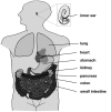
Figure 1
Figure 1
Distribution of Kv7.1. Kv7.1 is expressed in several tissues throughout the human body, including heart, lung, inner ear, kidney, and the gastrointestinal tract.
Figure 2
Figure 2
Characteristics of Kv7.1/KCNE-mediated currents. (A) Representative current traces of Kv7.1 homomers and Kv7.1/KCNE1 heteromers. Channels were expressed in Xenopus oocytes, and currents were elicited with 7 s pulses to potentials of −100 to +60 mV, applied in 20 mV increments from a holding potential of −80 mV. Tail currents were recorded at −120 mV. (B) Current-voltage relationships. At voltages between −60 and −40 mV KCNE1 suppresses currents, whereas it stimulates them at voltages above 0 mV. (C) Voltage dependence of channel activation determined by tail current analysis. Activation curves were fitted to a Boltzmann function. Note: Kv7.1/KCNE1 channels are not fully activated at +60 mV. (D) Effects of different KCNE subunits on Kv7.1 currents. “+” and “++” indicate increased and strongly increased effects, while “−” and “−−” indicate decreased and strongly decreased effects, respectively. *Effect shown in the calmodulin binding-deficient KCNE4L69–L72 mutant (Ciampa et al., 2011). The Rb+/K+ conductance tightly correlates with the partial inactivation, and KCNE5 slightly increases it, whereas KCNE1/3 decreases it compared to Kv7.1 (Seebohm et al., 2003c).
Figure 3
Figure 3
Topology and stoichiometry of Kv7.1/KCNE1 channels. (A) Kv7.1 α-subunits are made of six membrane-spanning segments S1–S6 and intracellular N- and C-terminal domains. The segments S1–S4 form the voltage-sensing domain (VSD), while the pore domain (PD) consists of segments S5 and S6. KCNE1 β-subunits contain a single transmembrane segment flanked by an extracellular N-terminus and a cytosolic C-terminus. (B) Four Kv7.1 α-subunits assemble to form a functional channel. The number of KCNE1 β-subunits associating with Kv7.1 tetramers is still a matter of extensive debate (Strutz-Seebohm et al., 2011).
Figure 4
Figure 4
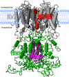
Structural model of the Kv7.1 channel. The transmembrane domain of the Kv7.1 channel bears the common Kv channel structure. Homology models using the solved crystal structural constraints allow for good model predictions (gray). For docking experiments the solved NMR-structural data of the full-length KCNE1 in lipid environments can be used (here only the transmembrane segment is shown in red; Tian et al., ; Kang et al., 2008). The structure of the Kv7.1 tetrameric assembly domain (A-domain) was solved and can be incorporated into a model (Xu and Minor, 2009). The region linking the S6 and the A-domain shows amino acid similarity to the structure 1VNS.pdb. No structures or even template coordinates for the Kv7.1 N-terminus and the distal C-terminus are available and thus homology modeling is currently problematic.
Figure 5
Figure 5
Dynamic model of gating transitions in Kv7.1/KCNE1 channels. The closed (C), pre-open (C*), and open (O) state models described by Smith et al. (2007) and Strutz-Seebohm et al. (2011) were used to generate dynamic models using simple morphing approaches. The gating of Kv7.1/KCNE1 channels can be approximated by a simplified circular gating model. Major gating steps are C ⇒ C* (A), C* ⇒ O (B), and O ⇒ C (C) (colors as indicated in the letters above the models). For clarity the central axes of the KCNE1 start and end models are marked by the yellow dashed lines, and the direction and extent of the proposed motion is indicated by the yellow arrow. In (A) the KCNE1 start model is shown in red and the end model in magenta. The morphs indicate larger motions of KCNE1 during channel gating in the gating cleft of Kv7.1 channels. A particularly large gating motion around Thr58 is seen from C* ⇒ O (B), suggesting a major energetic barrier. This may be the molecular basis of the dramatically slowed activation of Kv7.1 by association with KCNE1. However, calculation of energies on the models is highly speculative because specific interactions with surrounding membrane molecules [e.g., PI(4,5)P2] would be highly influential, and the current knowledge on lipid-channel interactions is not sufficient to allow for precise calculations.

References

    1. Abbott G. W., Sesti F., Splawski I., Buck M. E., Lehmann M. H., Timothy K. W., Keating M. T., Goldstein S. A. (1999). MiRP1 forms IKr potassium channels with HERG and is associated with cardiac arrhythmia. Cell 97, 175–18710.1016/S0092-8674(00)80728-X - DOI - PubMed
    1. Aggeli A., Bannister M. L., Bell M., Boden N., Findlay J. B., Hunter M., Knowles P. F., Yang J. C. (1998). Conformation and ion-channeling activity of a 27-residue peptide modeled on the single-transmembrane segment of the IsK (minK) protein. Biochemistry 37, 8121–813110.1021/bi972112h - DOI - PubMed
    1. Angelo K., Jespersen T., Grunnet M., Nielsen M. S., Klaerke D. A., Olesen S. P. (2002). KCNE5 induces time- and voltage-dependent modulation of the KCNQ1 current. Biophys. J. 83, 1997–200610.1016/S0006-3495(02)73961-1 - DOI - PMC - PubMed
    1. Arrighi I., Bloch-Faure M., Grahammer F., Bleich M., Warth R., Mengual R., Drici M. D., Barhanin J., Meneton P. (2001). Altered potassium balance and aldosterone secretion in a mouse model of human congenital long QT syndrome. Proc. Natl. Acad. Sci. U.S.A. 98, 8792–879710.1073/pnas.141233398 - DOI - PMC - PubMed
    1. Bähring R., Barghaan J., Westermeier R., Wollberg J. (2012). Voltage sensor inactivation in potassium channels. Front. Pharmacol. 3:100.10.3389/fphar.2012.00100 - DOI - PMC - PubMed