. 2012 Jul 2:3:143.
doi: 10.3389/fpls.2012.00143. eCollection 2012.

ER Import Sites and Their Relationship to ER Exit Sites: A New Model for Bidirectional ER-Golgi Transport in Higher Plants

Affiliations

ER Import Sites and Their Relationship to ER Exit Sites: A New Model for Bidirectional ER-Golgi Transport in Higher Plants

Alexander Lerich et al. Front Plant Sci. .

Abstract

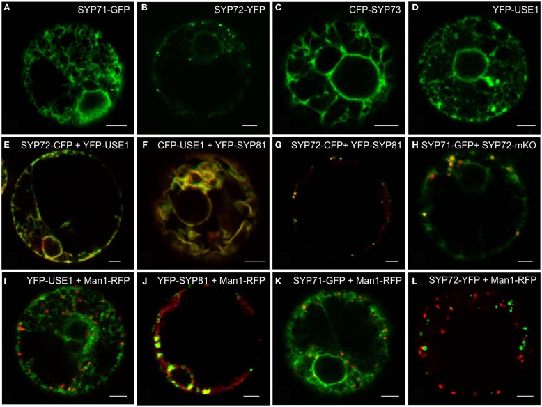
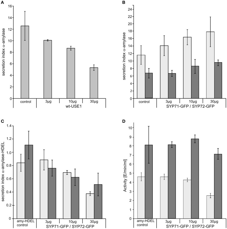
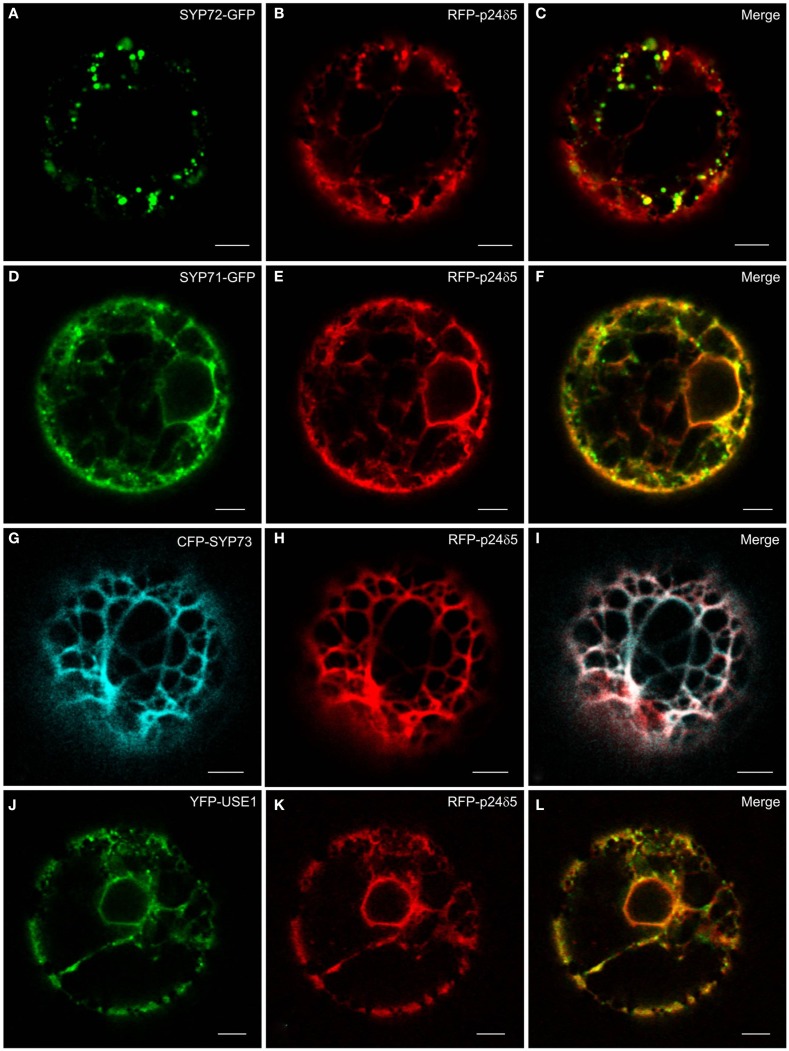
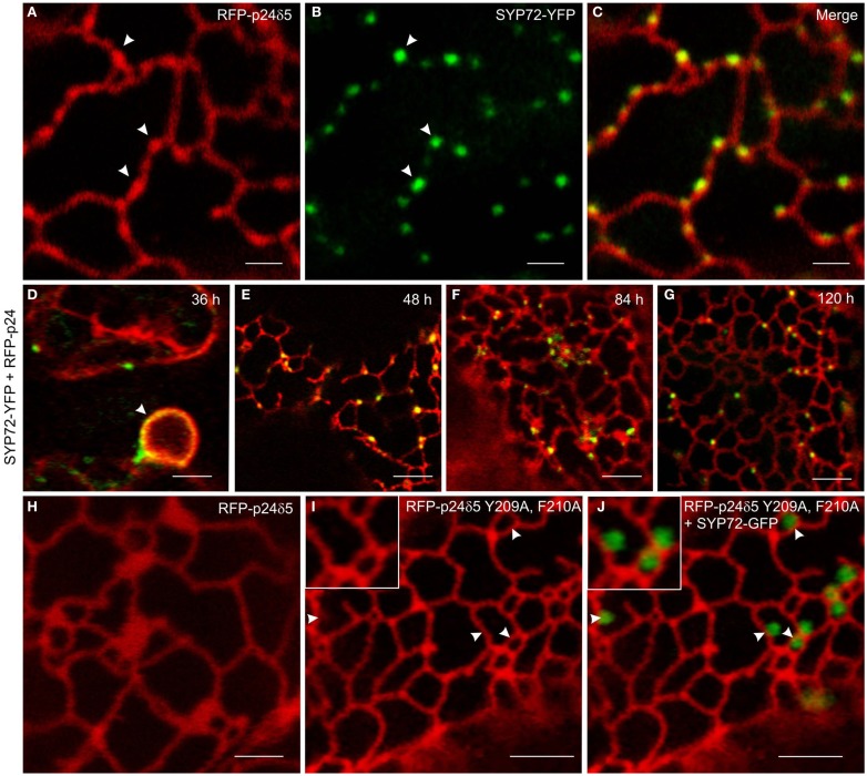
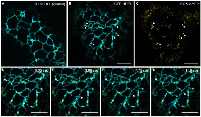
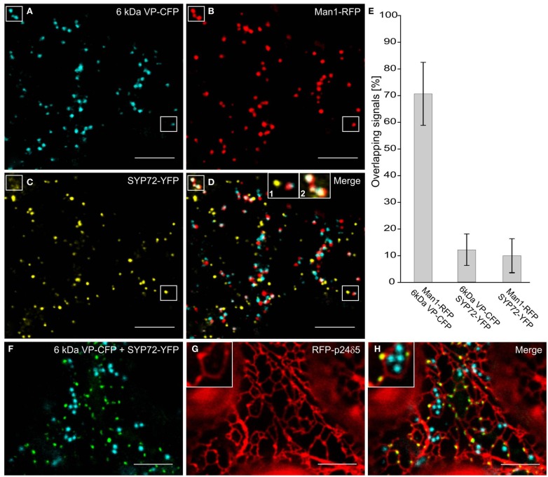
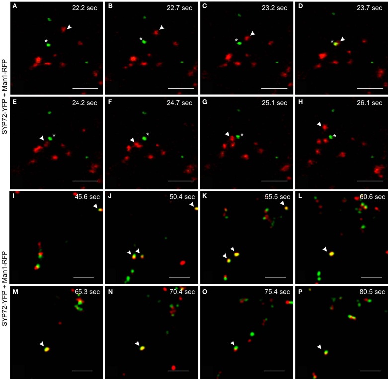
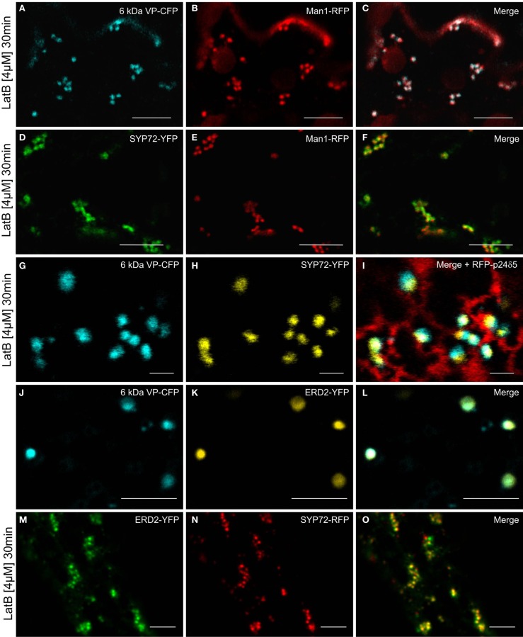
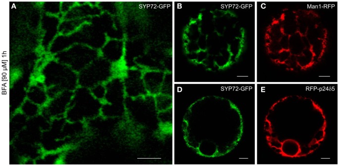
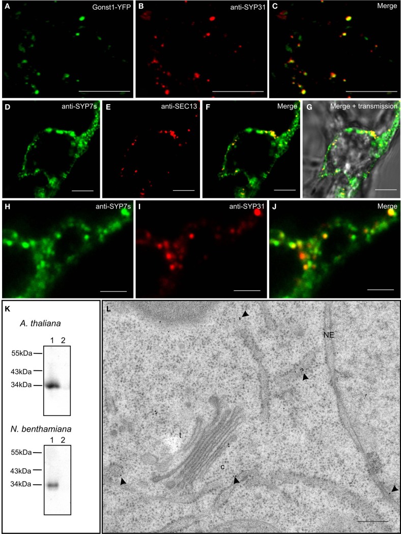
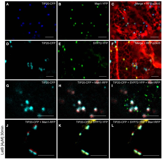
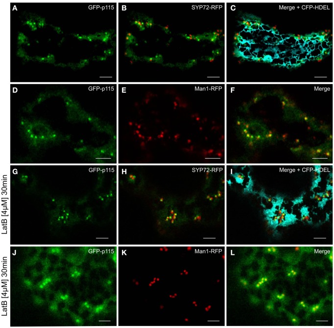
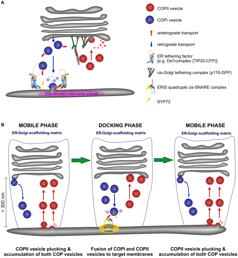
Per definition, ER exit sites are COPII vesiculation events at the surface of the ER and in higher plants are only visualizable in the electron microscope through cryofixation techniques. Fluorescent COPII labeling moves with Golgi stacks and locates to the interface between the ER and the Golgi. In contrast, the domain of the ER where retrograde COPI vesicles fuse, i.e., ER import sites (ERIS), has remained unclear. To identify ERIS we have employed ER-located SNAREs and tethering factors. We screened several SNAREs (SYP81, the SYP7 family, and USE1) to find a SNARE whose overexpression did not disrupt ER-Golgi traffic and which gave rise to discrete fluorescent punctae when expressed with an XFP tag. Only the Qc-SNARE SYP72 fulfilled these criteria. When coexpressed with SYP72-YFP, both the type I-membrane protein RFP-p24δ5 and the luminal marker CFP-HDEL whose ER localization are due to an efficient COPI-mediated recycling, form nodules along the tubular ER network. SYP72-YFP colocalizes with these nodules which are not seen when RFP-p24δ5 or CFP-HDEL is expressed alone or when SYP72-YFP is coexpressed with a mutant form of RFP-p24δ5 that cannot exit the ER. SYP72-YFP does not colocalize with Golgi markers, except when the Golgi stacks are immobilized through actin depolymerization. Endogenous SYP7 SNAREs, also colocalize with immobilized COPII/Golgi. In contrast, XFP-tagged versions of plant homologs to TIP20 of the Dsl1 COPI-tethering factor complex, and the COPII-tethering factor p115 colocalize perfectly with Golgi stacks irrespective of the motile status. These data suggest that COPI vesicle fusion with the ER is restricted to periods when Golgi stacks are stationary, but that when moving both COPII and COPI vesicles are tethered and collect in the ER-Golgi interface. Thus, the Golgi stack and an associated domain of the ER thereby constitute a mobile secretory and recycling unit: a unique feature in eukaryotic cells.

Keywords: ER export sites; ER import sites; Golgi motility; SNAREs; secretory unit; tobacco.


Figures

Figure 1
Figure 1
Screening for punctate ER-localized SNAREs in tobacco mesophyll protoplasts. Protoplasts were electroporated with plasmid DNA (up to 30 μg) encoding for SYP71-GFP (A,H), SYP72-(X)FP (B,E,G,H,K), CFP-SYP73 (C), or (X)FP-USE1 (D–F,I), either alone, in various combinations or electroporated together with the cis-Golgi marker Man1-RFP (I–K). CFP fluorescence is depicted in green (E–G), YFP in red (E–G), GFP in green (H), mKO in red (H). Man1-RFP signals are always shown in red (I–L). All images are of single optical sections. Magnification bars = 5 μm.
Figure 2
Figure 2
The effects of ER-SNARE overexpression on secretion and retrograde Golgi-ER transport in tobacco protoplasts. Protoplasts were co-electroporated with a constant amount of plasmid DNA encoding for α-amylase and increasing amounts of plasmid DNA encoding for full-length wtUSE1 (A), SYP71-GFP [(B) light gray], or SYP72-GFP [(B) dark gray]. SYP71-GFP [(C) light gray], and SYP72-GFP [(C) dark gray], respectively, were also coexpressed with α-amylase-HDEL which is normally retained in the ER by efficient COPI-mediated retrograde transport from the Golgi. Secreted and protoplast retained levels of α-amylase activity were measured and the secretion index calculated as described in Materials and Methods. The first column shows as a control the expression of the reporter alone, the other columns the coexpression with constructs in different concentrations as indicated below the axis. USE1 inhibits secretion (A) while SYP71, SYP72 do not (B). Expression of either SYP71-GFP or SYP72-GFP lowers the secretions index of α-amylase-HDEL indicating a more efficient retrograde Golgi-ER transport (C). Total activity for SYP71-GFP [(D) light gray] or SYP72-GFP [(D) dark gray] decreased only at very high amounts of electroporated DNA. Standard deviations are given as vertical lines in each column.
Figure 3
Figure 3
The ER marker RFP-p24δ5 forms nodules in the presence of SYP72. Tobacco mesophyll protoplasts were co-electroporated with RFP-p24δ5 (10 μg) and 15 μg of one ER-SNARE [SYP72-GFP (A–C), SYP71-GFP (D–F), CFP-SYP73 (G–I), or YFP-USE1 (J–L)]. In addition to a general staining of the ER network the red RFP-p24δ5 signal is also present as punctae which colocalize exactly with the punctate green SYP72-GFP signals. This phenomenon was partially recognizable in the case of SYP71 and USE1 but was not at all visible with SYP73. All images are of single optical sections. Magnification bars = 5 μm.
Figure 4
Figure 4
Overexpression of SYP72 enhances retrograde Golgi-ER traffic and leads to the formation of p24δ5 nodules in the ER. (A–C) Coexpression of RFP-p24δ5 and SYP72-YFP in agroinfiltrated tobacco leaf epidermal cells leads to production of nodules in the RFP-p24δ5 stained ER tubular network. These nodules label positively with SYP72-YFP (indicated by arrowheads). (D–G) SYP72-YFP and RFP-p24δ5 fluorescence are first detected after 36 h in the nuclear envelope (arrow) and persist for at least 120 h post-infiltration. For images (A–G), the YFP signal is presented in green for easier viewing. (H–J) Coexpression of SYP72-GFP with a mutant form of RFP-p24δ5 (which cannot be exported out of the ER – see Materials and Methods) in tobacco leaf epidermal cells. As with leaf cells expressing only RFP-p24δ5, nodules in the ER tubular network are not present [compare (H) with (I)], and the SYP72-GFP punctate signals are distributed randomly on the surface of the ER (J). Magnification bars = 2 μm (A–C,H); 5 μm (D–G,I,J).
Figure 5
Figure 5
CFP-HDEL forms nodules only when coexpressed with SYP72-YFP. (A–C) CFP-HDEL expressed alone labels the ER uniformly (A) but forms nodules when coexpressed with SYP72-YFP (B,C) in tobacco leaf epidermal cells. Arrows point to colocalization of nodules with SYP72-YFP punctae. (D–G) Frames of a movie (CFP-HDEL in cyan; SYP72-YFP in yellow) depicting a growing tubule (arrowhead; Movie S1 in Supplementary Material). Magnification bars = 5 μm (A–G).
Figure 6
Figure 6
Punctate SYP72-YFP signals seldom colocalize with ERES and Golgi markers in normal cells. Tobacco leaves were triple agroinfiltrated with 6 kDa VP-CFP (A), Man1-RFP (B), SYP72-YFP (C). Although a high degree of colocalization between the blue COPII (6 kDa VP-CFP) and red Golgi (Man1-RFP) markers was observed, there was little overlap between the signals for SYP72-YFP (yellow) and the COPII and Golgi markers (D–E). A case of colocalization between SYP72-YFP and Man1-RFP punctae is indicated in the rectangles in (A–C), and in inset 2 in (D) When tobacco leaves were triple agroinfiltrated with 6 kDa VP-CFP, SYP72-YFP, and RFP-p24δ5 a clear distinction can be made between the punctate SYP72-YFP signals which lie directly on the tubular ER network (see insets in (G,H) for nodules) and the Golgi-associated ERES marker [6 kDa VP-CFP; (F–H)]. Magnification bars = 10 μm (A–D,F–H).
Figure 7
Figure 7
Mobilities and transient colocalization of Man1-RFP and SYP72-YFP as observed in videos of untreated tobacco leaf epidermal cells. (A–H) Eight frames taken at 0.5–1 s intervals from a movie sequence of the SYP72-YFP (shown in green) and Man1-RFP (in red) images (movie available as Movie S2 in Supplementary Material). A Man1-RFP puncta (arrowhead) is seen moving toward and over a SYP72-YFP puncta (star). (I–P) SYP72-YFP (in green) and Man1-RFP (in red) are shown in these eight frames (5 s interval; movie available as Movie S3 in Supplementary Material). Several examples of temporary colocalization between SYP72-YFP and Man1-RFP signals are to be seen (arrowhead). Magnification bars = 5 μm (A–P).
Figure 8
Figure 8
Actin depolymerization causes SYP72, Golgi, and ERES signals to colocalize in clusters. Tobacco leaves were double agroinfiltrated with either 6 kDa VP-CFP and Man1-RFP (A–C), or with SYP72-YFP and Man1-RFP (D–F), or triple agroinfiltrated with 6 kDa VP-CFP, SYP72-YFP, and RFP-p24δ5 (G–I). ERD2-YFP and 6 kDa VP-CFP signals were observed in untreated cells (J–L) and ERD2-YFP and SYP72-RFP fluorescence after latrunculin B treatment (M–O). Segments of the agroinfiltrated leaves were cut out 2 days post-infiltration and incubated for 30 min with latrunculin B (4 μM) before observation in the CLSM. Magnification bars = 10 μm (A–F); 1 μm (G–I); 5 μm (J–O).
Figure 9
Figure 9
BFA prevents formation of SYP72 punctae. Pretreatment with BFA (90 μM; 1 h) leads to a general ER distribution of SYP72-GFP signals in leaf epidermal cells (A), and protoplasts (B–E) when coexpressed with the Golgi marker Man1-RFP (C) or the ER marker RFP-p24δ5 (E). Magnification bars (A–E) = 5 μm.
Figure 10
Figure 10
Endogenous SYP7 proteins colocalize with ERES and Golgi stacks. (A–C) Immunofluorescent localization of the syntaxin SYP31 to Golgi stacks in transgenic tobacco BY-2 cells expressing the Golgi marker GONST1-YFP. (D–J) Double immunofluorescence on tobacco BY-2 cells using antibodies against the SYP7 family and the COPII protein SEC13 (D–G) or the syntaxin SYP31 (H–J). (K) Western blots showing the specificity of the SYP7 antiserum in total membrane preparations (lanes 1) from Arabidopsis and tobacco leaves (lane 2 is the 100,000-g supernatant as a negative control). (L) Immunogold labeling with SYP7 antibodies of a thin section cut from a high pressure frozen Arabidopsis root tip. Arrowheads point to gold particles. c, cis-Golgi; t, trans-Golgi; NE, nuclear envelope. Magnification bars = 5 μm (A–G); 1 μm (H–J); 300 nm (L).
Figure 11
Figure 11
Golgi and ER-tethering factors colocalize to mobile Golgi stacks. Tobacco leaves were triple agroinfiltrated with either TIP20-CFP (A), Man1-YFP (B), and RFP-p24δ5 (C) or TIP20-CFP (D), SYP72-YFP (E), and RFP-p24δ5 (F). Triple infiltration of TIP20-CFP, Man1-RFP, and SYP72 was observed in untreated (G–I) and latrunculin B (4 μM, 30 min) treated cells (J–L). Magnification bars = 5 μm.
Figure 12
Figure 12
GFP-p115 colocalizes with a cis-Golgi marker but not with SYP72. In agroinfiltrated tobacco leaves GFP-p115 and SYP72-RFP only overlapped in latrunculin B treated cells [compare (A–C) with(G–I)]. A high degree of colocalization was detected between GFP-p115 and Man1-RFP before (D–F) and after latrunculin B treatment (J–L). Magnification bars = 5 μm.
Figure 13
Figure 13
Anterograde and retrograde traffic between the ER and the Golgi in higher plants is accomplished by a short-range secretory and recycling unit. (A) Cartoon showing the close proximity of ERES and ERIS in a domain of the ER which in size approximates the diameter of the Golgi stack. (B) This cartoon represents a new model for ER-Golgi transport in higher plants. Golgi stacks move intermittently over the surface of the ER. They are in tight association with the ER through a joint scaffolding matrix of tethering factors (dotted blue line). As Golgi stacks move they capture individual COPII vesicles released from ERES at the surface of the ER (“mobile phase”) and release COPI vesicles. Both types of vesicles accumulate in the ER-Golgi interface. When the Golgi stacks temporarily stop moving (“docking phase”) fusion of COPI and COPII vesicles to their respective target membranes occurs. Golgi stacks stop at ERIS as marked by the presence of “SYP72 nodules” on the ER.

References

    1. Allan B. B., Moyer B. D., Balch W. E. (2000). Rab1 recruitment of p115 into a cis-SNARE complex: programming budding COPII vesicles for fusion. Science 289, 444–44810.1126/science.289.5478.444 - DOI - PubMed
    1. Aoki T., Kojima M., Tani K., Tagaya M. (2008). Sec22b-dependent assembly of endoplasmic reticulum Q-SNARE proteins. Biochem. J. 410, 93–10010.1042/BJ20071304 - DOI - PubMed
    1. Appenzeller-Herzog C., Hauri H.-P. (2006). The ER-Golgi intermediate compartment (ERGIC): in search of its identity and function. J. Cell Sci. 119, 2173–218310.1242/jcs.03019 - DOI - PubMed
    1. Avisar D., Abu-Abied M., Belausov E., Sadot E., Hawes C., Sparkes I. A. (2009). A comparative study of the involvement of 17 Arabidopsis myosin family members on the motility of Golgi and other organelles. Plant Physiol. 150, 700–70910.1104/pp.109.136853 - DOI - PMC - PubMed
    1. Avisar D., Prokhnevsky A. I., Makarova K. S., Koonin E. V., Dolja V. V. (2008). Myosin XI-K is required for rapid trafficking of Golgi stacks, peroxisomes, and mitochondria in leaf cells of Nicotiana benthamiana. Plant Physiol. 146, 1098–110810.1104/pp.107.113647 - DOI - PMC - PubMed