

Host modulators of H1N1 cytopathogenicity
- PMID: 22876275
- PMCID: PMC3410888
- DOI: 10.1371/journal.pone.0039284
Host modulators of H1N1 cytopathogenicity
Abstract
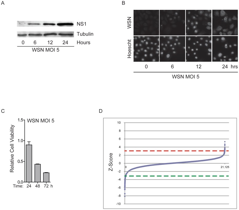
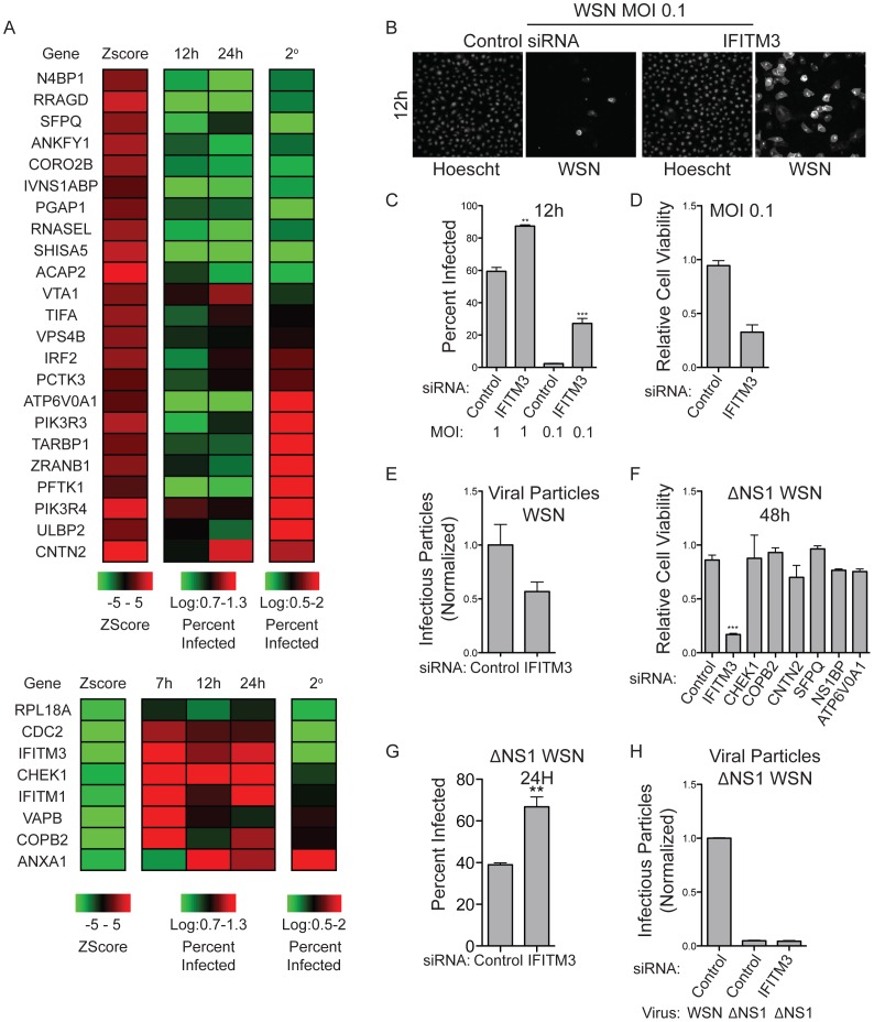
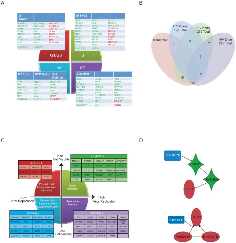
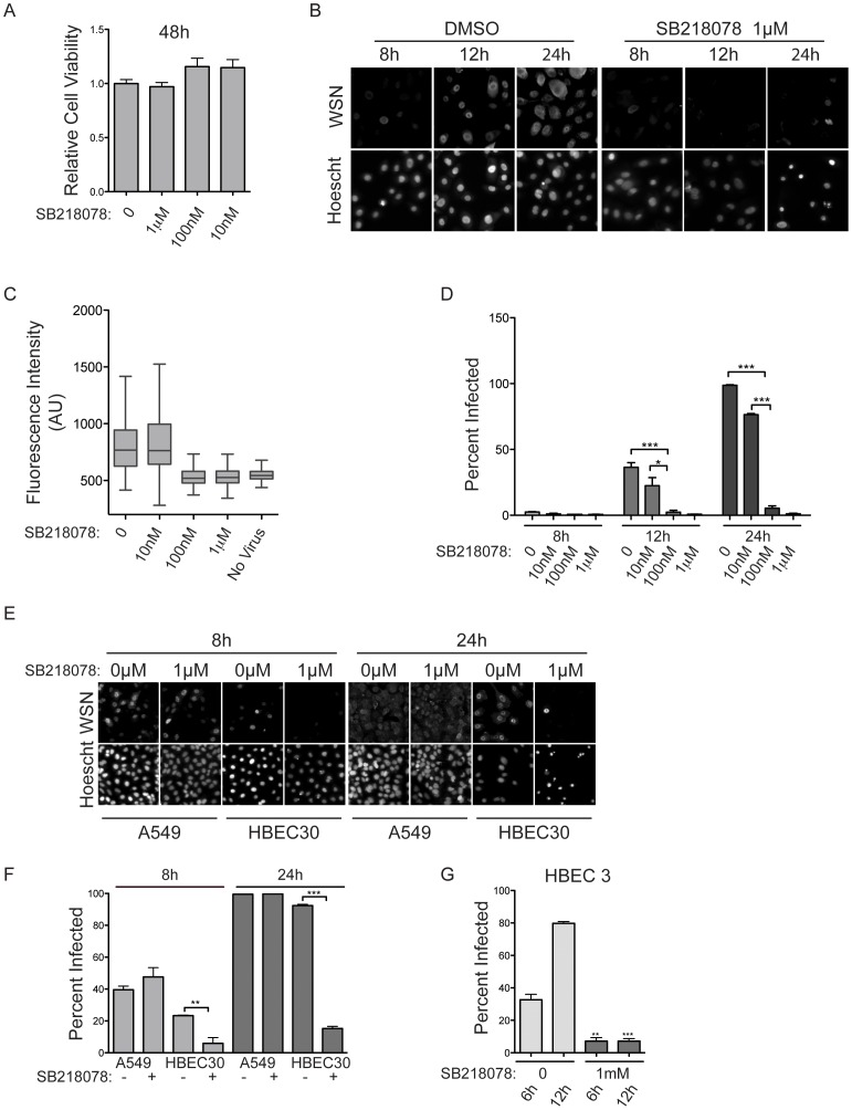
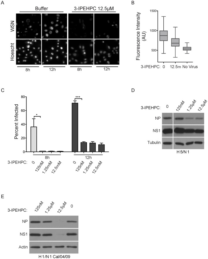
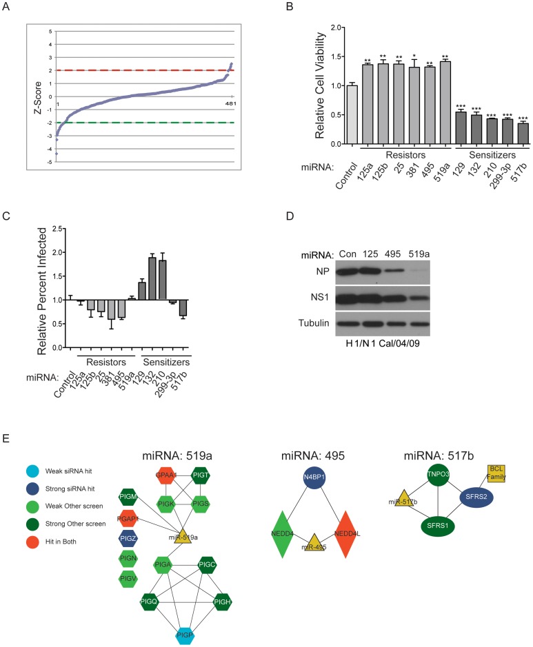
Influenza A virus infects 5-20% of the population annually, resulting in ~35,000 deaths and significant morbidity. Current treatments include vaccines and drugs that target viral proteins. However, both of these approaches have limitations, as vaccines require yearly development and the rapid evolution of viral proteins gives rise to drug resistance. In consequence additional intervention strategies, that target host factors required for the viral life cycle, are under investigation. Here we employed arrayed whole-genome siRNA screening strategies to identify cell-autonomous molecular components that are subverted to support H1N1 influenza A virus infection of human bronchial epithelial cells. Integration across relevant public data sets exposed druggable gene products required for epithelial cell infection or required for viral proteins to deflect host cell suicide checkpoint activation. Pharmacological inhibition of representative targets, RGGT and CHEK1, resulted in significant protection against infection of human epithelial cells by the A/WS/33 virus. In addition, chemical inhibition of RGGT partially protected against H5N1 and the 2009 H1N1 pandemic strain. The observations reported here thus contribute to an expanding body of studies directed at decoding vulnerabilities in the command and control networks specified by influenza virulence factors.
Conflict of interest statement
Figures
Similar articles
-
Genome-wide RNAi screen identifies human host factors crucial for influenza virus replication.Nature. 2010 Feb 11;463(7282):818-22. doi: 10.1038/nature08760. Epub 2010 Jan 17. Nature. 2010. PMID: 20081832
-
Highly Pathogenic H5N1 and Novel H7N9 Influenza A Viruses Induce More Profound Proteomic Host Responses than Seasonal and Pandemic H1N1 Strains.J Proteome Res. 2015 Nov 6;14(11):4511-23. doi: 10.1021/acs.jproteome.5b00196. Epub 2015 Oct 9. J Proteome Res. 2015. PMID: 26381135
-
Influenza H5N1 and H1N1 virus replication and innate immune responses in bronchial epithelial cells are influenced by the state of differentiation.PLoS One. 2010 Jan 15;5(1):e8713. doi: 10.1371/journal.pone.0008713. PLoS One. 2010. PMID: 20090947 Free PMC article.
-
Impact of Influenza A Virus Infection on the Proteomes of Human Bronchoepithelial Cells from Different Donors.J Proteome Res. 2017 Sep 1;16(9):3287-3297. doi: 10.1021/acs.jproteome.7b00286. Epub 2017 Aug 16. J Proteome Res. 2017. PMID: 28745058
-
Interplay of PA-X and NS1 Proteins in Replication and Pathogenesis of a Temperature-Sensitive 2009 Pandemic H1N1 Influenza A Virus.J Virol. 2017 Aug 10;91(17):e00720-17. doi: 10.1128/JVI.00720-17. Print 2017 Sep 1. J Virol. 2017. PMID: 28637750 Free PMC article.
Cited by
-
Common and species-specific molecular signatures, networks, and regulators of influenza virus infection in mice, ferrets, and humans.Sci Adv. 2022 Oct 7;8(40):eabm5859. doi: 10.1126/sciadv.abm5859. Epub 2022 Oct 5. Sci Adv. 2022. PMID: 36197970 Free PMC article.
-
Integrative gene network analysis identifies key signatures, intrinsic networks and host factors for influenza virus A infections.NPJ Syst Biol Appl. 2017 Dec 4;3:35. doi: 10.1038/s41540-017-0036-x. eCollection 2017. NPJ Syst Biol Appl. 2017. PMID: 29214055 Free PMC article.
-
Meta- and Orthogonal Integration of Influenza "OMICs" Data Defines a Role for UBR4 in Virus Budding.Cell Host Microbe. 2015 Dec 9;18(6):723-35. doi: 10.1016/j.chom.2015.11.002. Cell Host Microbe. 2015. PMID: 26651948 Free PMC article.
-
Molecular and genetic inflammation networks in major human diseases.Mol Biosyst. 2016 Jul 19;12(8):2318-41. doi: 10.1039/c6mb00240d. Mol Biosyst. 2016. PMID: 27303926 Free PMC article. Review.
-
Strain-Specific Contribution of Eukaryotic Elongation Factor 1 Gamma to the Translation of Influenza A Virus Proteins.Front Microbiol. 2018 Jun 29;9:1446. doi: 10.3389/fmicb.2018.01446. eCollection 2018. Front Microbiol. 2018. PMID: 30008712 Free PMC article.
References
-
- Knipe DM, Howley PM (2001) Fields Virology. Fourth Ed. Vol. 1 1487–1531.
-
- Micillo E, Bianco A, D'Auria D, Mazzarella G, Abbate GF (2000) Respiratory infections and asthma. Allergy 55 Suppl 6142–45. - PubMed
-
- Bright RA, Medina MJ, Xu X, Perez-Oronoz G, Wallis TR, et al. (2005) Incidence of adamantane resistance among influenza A (H3N2) viruses isolated worldwide from 1994 to 2005: a cause for concern. Lancet 366: 1175–1181. - PubMed
-
- Karlas A, Machuy N, Shin Y, Pleissner KP, Artarini A, et al. (2010) Genome-wide RNAi screen identifies human host factors crucial for influenza virus replication. Nature 463: 818–822. - PubMed
Publication types
MeSH terms
Substances
Grants and funding
- R01 AI089539/AI/NIAID NIH HHS/United States
- CA071443/CA/NCI NIH HHS/United States
- R21 AI083614/AI/NIAID NIH HHS/United States
- R01 CA129451/CA/NCI NIH HHS/United States
- P30 CA142543/CA/NCI NIH HHS/United States
- R00 AI095320/AI/NIAID NIH HHS/United States
- CA129451/CA/NCI NIH HHS/United States
- AI083614/AI/NIAID NIH HHS/United States
- R01 AI079110/AI/NIAID NIH HHS/United States
- HHSN266200700010C/AI/NIAID NIH HHS/United States
- K99 AI095320/AI/NIAID NIH HHS/United States
- AI095320/AI/NIAID NIH HHS/United States
- R01 CA071443/CA/NCI NIH HHS/United States
LinkOut - more resources
Full Text Sources
Other Literature Sources
Miscellaneous
