. 2012 Aug 9:12:348.
doi: 10.1186/1471-2407-12-348.

MicroRNA-196a promotes non-small cell lung cancer cell proliferation and invasion through targeting HOXA5

Affiliations

MicroRNA-196a promotes non-small cell lung cancer cell proliferation and invasion through targeting HOXA5

Xiang-hua Liu et al. BMC Cancer. .

Abstract

Background: MicroRNAs (miRNAs) are short, non-coding RNAs (~22 nt) that play important roles in the pathogenesis of human diseases by negatively regulating gene expression. Although miR-196a has been implicated in several other cancers, its role in non-small cell lung cancer (NSCLC) is unknown. The aim of the present study was to examine the expression pattern of miR-196a in NSCLC and its clinical significance, as well as its biological role in tumor progression.

Methods: Expression of miR-196a was analyzed in 34 NSCLC tissues and five NSCLC cell lines by quantitative reverse-transcription polymerase chain reaction (qRT-PCR). The effect of DNA methylation on miR-196a expression was investigated by 5-aza-2-deoxy-cytidine treatment and bisulfite sequencing. The effect of miR-196a on proliferation was evaluated by MTT and colony formation assays, and cell migration and invasion were evaluated by transwell assays. Analysis of target protein expression was determined by western blotting. Luciferase reporter plasmids were constructed to confirm the action of miR-196a on downstream target genes, including HOXA5. Differences between the results were tested for significance using Student's t-test (two-tailed).

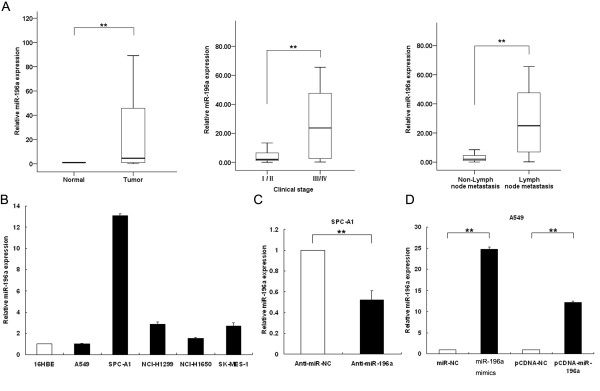
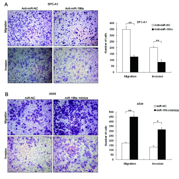
Results: miR-196a was highly expressed both in NSCLC samples and cell lines compared with their corresponding normal counterparts, and the expression of miR-196a may be affected by DNA demethylation. Higher expression of miR-196a in NSCLC tissues was associated with a higher clinical stage, and also correlated with NSCLC lymph-node metastasis. In vitro functional assays demonstrated that modulation of miR-196a expression affected NSCLC cell proliferation, migration and invasion. Our analysis showed that miR-196a suppressed the expression of HOXA5 both at the mRNA and protein levels, and luciferase assays confirmed that miR-196a directly bound to the 3'untranslated region of HOXA5. Knockdown of HOXA5 expression in A549 cells using RNAi was shown to promote NSCLC cell proliferation, migration and invasion. Finally, we observed an inverse correlation between HOXA5 and miR-196a expression in NSCLC tissues.

Conclusions: Our findings indicate that miR-196a is significantly up-regulated in NSCLC tissues, and regulates NSCLC cell proliferation, migration and invasion, partially via the down-regulation of HOXA5. Thus, miR-196a may represent a potential therapeutic target for NSCLC intervention.


Figures

Figure 1
Figure 1
qRT-PCR analysis of miR-196a in NSCLC tissues and matched normal tissues. (A) Determining miR-196a expression in NSCLC tissues and its clinical significance. miR-196a was detected in 34 pairs of NSCLC tissues by qRT-PCR. Data are presented as fold-changes in tumor tissues relative to normal tissues. miR-196a expression was significantly higher in patients with a higher pathological stage and lymph-node metastasis. (B) Analysis of miR-196a expression levels in NSCLC cell lines (A549, SPC-A1, NCI-H1650, NCI-H1299 and SK-MES-1) compared with the normal bronchial epithelial cell line (16HBE) by qRT-PCR. (C) Analysis of miR-196a expression following treatment of SPCA1 cells with anti-miR-NC or miR-196a inhibitors by qRT-PCR. (D) Analysis of miR-196a expression following treatment of A549 cells with miR-NC, miR-196a mimics, pCDNA-NC or pCDNA-miR-196a by qRT-PCR. All experiments were performed in biological triplicate with three technical replicates. *P < 0.05, **P < 0.01.
Figure 2
Figure 2
Analysis of the correlation between methylation status and expression of miR-196a. (A) Map of the CpG island position of miR-196a-1. Vertical ticks mark CpG sites. (B) The level of miR-196a expression in 16HBE cells after treatment of 5-aza-dC (0, 2, 5 μM). (C) The methylation status of the CpG island of miR-196a-1 was assessed by bisulfite sequencing before and after 5-aza-dC treatment in 16HBE cells. Open and filled squares denote unmethylated and methylated CpG sites, respectively. Each row represents a single clone. *P < 0.05; **P < 0.01.
Figure 3
Figure 3
Effect of miR-196a on cell proliferationin vitro. (A, B) SPC-A1 cells were transfected with miR-196a inhibitors or anti-miR-NC, and A549 cells were transfected with pCDNA/miR-196a or pCDNA/miR-NC. MTT assay was performed to determine the proliferation of SPC-A1 or A549 cells. Data represent the mean ± S.D. from three independent experiments. (C, D) Colony-forming growth assays were performed to determine the proliferation of SPC-A1 or A549 cells. The colonies were counted and captured. *P < 0.05, **P < 0.01.
Figure 4
Figure 4
Effect of miR-196a on cell migration and invasionin vitro. (A, B) SPC-A1 cells were transfected with miR-196a inhibitors or anti-miR-NC, and A549 cells were transfected with miR-196a mimics or miR-NC. Transwell assays were performed to investigate the migratory and invasive ability of NSCLC cells. *P < 0.05 and **P < 0.01.
Figure 5
Figure 5
miR-196a directly targets theHOXA5gene. (A) A human HOXA5 or FOXO1 3’ untranslated region (UTR) fragment containing the wild-type or mutant miR-196a binding sequence was cloned downstream of the luciferase reporter gene. (B) The luciferase reporter plasmid containing wild-type or mutant HOXA5 and FOXO1 3’UTR were co-transfected into HEK-293 T cells with pCDNA-miR-196a or pCDNA-miR-NC. Luciferase activity was determined using the dual luciferase assay and shown as the relative firefly activity normalized to renilla activity. (C) qRT-PCR analyses of HOXA5 mRNA level following treatment of SPCA1 cells with miR-196a inhibitors or A549 cells with miR-196a mimics. (D) Western blot analyses of HOXA5 protein levels following treatment of SPCA1 cells with miR-196a inhibitors or A549 cells with miR-196a mimics. GAPDH was used as control. *P < 0.05, **P < 0.01 and N.S. not significant.
Figure 6
Figure 6
Effect of HOXA5 on NSCLC cell proliferation, migration and invasion. (A) A549 cells were transfected with si-HOXA5 or si-NC, and HOXA5 mRNA and protein levels were assessed by qRT-PCR and western blot. (B) Transwell assays were performed to investigate the migratory and invasive ability of NSCLC cells. (C, D) MTT assay and colony-forming growth assays were performed to determine the proliferation of si-NC or si-HOXA5 A549 cells. (E) The level of HOXA5 mRNA in NSCLC tissues was analyzed by qRT-PCR. (F) Analysis of the relationship between miR-196a expression and HOXA5 mRNA levels. *P < 0.05, **P < 0.01.

Similar articles

Cited by

References

    1. Jemal A, Siegel R, Xu J, Ward E. Cancer statistics, 2010. CA Cancer J Clin. 2010;60(5):277–300. doi: 10.3322/caac.20073. - DOI - PubMed
    1. Verdecchia A, Francisci S, Brenner H, Gatta G, Micheli A, Mangone L, Kunkler I. Recent cancer survival in Europe: a 2000–02 period analysis of EUROCARE-4 data. Lancet Oncol. 2007;8(9):784–796. doi: 10.1016/S1470-2045(07)70246-2. - DOI - PubMed
    1. Farazi TA, Spitzer JI, Morozov P, Tuschl T. miRNAs in human cancer. J Pathol. 2011;223(2):102–115. doi: 10.1002/path.2806. - DOI - PMC - PubMed
    1. Bartel DP. MicroRNAs: genomics, biogenesis, mechanism, and function. Cell. 2004;116(2):281–297. doi: 10.1016/S0092-8674(04)00045-5. - DOI - PubMed
    1. Chitwood DH, Timmermans MC. Small RNAs are on the move. Nature. 2010;467(7314):415–419. doi: 10.1038/nature09351. - DOI - PubMed

Publication types