. 2012 Oct;13(11):1445-53.
doi: 10.2174/138945012803530215.

Deleted in Colorectal Cancer (DCC) pathfinding: axon guidance gene finally turned tumor suppressor

Affiliations

Deleted in Colorectal Cancer (DCC) pathfinding: axon guidance gene finally turned tumor suppressor

Molly Duman-Scheel. Curr Drug Targets. 2012 Oct.

Abstract

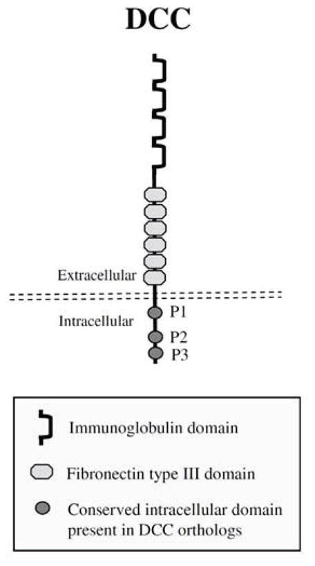
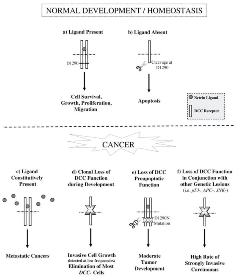
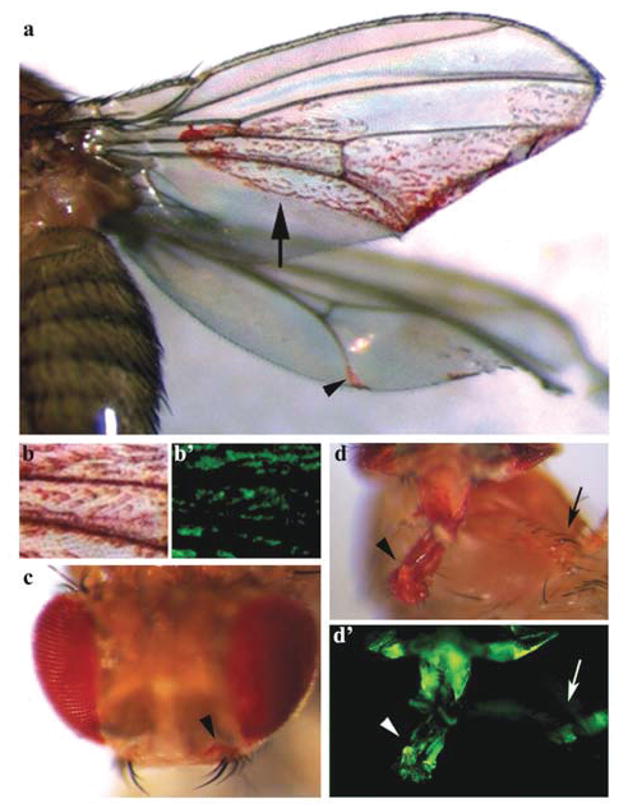
Loss of heterozygosity (LOH) at human chromosome 18q, which includes the gene Deleted in Colorectal Cancer (DCC), has been linked to colorectal and many other human cancers. DCC encodes the receptor for the axon guidance molecule Netrin (Net) and functions during neural development in a variety of organisms. However, since its discovery in the 1990s, the status of DCC as a tumor suppressor has been debated, primarily due to a lack of support for this hypothesis in animal models. A recent study from our laboratory capitalized on the genetic tractability of Drosophila melanogaster to demonstrate that this gene functions as an invasive tumor suppressor, thereby providing the first direct link between DCC loss and metastatic phenotypes in an animal model for cancer. Two subsequent studies from other laboratories have demonstrated that DCC suppresses tumor progression and metastasis in murine colorectal and mammary tumor models. Combined, these findings have prompted the rebirth of DCC as a tumor suppressor and highlighted the need for continued analysis of DCC function in animal models for human cancer.


Conflict of interest statement

CONFLICTS OF INTEREST

The authors confirm that this article content has no conflicts of interest.

Figures

Fig. 1
Fig. 1
DCC protein structure. A schematic representation of DCC with the location of the immunoglobulin, fibronectin type III, transmembrane, as well as the conserved intracellular P1, P2, and P3 domains noted. The structure depicted is adapted from reference [7].
Fig. 2
Fig. 2
DCC tumor suppressing functions. Top: DCC functions as a dependence receptor during normal development and homeostasis. a) In the presence of Net ligand, DCC induces a variety of cellular responses including survival, growth, proliferation, and migration. b) However, in the absence of ligand, DCC is cleaved at D1290 and promotes cell death. c) In human cancers (bottom), the constitutive presence of Net ligand promotes metastatic cancer. (df) Recent studies in D. melanogaster and murine cancer models suggest that loss of DCC function also promotes cancer. d) During D. melanogaster development, although most somatic fra/DCC loss of function clones are eliminated, DCC deficiency can result in excess cell growth and invasion. e) The D1290N mutation results in loss of the proapoptotic function of DCC and moderate tumor formation in mice. f) In conjunction with other genetic lesions (i.e. mutation of P53, APC, or JNK), loss of DCC function results in highly invasive carcinomas. Such metastatic cancers may result from loss of the proapoptotic functions of DCC and direct stimulation of growth, proliferation, and invasion in response to changes in cell signaling (i.e. activated Rho signaling) that occur when DCC function is compromised. See text for details and references to studies supporting this model.
Fig. 3
Fig. 3
Loss of fra/DCC function during development of D. melanogaster results in metastatic phenotypes. Metastatic phenotypes resulting from somatic loss of function fra/DCC clones generated in the developing eye are shown. P35-rescued fra/DCC mutant red-pigmented eye cells generated in the developing eye have invaded the wing (arrow and arrowhead in a), head (arrowhead in c), proboscis (arrowhead in d) and leg (arrow in d). A high magnification view of the region marked by the arrow in a is shown in b. GFP expression (b′ and d′) used to positively mark mutant cells permitted live imaging of these invasive cells during development [36]. Panels a, b, and b′ were originally published in reference [36].

Similar articles

Cited by

References

    1. Bos JL, Fearon ER, Hamilton SR, et al. Prevalence of ras gene mutations in human colorectal cancers. Nature. 1987;327:293–7. - PubMed
    1. Vogelstein B, Fearon ER, Hamilton SR, et al. Genetic alterations during colorectal-tumor development. N Engl J Med. 1988;319:525–32. - PubMed
    1. Fearon ER, Vogelstein B. A genetic model for colorectal tumorigenesis. Cell. 1990;61:759–67. - PubMed
    1. Roush W. Putative cancer gene shows up in development instead. Science. 1997;276:534–5. - PubMed
    1. Mehlen P, Fearon ER. Role of the dependence receptor DCC in colorectal cancer pathogenesis. J Clin Oncol. 2004;22:3420–8. - PubMed

Publication types

LinkOut - more resources