. 2012 Aug 9:9:157.
doi: 10.1186/1743-422X-9-157.

Low-level HIV infection of hepatocytes

Affiliations

Low-level HIV infection of hepatocytes

Ling Kong et al. Virol J. .

Abstract

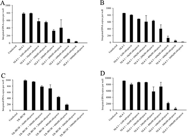
Background: There are only limited data on whether HIV infection occurs within the liver; therefore, we explored early and late stages of the HIV life cycle in two hepatocyte cell lines--Huh7.5 and Huh7.5JFH1--as well as in primary human hepatocytes.

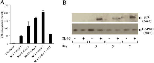
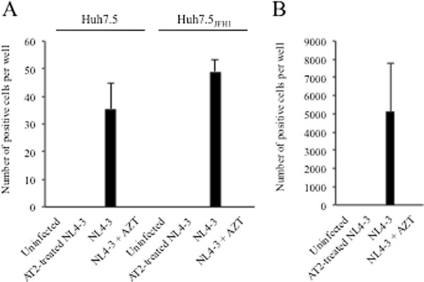
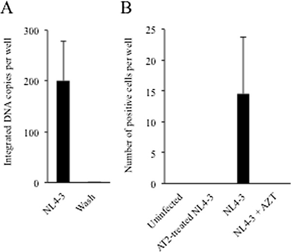
Results: Integrated HIV DNA was detected in Huh7.5 and Huh7.5JFH1 cells, as well as in primary hepatocytes, and was inhibited by the integrase inhibitor raltegravir in a dose-dependent manner. HIV p24 protein was also detected in cell culture supernatants at days 1, 3, 5, and 7 post-infection and was inhibited by AZT, although levels were modest compared to those in a lymphocyte cell line. Culture supernatants from HIV-infected hepatocytes were capable of infecting a non-hepatic HIV indicator cell line.

Conclusions: These results indicating low-level HIV replication in hepatoctyes in vitro complement evidence suggesting that HIV has deleterious effects on the liver in vivo.


Figures

Figure 1
Figure 1
Detection of integrated HIV DNA in hepatocytes. Huh7.5 (A), Huh7.5JFH1(B), or Jurkat (D) cells were incubated in the presence of 0, 1, 10, 100, or 1000 uM of the integrase inhibitor raltegravir for one hour prior to and during infection with DNase-treated HIVNL4-3. Huh7.5 (C) cells were incubated in the presence of raltegravir for one hour prior to and during infection with DNase-treated HIVYK-JRCSF. After 24 hours, integrated HIV DNA was quantified in cell lysates by nested real-time PCR. Error bars represent the standard deviation between duplicates.
Figure 2
Figure 2
HIV protein expression in hepatocytes. (A) HIV p24 protein levels were quantified by ELISA in culture supernatants from Huh7.5 cells infected with HIVNL4-3 at days 1, 3, 5, and 7 post-infection. Infection was also performed in the presence of 100 uM AZT for one hour prior to and during infection. Error bars represent the standard deviation between duplicates. (B) Huh7.5JFH1 cells were incubated with HIVNL4-3. At days 1, 3, 5, and 7 post-infection, cells were harvested, lysed, and subjected to Western Blot analysis using a rabbit monoclonal Ab (Epitomics) as the primary Ab and a rabbit polyclonal Ab to mouse IgG from (Abcam) as the secondary Ab. As a loading control, GAPDH was detected using a rabbit polyclonal Ab (Santa Cruz Biotechnology).
Figure 3
Figure 3
Infectious HIV production in hepatocytes. TZM-bl indicator cells were incubated with culture supernatants collected at day 3 post-infection from Huh7.5 or Huh7.5JHF1 cells (A) or Jurkat cells (B) infected with HIVNL4-3 or 100 ng AT-treated HIVNL4-3and tested for β-galactosidase activity. The mean number of positive TZM-bl cells per well is shown with the error bars representing the standard deviation between duplicates. β-gal expression was also evaluated in the presence of 100 uM AZT for one hour prior to and during infection.
Figure 4
Figure 4
HIV infection of primary hepatocytes. (A) Primary human hepatocytes were incubated with DNase-treated HIVNL4-3 for two hours. After 24 hours, integrated HIV DNA was quantified in cell lysates by nested real-time PCR. (B)TZM-bl indicator cells were incubated with culture supernatants collected at day 3 post-infection from primary human hepatocytes infected with HIVNL4-3 or 100 ng AT-2 treated HIVNL4-3 and tested for β-galactosidase activity. The mean number of positive TZM-bl cells per well is shown with the error bars representing the standard deviation between duplicates. β-gal expression was also evaluated in the presence of 100 uM AZT for one hour prior to and during infection.

References

    1. Keaveny AP, Karasik M. Hepatobiliary and pancreatic infections in AIDS: Part one. AIDS Patient Care and STDs. 1998;12(5):347–357. doi: 10.1089/apc.1998.12.347. - DOI - PubMed
    1. Kahn JO, Walker B. Acute human immunodeficiency virus type 1 infection. N Engl J Med. 1998;339(1):33–39. doi: 10.1056/NEJM199807023390107. - DOI - PubMed
    1. Bonacini M. Hepatobiliary complications in patients with human immunodeficiency virus infection. Am J Med. 1992;92:404–411. doi: 10.1016/0002-9343(92)90271-C. - DOI - PubMed
    1. Ingiliz P. et al.Liver damage underlying unexplained transaminase elevation in human immunodeficiency virus-1 mono-infected patients on antiretroviral therapy. Hepatology. 2009;49(2):436–442. doi: 10.1002/hep.22665. - DOI - PubMed
    1. Lefkowitch J. Pathology of AIDS-related liver disease. Dig Dis. 1994;12(6):321–330. doi: 10.1159/000171468. - DOI - PubMed

Publication types