Review
. 2012:298:1-31.
doi: 10.1016/B978-0-12-394309-5.00001-8.

Cytoskeleton assembly in Toxoplasma gondii cell division

Affiliations
Review

Cytoskeleton assembly in Toxoplasma gondii cell division

Brooke Anderson-White et al. Int Rev Cell Mol Biol. 2012.

Abstract

Cell division across members of the protozoan parasite phylum Apicomplexa displays a surprising diversity between different species as well as between different life stages of the same parasite. In most cases, infection of a host cell by a single parasite results in the formation of a polyploid cell from which individual daughters bud in a process dependent on a final round of mitosis. Unlike other apicomplexans, Toxoplasma gondii divides by a binary process consisting of internal budding that results in only two daughter cells per round of division. Since T. gondii is experimentally accessible and displays the simplest division mode, it has manifested itself as a model for apicomplexan daughter formation. Here, we review newly emerging insights in the prominent role that assembly of the cortical cytoskeletal scaffold plays in the process of daughter parasite formation.


Figures

Figure 1
Figure 1
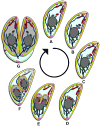
Toxoplasma divides by internal daughter budding. (A) Mature parasite in G1. Red are MT (conoid, subpellicular, and spindle pole), yellow are the alveoli, bright green is the IMC meshwork, brown are secretory vesicles, dark blue line is the mitochondrion, purple is the apicoplast, blue is the centrosome, black is the Golgi apparatus, dark green is the ER, grey is the nucleus, and pink is the posterior cup or basal complex. (B) Mitosis is initiated at 1.2N with the duplication of the Golgi apparatus followed by the duplication of the centrosomes. (C) Budding is initiated with the appearance of early components of the cytoskeleton. (D) The spindle pole duplicates and the apicoplast moves below the centrosomes, elongating as the centrosomes separate. (E) The organelles begin to partition as the daughter buds elongate. The components of the basal complex accumulate at the leading edge of the bud. (F) The daughter buds contract and all the organelles are partitioned except for the mitochondrion. The secretory vesicles and cytoskeleton of the mother begin to degrade. (G) Daughter buds emerge and the plasma membrane from the mother is incorporated onto the daughters. The mother falls away as a residual body.
Figure 2
Figure 2
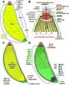
Schematic representation of the structures in the Toxoplasma cytoskeleton. (A) Directly under the plasma membrane lie the alveolar vesicles, shown in yellow. The most unique alveolar vesicle called the apical cap forms a cone around the parasite apex and three bands of rectangular, elongated vesicles fill in the remainder of the IMC below this cap. As marked, different proteins localize to different sections of the alveolar vesicles. (B) Representation of the most apical end of the cytoskeleton. The alveolar vesicles and IF-like protein filament meshwork have been removed to expose the 22 sub-pellicular microtubules and the conoid. A series of three cytoskeletal rings are located at the apex (grey). Components known to localize to these structures are indicated. Other structures are marked and named in the panel. (C) Representation of the Intra Membrane Particles (IMPs) lying within the flattened alveolar vesicles. Their molecular nature is unknown. At the basal end is the posterior cup (green), which contains TgCentrin2. (D) Representation wherein the alveolar vesicles have been removed from the top, exposing the IMC protein meshwork (green) containing the IF-filament IMC proteins. Other proteins localizing to the same region on either side of the alveoli are indicated here as well. Proteins localizing to this section of the parasite are shown in green (note that not all these proteins are part of the same structure). TgCentrin2 annuli are shown at the boundary between the most apical alveolar vesicle and the other vesicles (see panel A). The basal inner ring (BIR) and the basal inner complex (BIC) are located at the basal end of the alveoli. Proteins occupying several regions and appearing in different colors are indicated with an asterisk (*). Proteins that exhibit localization only in mature parasites are indicated with a plus (+). Figure inspired by (Nichols, Chiappino, 1987).
Figure 3
Figure 3
IMC15 and Rab11B precede MORN1 into the initial daughter bud. (A-B) Parasites expressing YFP-IMC15 (green) and DDmyc-Rab11B (red) in the presence of Shield1 colocalize at the centrosomes (A) and then expand into the forming daughter buds (B). (C) DDmyc-Rab11B (red) co-stained with anti-MORN1 (green). Arrow indicates an unduplicated spindle pole. Both constructs are driven by the ptub promoter.
Figure 4
Figure 4
Timing of the recruitment of ISP proteins 1-3 relative to other assembly markers. (A) IMC3 is present in amorphous accumulations near the buds clearly indicated by ISP1. IMC3 does not fully associate with the buds until after ISP1 arrives at the buds; both proteins are clearly established in the daughters at an early stage (B). (C) ISP2-HA precedes IMC3 into the daughter buds as well. ISP2-HA is under the control of its native promoter. (D,E) Parasites expressing ISP3-YFP (green) are co-stained with anti-MORN1 (red) showing no ISP3 at the recently divided spindle poles (D) and then colocalization of ISP3 with the early MORN1 rings (E). ISP3-YFP parasites present with numerous inclusion bodies; therefore, the arrows indicate the bud-associated ISP3.
Figure 5
Figure 5
The basal and cortical IMCs associate with the buds concurrently. Parasites expressing YFP-IMC8 (green) and cherry-IMC3 (red) show concurrent localization to the daughter buds at an early stage. Both constructs are driven by the ptub promoter.
Figure 6
Figure 6
TgCAM1 enters the conoid near the midpoint of budding just after recruitment of PhIL1. (A-B) Parasites expressing TgCAM1-YFP (green) co-stained with anti-IMC3 (red) show an absence of TgCAM1 in the early bud (A). TgCAM1 enters the conoid around the midpoint of budding as indicated by the arrows (B). (C-D) Parasites expressing YFP-IMC8 (green) support the entrance of TgCAM1 to the conoid at the midpoint since TgCAM1 is again absent from the early bud as indicated by YFP-IMC8 (C), but appears as YFP-IMC8 transitions to the growing edge of the midbud (D). Arrows indicate localization of TgCAM1 to the conoid. TgCAM1-YFP, TgCAM1-RFP, and YFP-IMC8 are driven by the ptub promoter. (E-F) PhIL1 enters the daughter buds prior to the midpoint. Parasites expressing PhIL1-YFP (green) and TgCAM1-RFP (red) show PhIL1-YFP in the early bud prior to the appearance of TgCAM1-RFP (E). At the midpoint of budding, as indicated by TgCAM1-RFP, PhIL1-YFP is well established in the daughter buds (F). PhIL1-YFP, YFP-IMC8, and TgCAM1-RFP are driven by the ptub promoter.
Figure 7
Figure 7
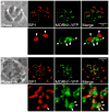
ISP1 and MORN1-YFP localization (A) during early budding in normally dividing parasites or (B) after 48 hours growth in 0.5 µM oryzalin. In normal division, a pair of MORN1 rings marks the growing ends of the two daughter buds (arrows) and ISP1 labels each apical cap (arrowheads). MORN1 is also present in the spindle pole (double arrowhead) as well as in the maternal basal complex. (B) In the presence of oryzalin, subpellicular MT polymerization is blocked, preventing cytokinesis and resulting in a large amorphous cell. Numerous early bud rings are labeled by ISP1 (arrowhead). A number of bright MORN1 puncta (double arrowhead) are centrally located in the cell and may correspond with spindle poles (>2 spindle poles are expected as DNA replication and mitosis are not inhibited under these conditions). These puncta are surrounded by less signal intense MORN1 structures (arrows), a few of which co-localize with an ISP1-positive early bud ring, but most of which do not.
Figure 8
Figure 8
Timeline of early budding activity. Time progresses from panel A through F. (A) Inter phase represents G1 without any budding. Bud components correspond to the text colors below the panels. Included components are those whose timing has been verified by comparative IFA.

Similar articles

Cited by

References

    1. Agop-Nersesian C, Egarter S, Langsley G, Foth BJ, Ferguson DJ, Meissner M. Biogenesis of the inner membrane complex is dependent on vesicular transport by the alveolate specific gtpase rab11b. PLoS Pathog. 2010;6:e1001029. - PMC - PubMed
    1. Agop-Nersesian C, Naissant B, Ben Rached F, Rauch M, Kretzschmar A, Thiberge S, Menard R, Ferguson DJ, Meissner M, Langsley G. Rab11a-controlled assembly of the inner membrane complex is required for completion of apicomplexan cytokinesis. PLoS Pathog. 2009;5:e1000270. - PMC - PubMed
    1. Anderson-White BR, Ivey FD, Cheng K, Szatanek T, Lorestani A, Beckers CJ, Ferguson DJ, Sahoo N, Gubbels MJ. A family of intermediate filament-like proteins is sequentially assembled into the cytoskeleton of toxoplasma gondii. Cell Microbiol. 2011;13:18–31. - PMC - PubMed
    1. Barkhuff WD, Gilk SD, Whitmarsh R, Tilley LD, Hunter C, Ward GE. Targeted disruption of tgphil1 in toxoplasma gondii results in altered parasite morphology and fitness. PLoS ONE. 2011;6:e23977. - PMC - PubMed
    1. Beck JR, Rodriguez-Fernandez IA, Cruz de Leon J, Huynh MH, Carruthers VB, Morrissette NS, Bradley PJ. A novel family of toxoplasma imc proteins displays a hierarchical organization and functions in coordinating parasite division. PLoS Pathog. 2010;6:e1001094. - PMC - PubMed

Publication types

LinkOut - more resources