

doi: 10.1038/embor.2012.119.
Epub 2012 Aug 10.
R-loops cause replication impairment and genome instability during meiosis
Affiliations
- PMID: 22878416
- PMCID: PMC3463965
- DOI: 10.1038/embor.2012.119
Item in Clipboard
R-loops cause replication impairment and genome instability during meiosis
EMBO Rep.
2012 Oct.
Abstract
R-loops are harmful structures with a negative impact on transcription and recombination during mitosis, but no information exists for meiosis. We used Saccharomyces cerevisiae and Caenorhabditis elegans THO mutants as a tool to determine the consequences of R-loops in meiosis. We found that both S. cerevisiae and C. elegans THO mutants show defective meiosis and an impairment of premeiotic replication as well as DNA-damage accumulation. Importantly, RNase H partially suppressed the replication impairment and the DNA-damage accumulation. We conclude that R-loops can form during meiosis causing replication impairment with deleterious results.
Conflict of interest statement
The authors declare that they have no conflict of interest.
Figures
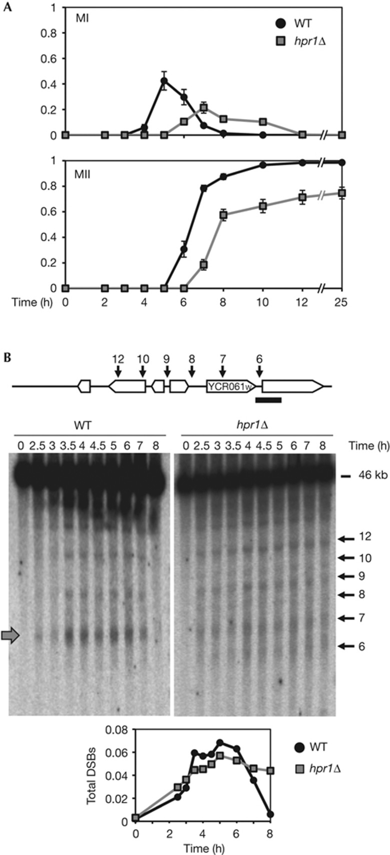
R-loop formation impairs meiosis progression. (A) Fraction of cells that completed meiosis I (MI) and II (MII) in HPR1 and hpr1Δ homozygous diploids. The means of cells±s.d. are shown. (B) Meiotic DSB formation in HPR1 and hpr1Δ diploids. Genomic DNA was taken after transfer to sporulation media and DSBs were scored by Southern probing with YCR065w. Numbered arrows indicate the position of described meiotic DSBs for this region (upper diagram). The grey arrow indicates a Spo11 DSB hotspot. Quantification of total DSBs activated during meiosis. Independent experiments were repeated three times with similar profiles. One is shown. DSB, double-strand break; WT, wild-type.
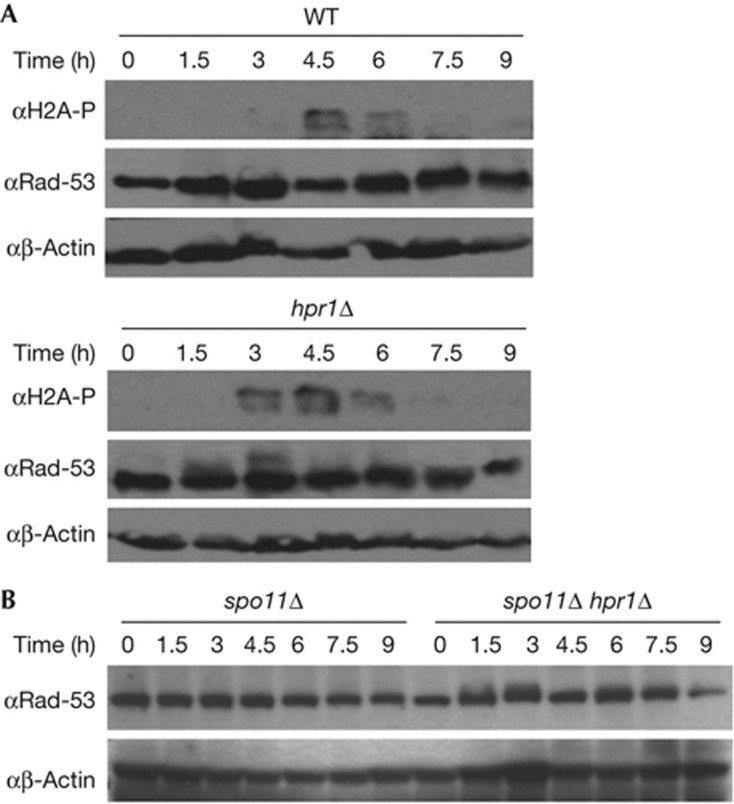
Meiotic DNA-damage and checkpoint activation in hpr1Δ cells. (A) Western analysis using anti-phosphorylation-H2A, anti-Rad53 and anti-β-actin antibodies for HPR1 and hpr1Δ homozygous diploids after transfer to sporulation media. (B) Western analysis using anti-Rad53 and anti-β-actin antibodies after transfer to sporulation media in spo11Δ and spo11Δ hpr1Δ homozygous diploids after transfer to sporulation media. WT, wild-type.
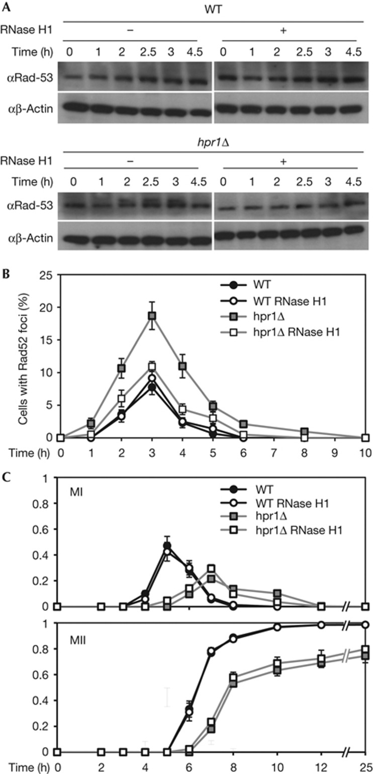
Meiotic DNA-damage and checkpoint activation are R-loop dependent. (A) Western analysis using anti-Rad53 and anti-β-actin antibodies for HPR1 and hpr1Δ homozygous diploids after transfer to sporulation media with and without RNase H1 overexpression. (B) Quantification of Rad52 foci for HPR1 and hpr1Δ homozygous diploids after transfer to sporulation media with and without RNase H1 overexpression. The means of cells with foci±s.d. are shown. (C) Fraction of cells that completed meiosis I (MI) and II (MII) in HPR1 and hpr1Δ homozygous diploids with and without RNase H1 overexpression. The means of cells±s.d. are shown.
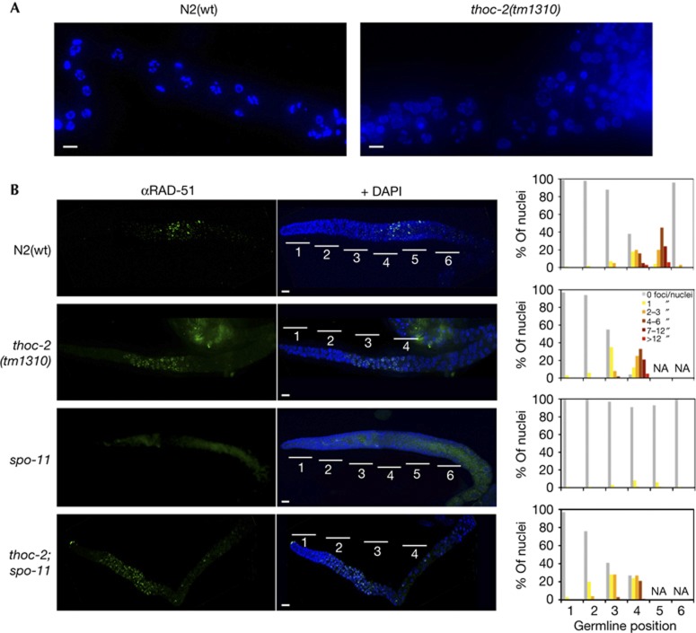
C. elegans thoc-2 mutants show meiosis failure and increased levels of SPO-11-independent RAD-51 foci. (A) Representative images of diplotene-diakinesis germline region of fixed germlines counterstained with DAPI. (B) Representative images of fixed germlines immunostained with anti-RAD-51 antibodies and counterstained with DAPI, and quantification of RAD-51 foci of germline nuclei. Zones 1–2 (mitosis), zone 3 (transition zone), zones 4–5–6 (pachytene). The number of nuclei was scored in 1-day post-L4 stage animals. At least 10 germlines per strain were scored for each experiment (n=30). Scale bar, 10 μm. DAPI, 4,6-Diamidino-2-phenylindole; NA, not applicable; WT, wild-type.
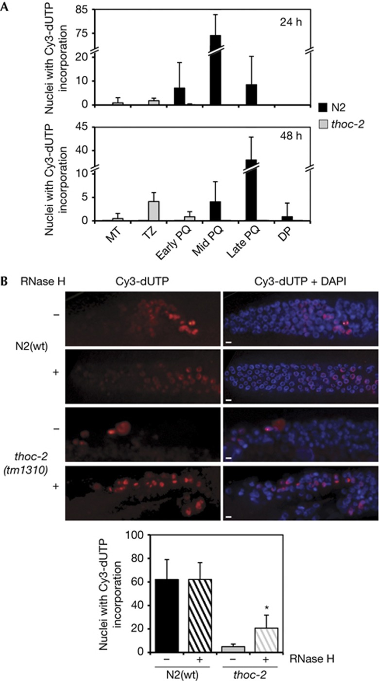
Replication is impaired in C. elegans thoc-2 germlines and partially alleviated by RNase H microinjection. (A) Quantification of Cy3-dUTP incorporation 24 and 48 h after microinjection in the germline regions of mitosis (MT), transition zone (TZ), early-, mid- and late-pachytene (PQ) and diplotene (DT). Error bars indicate standard error of mean from at least 10 germlines for each experiment (n=30). (B) Representative images of fixed germlines of the indicated genotype of adult hermaphrodites 16 h after microinjection with Cy3-dUTP and with or without RNase H and quantification of Cy3-dUTP incorporation. Error bars indicate standard error of mean from at least 10 germlines for each experiment (n=15). Statistically significant differences with respect to N2(wt) (P<0.001) are indicated by an asterisk ‘*’ (Student’s t-test). Scale bar, 5 μm. DAPI, 4,6-Diamidino-2-phenylindole; WT, wild-type.
References
Publication types
MeSH terms
Substances
LinkOut - more resources
Full Text Sources
Molecular Biology Databases
Research Materials
