. 2013 Jul;20(7):2204-12.
doi: 10.1245/s10434-012-2570-5. Epub 2012 Aug 10.

Expression of the RET proto-oncogene is regulated by TFAP2C in breast cancer independent of the estrogen receptor

Affiliations

Expression of the RET proto-oncogene is regulated by TFAP2C in breast cancer independent of the estrogen receptor

Philip M Spanheimer et al. Ann Surg Oncol. 2013 Jul.

Abstract

Background: The RET proto-oncogene is expressed as part of the estrogen receptor (ER) cluster in breast cancer. We sought to determine if TFAP2C regulates Ret expression directly or indirectly through ER.

Methods: Chromatin immunoprecipitation sequencing (ChIP-Seq) and gel-shift assay were used to identify TFAP2C binding sites in the RET promoter in four breast cancer cell lines. Ret mRNA and protein levels were evaluated in ER-positive and ER-negative breast cancer cell lines after knockdown of TFAP2C. Luciferase expression assay was performed to assess expression from two of the identified sites.

Results: ChIP-Seq identified five main binding peaks for TFAP2C in the RET promoter at -101.5 kb, -50.7 kb, -32.5 kb, +5.0 kb, and +33.6 from the RET transcriptional start site. Binding at three of the AP-2 sites was conserved across all four cell lines, whereas the RET -101.5 and RET +33.6 sites were each found to be unbound by TFAP2C in one cell line. A TFAP2C consensus element was confirmed for all five sites. Knockdown of TFAP2C by siRNA in ER-positive MCF-7 cells resulted in significant down regulation of Ret mRNA compared to nontargeting (NT) siRNA (0.09 vs. 1.0, P < 0.001). Knockdown of TFAP2C in ER-negative MDA-MB-453 cells also led to a significant reduction in Ret mRNA compared to NT siRNA (0.16 vs. 1.0, P < 0.001). In MCF-7 cells, knockdown of TFAP2C abrogated Ret protein expression (0.02 vs. 1.0, P < 0.001) before reduction in ER.

Conclusions: TFAP2C regulates expression of the RET proto-oncogene through five AP-2 regulatory sites in the RET promoter. Regulation of Ret by TFAP2C occurs independently of ER expression in breast carcinoma.


Figures
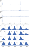
FIG. 1
FIG. 1
Genomic binding of TFAP2C in the RET locus. ChIP-Seq data graphical representation from the University of California–Santa Cruz, genome browser demonstrating locations of TFAP2C binding in the RET promoter. Five binding peaks are identified in MCF-7, BT-474, MDA-MB-453, and SKBR-3 at locations −101.5 kb, −50.7 kb, −32.5 kb, +5.0 kb, and +33.6 kb from the RET transcriptional start site. Bottom: Higher-resolution images around each peak. All peaks are conserved across the cell lines, except the RET −101.5 peak is absent in the BT-474 and the RET +33.6 peak is absent in MDA-MB-453
FIG. 2
FIG. 2
Localization of AP-2 sites corresponds to ChIP-Seq peaks. Gel-shift assay confirming binding of synthesized TFAP2C protein at each binding site identified by ChIP-Seq. Upper left: ChIP-Seq data from MCF-7 demonstrating the location of the gel-shift probe with respect to the ChIP-Seq peak. Shift of the probe is demonstrated at all sites confirming TFAP2C binding in that region, and supershift was performed with TFAP2C antibody to confirm specificity of the gel-shift complex. Beneath each gel-shift image is a graphical representation of the probe and competition oligonucleotides. For oligonucleotides competing for binding of synthesized TFAP2C protein the sequence is provided, with the consensus AP-2 family binding sequence underlined. For the RET −32.5 peak the AP-2 family site was split between competitor 3 and competitor 4, with competition for binding demonstrated by joining 3 and 4 as well as creating an oligonucleotide of the end of 3 and beginning of 4. The binding sequence at the RET +5.0 site was confirmed when mutation of 2 base pairs in the AP-2 binding sequence (boxed 4*) failed to compete for TFAP2C binding. Partial competition occurred with competitors 1 and 5 at the RET +33.6 kb site; complete competition occurred with a competitor composed of 1 + 5 linked and is indicative of deviation of these 2 sites from an optimal AP-2 consensus element
FIG. 3
FIG. 3
Knockdown of TFAP2C abrogates RET expression. RT-PCR demonstrated the effects of siRNA knockdown of Ret and TFAP2C on Ret and TFAP2C expression in ER-positive MCF-7 and ER-negative MDA-MB-453 breast cancer cell lines. mRNA levels were normalized to human beta-2-microglobulin and to nontargeting (NT) transfection. RT-PCR was performed in triplicate and averages and standard deviations were calculated from 3 biologic replicates. Asterisks indicate P < 0.001 compared to NT transfection
FIG. 4
FIG. 4
Knockdown of TFAP2C affects Ret protein before effect on ER. Western blot test demonstrated the effects of siRNA knockout of Ret and TFAP2C on Ret, TFAP2C, and ERα protein expression. Protein samples were collected 72 h and 120 h after transfection. Samples collected at 72 h demonstrated knock down of TFAP2C protein resulted in a significant reduction in Ret protein without reduction in ERα protein. At 120 h, knockout of TFAP2C led to significant reduction in Ret, TFAP2C, and ERα protein. Endogenous control is GAPDH. Graphical representation of protein quantification is shown with averages and standard deviations calculated from 3 biologic replicates and asterisks correspond to P < 0.001 compared to nontargeting (NT) transfection
FIG. 5
FIG. 5
Expression of RET is induced by TFAP2C at RET −50.7 and RET +33.6. Luciferase reporter constructs with AP-2 regulatory regions cloned into TATA-LUC minimal promoter were assayed in MCF-7 and MDA-MB-453 cells. Empty vector (EV) was performed with transfection of pGL3. Construct with RET −50.7 region was active in both cell lines and expression was abolished by mutation of the AP-2 site (mutAP2). Relative expression in MCF-7 cells demonstrated expression of the construct with the AP-2 regulatory region corresponding to the RET +33.6 site and significant reduction of expression by mutation of the AP-2 site. In MDA-MB-453, relative expression from the RET +33.6 site was significantly reduced with minimal effect noted with mutation of the AP-2 site

References

    1. Tozlu S, Girault I, Vacher S, et al. Identification of novel genes that co-cluster with estrogen receptor alpha in breast tumor biopsy specimens, using a large-scale real-time reverse transcription–PCR approach. Endocr Relat Cancer. 2006;13:1109–1120. - PubMed
    1. Esseghir S, Todd SK, Hunt T, et al. A role for glial cell derived neurotrophic factor induced expression by inflammatory cytokines and RET/GFR alpha 1 receptor up-regulation in breast cancer. Cancer Res. 2007;67:11732–11741. - PubMed
    1. Boulay A, Breuleux M, Stephan C, et al. The Ret receptor tyrosine kinase pathway functionally interacts with the ERalpha pathway in breast cancer. Cancer Res. 2008;68:3743–3751. - PubMed
    1. Plaza-Menacho I, Morandi A, Robertson D, et al. Targeting the receptor tyrosine kinase RET sensitizes breast cancer cells to tamoxifen treatment and reveals a role for RET in endocrine resistance. Oncogene. 2010;29:4648–4657. - PubMed
    1. Kao J, Salari K, Bocanegra M, et al. Molecular profiling of breast cancer cell lines defines relevant tumor models and provides a resource for cancer gene discovery. PLoS One. 2009;4:e6146. - PMC - PubMed

Publication types

MeSH terms