. 2012 Oct-Dec;6(4):234-9.
doi: 10.4161/fly.21160. Epub 2012 Aug 10.

EGFR-dependent downregulation of Capicua and the establishment of Drosophila dorsoventral polarity

Affiliations

EGFR-dependent downregulation of Capicua and the establishment of Drosophila dorsoventral polarity

María José Andreu et al. Fly (Austin). 2012 Oct-Dec.

Abstract

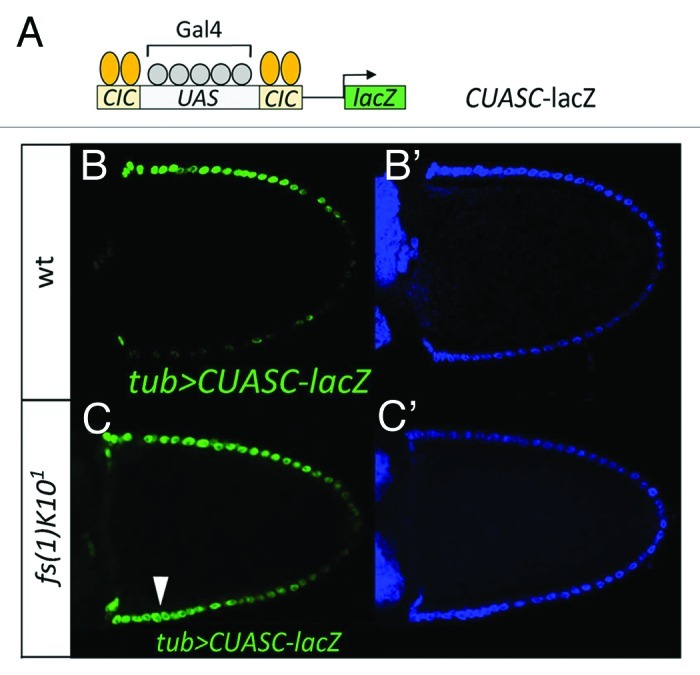
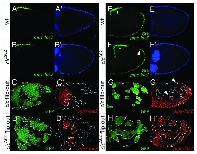
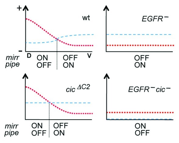
Dorsoventral (DV) axis formation in Drosophila begins during oogenesis through the graded activation of the EGF receptor (EGFR)-Ras-MAPK signaling pathway in the follicle cell layer of the egg chamber. EGFR signaling, which is higher in dorsal follicle cells, represses expression of the sulfotransferase-encoding gene pipe, thereby delimiting a ventral domain of Pipe activity that is critical for the subsequent induction of ventral embryonic fates. We have characterized the transcriptional circuit that links EGFR signaling to pipe repression: in dorsal follicle cells, the homeodomain transcription factor Mirror (Mirr), which is induced by EGFR signaling, directly represses pipe transcription, whereas in ventral follicle cells, the HMG-box protein Capicua (Cic) supports pipe expression by repressing mirr. Although Cic is under negative post-transcriptional regulation by Ras-MAPK signaling in different contexts, the relevance of this mechanism for the interpretation of the EGFR signal during DV pattern formation remains unclear. Here, we consider a model where EGFR-mediated downregulation of Cic modulates the spatial distribution of Mirr protein in lateral follicle cells, thereby contributing to define the position at which the pipe expression border is formed.


Figures

None
Figure 1. EGFR signaling downregulates Cic repressor activity in dorsal follicle cells. (A) Diagram of the CUASC-lacZ reporter construct containing five Gal4/UAS binding sites flanked by two Cic binding sites on either side. (B and C) Lateral views of stage-10 wild-type (B and B’) and fs(1)K10 (C and C’) egg chambers carrying tubulin-Gal4 and CUASC-lacZ insertions. Double stainings with anti−β−galactosidase antibody (B and C, green) and DAPI (B’ and C’, blue) are shown. Dorsal is up as indicated by the dorsal position of the oocyte nucleus under Nomarski optics (not shown). CUASC-lacZ expression shows preferential dorsal expression in a wild-type background, and is ectopically expressed in ventral regions of the fs(1)K10 egg chamber (arrowhead, C).
None
Figure 2. EGFR signaling modulates Cic-mediated repression of mirr. (A and B) Lateral views of stage-10 wild-type (A and A’) and cicΔC2 (B and B’) egg chambers carrying the mirrP2 (mirr-lacZ; ref. 33) enhancer trap. Double stainings with anti-β-galactosidase (A and B, green) and DAPI (A’ and B’, blue) are shown. (C and D) Dorsal views of stage-10 mosaic egg chambers expressing Cic (C and C’) and CicΔC2 (D and D’) proteins in clones marked by expression of GFP (C and D, green). (C’ and D’) show anti-β-galactosidase stainings of mirr-lacZ expression (red). Only CicΔC2 causes full repression of mirr-lacZ. (E and F) Lateral views of stage-10 wild-type (E and E’) and cicΔC2 (F and F’) egg chambers carrying the M2-lacZ (pipe-lacZ; ref. 12) reporter. Stainings with anti-β-galactosidase and anti-Gurken antibodies (E and F, green) and DAPI (E’ and F’, blue) are shown; the Gurken signals are indicated with asterisks. A single genomic cicΔC2 transgene (see ref. 22) leads to expanded pipe-lacZ expression by an average of 1.3 cells (n = 20) in the dorsal-posterior region (arrowhead in panel F). (G and H) Lateral views of stage-10 mosaic egg chambers expressing Cic (G and G’) and CicΔC2 (H and H’) proteins in clones marked by expression of GFP (G and H, green). G’ and H’ show anti-β-galactosidase stainings of M1-lacZ (pipe-lacZ; ref. 12) expression (red). Note that Cic-expressing clones show partial derepression of pipe-lacZ close to the endogenous pipe domain (arrowheads, G’), whereas CicΔC2 causes robust derepression of the reporter in all positions.
None
Figure 3. Model for the regulation of mirr and pipe expression in response to EGFR and Cic activities. Each diagram represents the regulatory inputs that control mirr expression in four different genetic backgrounds. For simplicity, only the anterior region of the egg chamber is represented along the DV axis (D, dorsal; V, ventral). The red dotted line represents the combined activation inputs from EGFR signaling and anterior positional cues that may depend on Dpp. The blue dashed line indicates the level of Cic-mediated repression. These inputs are proposed to define the position at which Mirr levels are sufficient to repress pipe (vertical dotted line), thereby establishing its border of expression. In cicΔC2 egg chambers, this position is shifted dorsally as a result of uniform Cic activity throughout the follicular epithelium. Note also that EGFR mutant backgrounds retain a weak activation input from anterior positional cues; this input is insufficient to drive mirr expression unless Cic repressor activity is absent.

Comment on

  • Andreu MJ, Gonzalez-Perez E, Ajuria L, Samper N, Gonzalez-Crespo S, Campuzano S, et al. Mirror represses pipe expression in follicle cells to initiate dorsoventral axis formation in Drosophila. Development. 2012;139:1110–4. doi: 10.1242/dev.076562. doi: 10.1242/dev.076562

References

    1. Moussian B, Roth S. Dorsoventral axis formation in the Drosophila embryo--shaping and transducing a morphogen gradient. Curr Biol. 2005;15:R887–99. doi: 10.1016/j.cub.2005.10.026. - DOI - PubMed
    1. Nilson LA, Schüpbach T. EGF receptor signaling in Drosophila oogenesis. Curr Top Dev Biol. 1999;44:203–43. doi: 10.1016/S0070-2153(08)60471-8. - DOI - PubMed
    1. Hong JW, Hendrix DA, Papatsenko D, Levine MS. How the Dorsal gradient works: insights from postgenome technologies. Proc Natl Acad Sci U S A. 2008;105:20072–6. doi: 10.1073/pnas.0806476105. - DOI - PMC - PubMed
    1. Sen J, Goltz JS, Stevens L, Stein D. Spatially restricted expression of pipe in the Drosophila egg chamber defines embryonic dorsal-ventral polarity. Cell. 1998;95:471–81. doi: 10.1016/S0092-8674(00)81615-3. - DOI - PubMed
    1. Zhang Z, Stevens LM, Stein D. Sulfation of eggshell components by Pipe defines dorsal-ventral polarity in the Drosophila embryo. Curr Biol. 2009;19:1200–5. doi: 10.1016/j.cub.2009.05.050. - DOI - PMC - PubMed

Publication types

MeSH terms

LinkOut - more resources