. 2012 Dec;470(12):3390-7.
doi: 10.1007/s11999-012-2468-3.

Is posterior hip instability associated with cam and pincer deformity?

Affiliations

Is posterior hip instability associated with cam and pincer deformity?

Aaron J Krych et al. Clin Orthop Relat Res. 2012 Dec.

Abstract

Background: Posterior hip instability is an increasingly recognized injury in athletes; however, the function of patients after these injuries and an understanding of the pathoanatomy and underlying mechanism are currently unclear.

Questions/purposes: We determined (1) the function of patients after these hip injuries using validated, self-reported outcome instruments and (2) the specific pathoanatomy sustained in these events to better understand the mechanism of posterior hip instability.

Methods: We reviewed the records of all 22 athletes presenting to our clinics with a posterior acetabular rim fracture confirming a posterior hip instability episode. Radiograph, CT, and MRI findings were documented in all patients. Intraoperative findings were recorded in patients undergoing surgery. There were 19 males and three females with an average age of 22 years (range, 13-31 years). Minimum followup was 2 years (average, 4 years; range, 2-16 years).

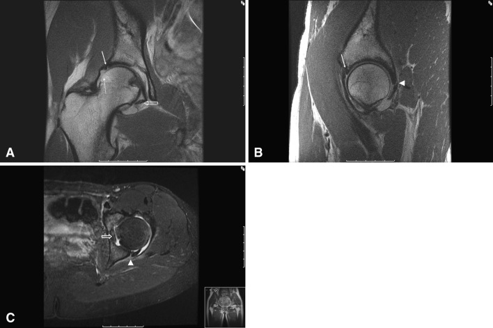
Results: The mean modified Harris hip score was 94, Hip Outcome Scores for Activities of Daily Living and Sport were 99 and 87, respectively, and 20 of 22 athletes returned to sport. The most common constellation of pathoanatomy was a posterior labral tear with rim fracture, anterior labral tear, capsular tear, ligamentum teres avulsion, and chondral injury of the femoral head with loose bodies. Sixteen of the 18 patients with femoroacetabular impingement (FAI) had a twisting or noncontact mechanism of injury.

Conclusions: When posterior hip subluxation is recognized and avascular necrosis avoided, these athletes generally have high functional outcome scores and high rates of return to sport. There is an apparent association between the occurrence of posterior hip instability and the presence of structural abnormalities often associated with FAI, which may contribute to a mechanism of FAI-induced posterior subluxation.

Level of evidence: Level IV, therapeutic study. See the Instructions for Authors for a complete description of levels of evidence.


Figures

Fig. 1A–C
Fig. 1A–C
(A) Coronal, (B) sagittal, and (C) axial MR images demonstrate typical findings, including anterior labral injury (arrow), posterior bony Bankart lesion (arrowhead), ligamentum teres avulsion (open arrow), and chondral injury to the femoral head (dotted arrow).
Fig. 2A–H
Fig. 2A–H
Intraoperative arthroscopy photographs through a lateral viewing portal demonstrate (A) synovitis of the hip, (B) anterior labral crush injury, (C) femoral head chondral injury, (D) loose chondral body, (E) ligamentum teres avulsion, (F) posterior labral tear with bony Bankart component (arrows), and (G) posterior labral status postrepair. (H) A view of the peripheral compartment demonstrates chronic cam lesion of the femoral head-neck junction with a view of posterior labral repair. FH = femoral head; A = acetabulum; LT = ligamentum teres; PL = posterior labrum; AL = anterior labrum.
Fig. 3A–B
Fig. 3A–B
(A) An AP pelvic radiograph demonstrates a subtle posterior wall rim fracture (arrowhead) and underlying cam-type FAI (arrows). (B) An obturator oblique radiograph of the left hip better demonstrates the posterior wall rim fracture (arrows).
Fig. 4
Fig. 4
A three-dimensional CT scan demonstrates a typical posterior acetabular wall rim fracture (arrows).
Fig. 5
Fig. 5
A diagram illustrates a theorized mechanism of injury for patients with FAI and posterior hip instability. Top row: With normal osseous hip anatomy, internal rotation is not impeded. Bottom row: In patients with cam-type FAI, internal rotation is limited. Attempts at achieving internal rotation cause anterior impingement and levering, which drives the femoral head posteriorly (red arrow).

References

    1. Beck M, Kalhor M, Leunig M, Ganz R. Hip morphology influences the pattern of damage to the acetabular cartilage: femoroacetabular impingement as a cause of early osteoarthritis of the hip. J Bone Joint Surg Br. 2005;87:1012–1018. doi: 10.1302/0301-620X.87B7.15203. - DOI - PubMed
    1. Blount WP. Don’t throw away the cane. J Bone Joint Surg Am. 1956;38:695–708. - PubMed
    1. Chudik S, Lopez V. Hip dislocations in athletes. Sports Med Arthrosc Rev. 2002;10:123–133. doi: 10.1097/00132585-200210020-00004. - DOI
    1. Clohisy JC, Carlisle JC, Beaulé PE, Kim YJ, Trousdale RTJ, Sierra R, Leunig M, Schoenecker PL, Millis MB. A systematic approach to the plain radiographic evaluation of the young adult hip. J Bone Joint Surg Am. 2008;90:47–66. doi: 10.2106/JBJS.H.00756. - DOI - PMC - PubMed
    1. Clohisy JC, Knaus ER, Hunt DM, Lesher JM, Harris-Hayes M, Prather H. Clinical presentation of patients with symptomatic anterior hip impingement. Clin Orthop Relat Res. 2009;467:638–644. doi: 10.1007/s11999-008-0680-y. - DOI - PMC - PubMed

MeSH terms