. 2012 Sep 18;126(12):1484-94.
doi: 10.1161/CIRCULATIONAHA.112.103051. Epub 2012 Aug 9.

Targeting mannose-binding lectin confers long-lasting protection with a surprisingly wide therapeutic window in cerebral ischemia

Affiliations

Targeting mannose-binding lectin confers long-lasting protection with a surprisingly wide therapeutic window in cerebral ischemia

Franca Orsini et al. Circulation. .

Abstract

Background: The involvement of the complement system in brain injury has been scarcely investigated. Here, we document the pivotal role of mannose-binding lectin (MBL), one of the recognition molecules of the lectin complement pathway, in brain ischemic injury.

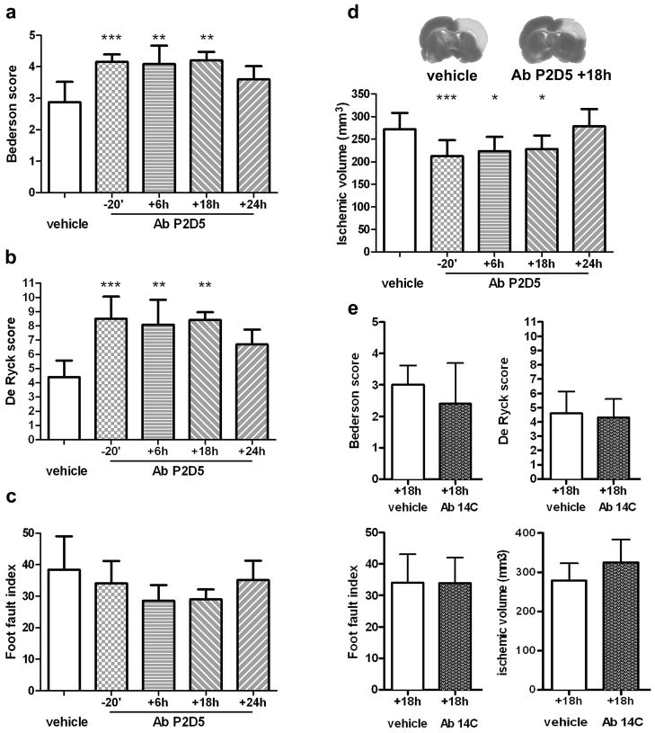
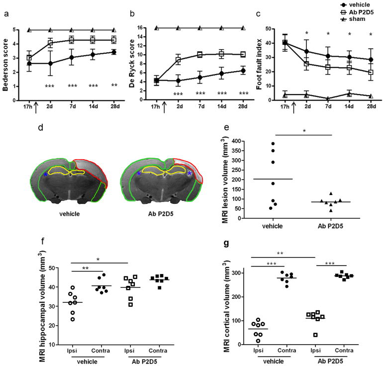
Methods and results: Focal cerebral ischemia was induced in mice (by permanent or transient middle cerebral artery occlusion) and rats (by 3-vessel occlusion). We first observed that MBL is deposited on ischemic vessels up to 48 hours after injury and that functional MBL/MBL-associated serine protease 2 complexes are increased. Next, we demonstrated that (1) MBL(-/-) mice are protected from both transient and permanent ischemic injury; (2) Polyman2, the newly synthesized mannosylated molecule selected for its binding to MBL, improves neurological deficits and infarct volume when given up to 24 hours after ischemia in mice; (3) anti-MBL-A antibody improves neurological deficits and infarct volume when given up to 18 hours after ischemia, as assessed after 28 days in rats.

Conclusions: Our data show an important role for MBL in the pathogenesis of brain ischemic injury and provide a strong support to the concept that MBL inhibition may be a relevant therapeutic target in humans, one with a wide therapeutic window of application.


Conflict of interest statement

Conflict of Interest Disclosures: None.

Figures

Figure 1
Figure 1
MBL is deposited on ischemic vessels after tMCAo and pMCAo. Representative images of staining for MBL-A or MBL-C (red), vessels (IB4, green) and nuclei (Hoechst, blue) in the ipsilateral cortex 12h after tMCAo (a,c) or pMCAo (b,d). After tMCAo, both MBL-A (a) and MBL-C (c) show deposition on ischemic vessels. Single xy plane views with z projections of the white boxes in a and c confirm that both isoforms were located in the luminal space of vessels (a′, c′). 3D renderings (a″, c″) and clipped volumes (a‴, c‴: clipping in correspondence of the yellow planes) further demonstrate intraluminal deposition of MBL-A and MBL-C. After pMCAo MBL-A and MBL-C show similar intraluminal deposition by fluorescence (b, b′, d, d′), 3D renderings (b″, d″) and clipped volumes (b‴, d‴). Confocal analysis, scale bar 20μm (a, b, c, d) and 5μm (a′, b′, c′, d′). Semiquantitative evaluation of MBL-A and MBL-C staining in ipsilateral cortices at different time points after tMCAo (e) or pMCAo (f), n=3. Scores were assigned blindly as follows: − = no positivity, + = low positivity, ++ = intermediate positivity, +++ = high positivity.
Figure 2
Figure 2
The lectin pathway is involved in pMCAo injury. Functional MBL/MASP-2 complexes in mice plasma samples collected 30min (a) or 24h (b) after sham surgery or pMCAo. C3 complement activation fragments in plasma samples collected 24h after sham surgery or pMCAo (c). Infarct volume assessed 48h after pMCAo in WT and MBL−/− mice (d). Data are reported as scatter dot plots and mean (bars), n=8–10 mice per group, unpaired t-test; *P<0.05,**P<0.01,***P<0.001.
Figure 3
Figure 3
Binding of mannosylated dendrimers to MBL evaluated by SPR. MBL was immobilized on the surface of sensor chip whereas the dendrimers were injected for 3 min at a flow rate of 100 μl/min. Left panel (a) shows the maximal binding (in Resonance Units, RU) obtained when injecting 100 μM of dendrimers with different structures. The data shown are from a single experimental session but the same rank order was obtained in independent injections, using different concentrations or different flow rates. No binding was observed on a parallel surface immobilizing BSA. Right panel (b) shows the sensorgrams obtained injecting four different concentrations of Polyman2 (black lines), together with the corresponding fittings (white lines). The analysis of these sensorgrams allowed estimation of the binding constants shown below.
Figure 4
Figure 4
Polyman2 protects ischemic mice from tMCAo injury with a wide therapeutic window. Haemolytic activity of Polyman2 assayed 1h after iv administration to non ischemic mice. Data are the mean of 3 mice per group and each plasma sample was an average of 3 determinations (a). Composite neurological score (b) and infarct volume (c) assessed 48h from tMCAo in mice receiving a single iv administration of vehicle (n=17, administered at each time point and gathered in a single control group, details in supplemental Fig. 7a) or Polyman2, given 3, 6, 12, 18, 24 or 30h from injury, (n=7, 6, 8, 8, 8 and 5, respectively). Representative images of MBL-A staining in the ipsilateral cortex 24h after tMCAo in a typical vehicle (left panel) or Polyman2 (post-ischemia treatment time: 6h, right) treated mouse. Nuclei, vessels and MBL stainings in blue, green and red, respectively, n=3 (d). C3 complement activation fragments in plasma samples assessed 48h from surgery in sham or tMCAo mice (e, n=8–10 per group) or in tMCAo mice treated with vehicle, Polyman2, 3 or 6h after injury (f, n=8–10 per group). Composite neurological score (g) and infarct volume (h) assessed 48h from tMCAo in MBL−/− mice receiving vehicle or Poyman2, given 6h from injury, (n=7 per group). Data are expressed as mean+SD (b,c) and as scatter dot plots and mean (bars) (e, f, g, h). One-way ANOVA followed by Dunnett post-hoc test; *P<0.05,**P<0.01,***P<0.001 versus vehicle (b, c, f) and unpaired t-test ****P<0.0001 (e).
Figure 5
Figure 5
Anti-MBL-A antibody selectively protects against functional and histopathological consequences of ischemia with a wide therapeutic window. Effect of anti-MBL-A mAb treatment (clone P2D5, 1 mg/Kg, iv) on neurological deficits and infarct volume assessed 48h after injury in rats receiving a single administration of vehicle (n=12, administered at each time point and gathered in a single control group, details in supplemental Fig. 7b) or mAb 20 min before, or 6, 18, 24h (n= 10, 6, 6 and 6, respectively) after 3-vo: postural-reflex deficit, (a, Bederson’s test, normal score =5); sensorimotor deficit (b, De Ryck’s test, normal score=16); integration of motor responses (c, foot-fault test, normal score=2); anatomical damage (d, infarct volume). The administration of an isotype control mAb (clone 14C, 1 mg/Kg iv) did not affect any of these parameters (e, n=10–11 per group). Data are expressed as mean+SD. Kruskal-Wallis test followed by Dunn’s test (a,b,c), one-way ANOVA followed by Dunnett post-hoc test (d) and unpaired t-test (e);*P<0.05,**P<0.01,***P<0.001 versus vehicle.
Figure 6
Figure 6
Anti-MBL-A antibody protection form ischemic injury is long lasting. Anti-MBL-A mAb (P2D5 clone, 1 mg/kg, iv) was given 18h after 3-vo (at arrow). Postural-reflex deficit (a, Bederson’s test; normal score=5), sensorimotor deficit (b, De Ryck’s test; normal score=16) and integration of motor responses (c, foot-fault test; normal score=2), were assessed 17h (pre-treatment time), 2, 7, 14 and 28d after 3-vo in vehicle or anti-MBL-A antibody treated rats or in sham-operated rats. Data are reported as mean±SD, n=7 per group. Two-way ANOVA for repeated measures followed by Bonferroni post-hoc test (a,b,c): *P<0.05, **P<0.01,***P<0.001, Ab P2D5 versus vehicle-treated rats at each given time. Structural MRI data analysis of brain volumes (T2w) in ischemic rats treated with vehicle or with anti-MBL-A antibody: total lesion volume (e: measured as the whole T2w hyperintense tissue across brain areas, red ROI in d) and atrophy (measured as residual T2w hypointense tissue) in hippocampus (f, yellow ROI in d) and cortex (g, green ROI in d) for ipsi- and contra- lateral areas. Asterisks (blue) identify ventricles. Data are reported as scatter dot plots and mean (bar), n=7 per group. Two-way ANOVA for mached values (ipsi and contralateral side) followed by Bonferroni post-hoc test (f,g) and unpaired t-test with Welch’s correction (e); *P<0.05,**P<0.01,***P<0.001, comparisons as indicated by horizontal lines.

References

    1. Lo EH. A new penumbra: transitioning from injury into repair after stroke. Nat Med. 2008;14:497–500. - PubMed
    1. Lees JS, Mishra NK, Saini M, Lyden PD, Shuaib A. Low body temperature does not compromise the treatment effect of alteplase. Stroke. 2011;42:2618–2621. - PubMed
    1. Fonarow GC, Smith EE, Saver JL, Reeves MJ, Bhatt DL, Grau-Sepulveda MV, Olson DM, Hernandez AF, Peterson ED, Schwamm LH. Timeliness of tissue-type plasminogen activator therapy in acute ischemic stroke: patient characteristics, hospital factors, and outcomes associated with door-to-needle times within 60 minutes. Circulation. 2011;123:750–758. - PubMed
    1. Iadecola C, Anrather J. The immunology of stroke: from mechanisms to translation. Nat Med. 2011;17:796–808. - PMC - PubMed
    1. Ricklin D, Hajishengallis G, Yang K, Lambris JD. Complement: a key system for immune surveillance and homeostasis. Nat Immunol. 2010;11:785–797. - PMC - PubMed

Publication types

MeSH terms