. 2012 Aug 28;109(35):14170-5.
doi: 10.1073/pnas.1208916109. Epub 2012 Aug 9.

The role of intracellular calcium phosphate in osteoblast-mediated bone apatite formation

Affiliations

The role of intracellular calcium phosphate in osteoblast-mediated bone apatite formation

Suwimon Boonrungsiman et al. Proc Natl Acad Sci U S A. .

Abstract

Mineralization is a ubiquitous process in the animal kingdom and is fundamental to human development and health. Dysfunctional or aberrant mineralization leads to a variety of medical problems, and so an understanding of these processes is essential to their mitigation. Osteoblasts create the nano-composite structure of bone by secreting a collagenous extracellular matrix (ECM) on which apatite crystals subsequently form. However, despite their requisite function in building bone and decades of observations describing intracellular calcium phosphate, the precise role osteoblasts play in mediating bone apatite formation remains largely unknown. To better understand the relationship between intracellular and extracellular mineralization, we combined a sample-preparation method that simultaneously preserved mineral, ions, and ECM with nano-analytical electron microscopy techniques to examine osteoblasts in an in vitro model of bone formation. We identified calcium phosphate both within osteoblast mitochondrial granules and intracellular vesicles that transported material to the ECM. Moreover, we observed calcium-containing vesicles conjoining mitochondria, which also contained calcium, suggesting a storage and transport mechanism. Our observations further highlight the important relationship between intracellular calcium phosphate in osteoblasts and their role in mineralizing the ECM. These observations may have important implications in deciphering both how normal bone forms and in understanding pathological mineralization.


Conflict of interest statement

The authors declare no conflict of interest.

Figures
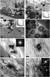
Fig. 1.
Fig. 1.
Bright-field TEM images outlining intracellularly produced, vesicle-mediated mineralization in mouse osteoblast cultures. (A) Osteoblast (OB) embedded within a mineralized nodule. Fibrous extracellular matrix (C) with banding typical of mammalian collagen (Inset; Scale bar, 200 nm) surrounds the cell. Electron dense particles of bone-like mineral are evident in the extracellular space. Sample was prepared via the chemical fixation protocol. (Scale bar, 1 μm.) (B) A vesicle (arrow) containing electron dense material inside an osteoblast abutting a heavily mineralized (M) area of a nodule. EDX analysis of the material within the vesicle demonstrates the presence of calcium and phosphorus (Inset). Sample was prepared via the chemical fixation protocol. (Scale bar, 0.5 μm.) (C) Electron-dense vesicles within a membrane invagination (arrow) of an osteoblast. EDX analysis of the vesicle demonstrates the presence of calcium and phosphorus (Inset). Sample was prepared via the chemical fixation protocol. (Scale bar, 0.2 μm.) (D) Calcium phosphate-containing vesicles in the extracellular space surrounding an osteoblast. Sample was prepared via the chemical fixation protocol. (Scale bar, 0.2 μm.) (E) Confined calcium phosphate aggregates (membranes are not distinguishable in anhydrously prepared specimens) in a mineralized nodule prepared via the anhydrous fixation protocol. Selected area electron diffraction of an aggregate (*) lacks a textured crystalline diffraction pattern, suggesting the amorphous nature of the material (Inset). (Scale bar, 0.5 μm.) (F) A dense calcium phosphate aggregate (*) associated with collagen fibrils in the extracellular space. Sample was prepared via the chemical fixation protocol. (Scale bar, 0.2 μm.) (G) Mineral (arrows) emanating from the dense focus of a mineral aggregate associated with the collagenous extracellular matrix (C). Sample was prepared via the chemical fixation protocol. (Scale bar, 0.2 μm.) (H) Extensive mineralization (M) on collagen fibrils (C) in the extracellular space of a mineralized nodule formed from osteoblasts. Sample was prepared via the chemical fixation protocol. Selected area electron diffraction of a similarly mineralized area processed via the anhydrous fixation method displayed a crystalline diffraction pattern which corresponded to the 002, 112, 211, and 300 planes of a crystalline hydroxyapatite standard (Inset). (Scale bar, 0.2 μm.)
Fig. 2.
Fig. 2.
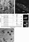
Analytical electron microscopy evidence of calcium- and phosphorus-containing mineral aggregates in osteoblast mitochondria. (A) Bright-field TEM image demonstrating electron dense granules (circled) within the mitochondria (white arrows) of an osteoblast within a mineralized nodule. Mitochondria are readily identified by their characteristic cristae (black arrows). Sample was prepared by HPF-FS. (Scale bar, 0.5 μm.) (B) HAADF scanning TEM image of an osteoblast within a mineralized nodule. Dense granule-containing mitochondria are evident throughout the cell. Mitochondria indicated as 1 and 2 are analyzed further in C. Sample was prepared by HPF-FS protocol. (Scale bar, 0.5 μm.) (C) EELS of specified areas within mitochondria of a mineralizing osteoblast. Images 1 and 2 indicate the positions at which spectra were collected and highlight the presence of calcium and phosphorus in dense granules (MG1 and MG2) with characteristic phosphorus L2,3 and calcium L2,3 edges at 132 and 346 eV, respectively. The phosphorus L2,3 edge contains characteristic double peaks (separated by 8.8 eV) followed by a more intense broad peak, which correlates with the phosphorus L2,3 edge of analogous X-ray adsorption near edge structure (XANES) spectra acquired for phosphate compounds (22). Spectra collected within the less electron-dense areas of the mitochondrial matrix lack characteristic phosphorus edges (MM1 and MM2); however, MM2 produced an edge at 346 eV, indicative of calcium. All spectra contain distinctive carbon K edges at 285 eV. (D) Bright-field TEM image of mitochondrial granules within an osteoblast. Note that the granules consist of globular accumulations of mineral with a disordered morphology. Sample was prepared by high pressure freezing and freeze substitution protocol. (Scale bar, 50 nm.)
Fig. 3.
Fig. 3.
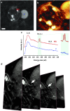
Analytical electron microscopy evidence of vesicle-mitochondrial interactions in mineralizing osteoblasts. (A) HAADF scanning TEM image of a dense granule-containing mitochondrion associating with a vesicle within an osteoblast in a mineralized nodule. The sample was prepared by HPF-FS. (Scale bar, 200 nm.) (B) Voltex projection of a 3D tomography reconstruction showing a mitochondrion conjoined with a vesicle. Dense granules are evident within the mitochondrion. See Movie S1 for the full reconstruction demonstrating a discontinuity in the mitochondrial membrane where it conjoins the vesicle. Sample was prepared by HPF-FS. (C) EELS of specified areas within the mitochondrion and vesicle in A. The mitochondrial granule and vesicle show characteristic calcium L2,3 edges at 346 eV. All spectra display carbon K edges. (D) Orthoslices at 10-nm intervals through the tomography reconstruction showing the mitochondrion-vesicle interface. The mitochondrial membrane is discontinuous where it conjoins the vesicle (arrows).
Fig. 4.
Fig. 4.
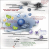
Diagram outlining current models and our proposed mechanism for bone mineral formation. Bone apatite formation likely proceeds via a number of cooperative/redundant mechanisms. Current hypotheses include the utilization of: (i) Matrix vesicles which bud from the plasma membrane and accumulate calcium (Ca2+) and phosphate (PO43−) ions extracellularly before associating with the collagenous ECM (2); (ii) Noncollagenous proteins associated with the gap zones in collagen, which mediate mineral nucleation and foster its propagation within and along collagen fibrils (1); and (iii) Our suggested model, by which amorphous calcium phosphate and ionic calcium stored in mitochondria is transported via vesicles to the ECM before converting to more crystalline apatite and propagating from dense foci. In the cartoon, “matrix vesicles” are purple, and collagen-mediated mineralization is depicted in the bottom left corner with calcium and phosphate ions highlighted in yellow and red. Mitochondria are shown in green, and vesicles are orange and blue, with and without mineral/ions, respectively. “N” identifies the cell nucleus.

References

    1. Glimcher MJ. Recent studies of the mineral phase in bone and its possible linkage to the organic matrix by protein-bound phosphate bonds. Philos Trans R Soc Lond B Biol Sci. 1984;304:479–508. - PubMed
    1. Anderson HC. Molecular biology of matrix vesicles. Clin Orthop Relat Res. 1995;(314):266–280. - PubMed
    1. Mahamid J, Sharir A, Addadi L, Weiner S. Amorphous calcium phosphate is a major component of the forming fin bones of zebrafish: Indications for an amorphous precursor phase. Proc Natl Acad Sci USA. 2008;105:12748–12753. - PMC - PubMed
    1. Boskey AL. Biomineralization: Conflicts, challenges, and opportunities. J Cell Biochem Suppl. 1998;30–31:83–91. - PubMed
    1. Mahamid J, et al. Mapping amorphous calcium phosphate transformation into crystalline mineral from the cell to the bone in zebrafish fin rays. Proc Natl Acad Sci USA. 2010;107:6316–6321. - PMC - PubMed

Publication types

MeSH terms

LinkOut - more resources