. 2012 Aug 10;337(6095):746-9.
doi: 10.1126/science.1223616.

Fate-restricted neural progenitors in the mammalian cerebral cortex

Affiliations

Fate-restricted neural progenitors in the mammalian cerebral cortex

Santos J Franco et al. Science. .

Abstract

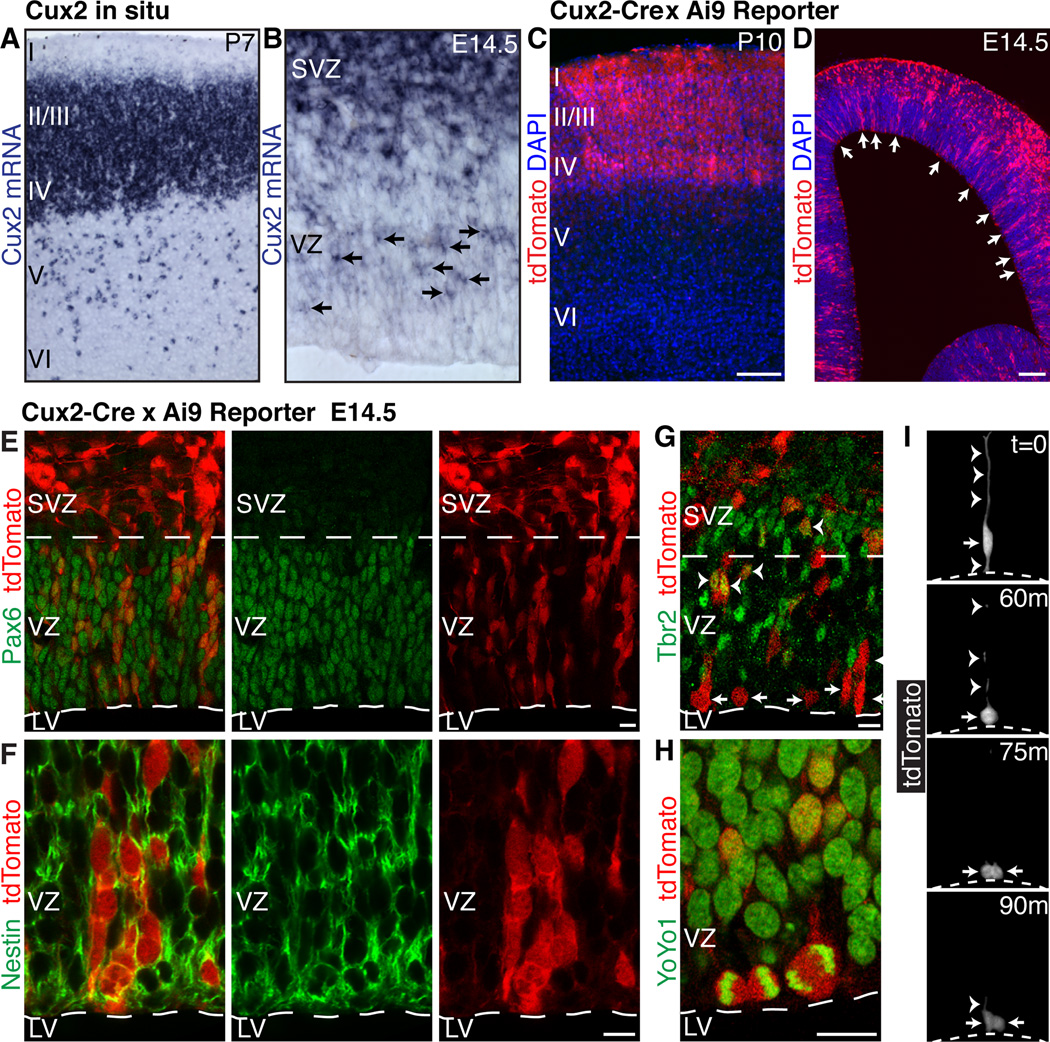
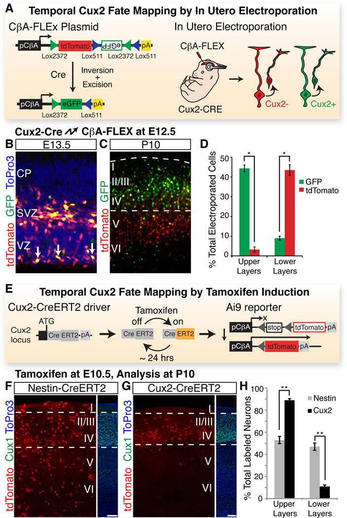
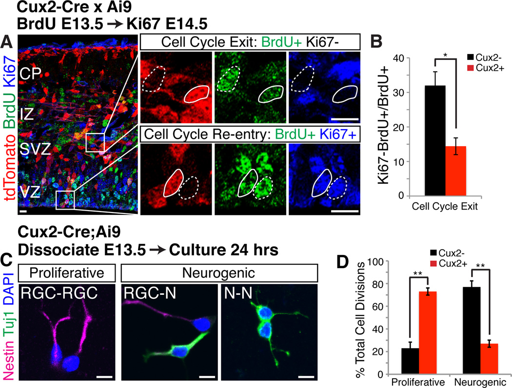
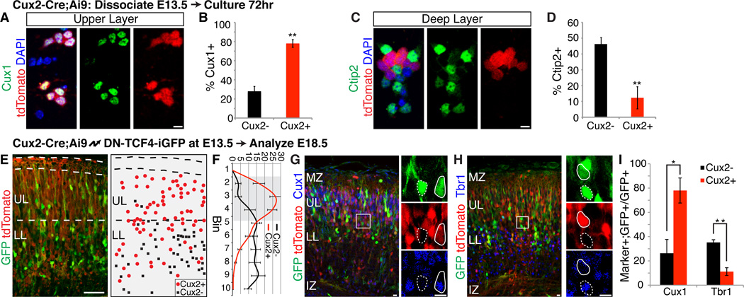
During development of the mammalian cerebral cortex, radial glial cells (RGCs) generate layer-specific subtypes of excitatory neurons in a defined temporal sequence, in which lower-layer neurons are formed before upper-layer neurons. It has been proposed that neuronal subtype fate is determined by birthdate through progressive restriction of the neurogenic potential of a common RGC progenitor. Here, we demonstrate that the murine cerebral cortex contains RGC sublineages with distinct fate potentials. Using in vivo genetic fate mapping and in vitro clonal analysis, we identified an RGC lineage that is intrinsically specified to generate only upper-layer neurons, independently of niche and birthdate. Because upper cortical layers were expanded during primate evolution, amplification of this RGC pool may have facilitated human brain evolution.


Figures

Fig. 1
Fig. 1. Expression of Cux2 in a subset of RGCs suggests multiple progenitor types
(A and B) In situ hybridization shows Cux2 mRNA at P7 (A) and E14.5 (B). Arrows indicate expression in a subset of ventricular zone cells. (C and D) Cumulative fate mapping of the Cux2 lineage in Cux2-Cre;Ai9 mice at P10 (C) and E14.5 (D). Arrows indicate recombination in a subset of ventricular zone cells. (E and F) Recombination in Pax6+ (E) and nestin+ (F) RGCs. (G) Recombination in Tbr2+ intermediate progenitors (arrowheads) and Tbr2 RGCs (arrows) in the ventricular zone. (H) Recombined mitotic cells dividing at the ventricular surface. (I) Live imaging of a slice culture from a Cux2-Cre;Ai9 embryo showing a recombined RGC (arrow) undergoing interkinetic nuclear migration and dividing at the ventricular surface (dotted line). Arrowheads, apical and basal radial processes. LV, lateral ventricle; SVZ, subventricular zone; VZ, ventricular zone. Scale bars, 100 µm (C and D) and 10 µm (E–H).
Fig. 2
Fig. 2. RGCs of the Cux2 lineage are fated to generate upper layer neurons
(A) Temporal fate mapping by in utero electroporation. Upon in utero electroporation into Cux2-Cre embryos, CβA-FLEx drives differential expression of tdTomato and eGFP in Cux2 and Cux2+ cells, respectively. (B) Electroporation of CβA-FLEx at E12.5, analysis at E13.5. Arrows identify cells in the ventricular zone that have recombined the plasmid. (C) Electroporation at E12.5, analysis at P10. (D) Percentage (± SEM) of electroporated GFP+ and tdTomato+ neurons in upper vs. lower layers at P10. *, P < 5E-4. (E) Temporal fate mapping by tamoxifen induction. Cux2- CreERT2 mice allow temporary activation of CreERT2 in the Cux2+ lineage within 6–24 hr after tamoxifen injection. CreERT2 activates the Ai9 reporter to allow permanent tdTomato labeling during this window, but not before or after. (F) E10.5 induction, P10 analysis of a Nestin- CreERT2 line, which drives recombination in Cux2+ and Cux2 RGCs. Dotted lines frame Cux1 expression in upper layers. (G) E10.5 induction, P10 analysis of the Cux2-CreERT2 line. (H) Percentage (± SEM) of recombined neurons in upper vs. lower layers for each driver. *, P < 1E-10. CP, cortical plate. Scale bars, 100 µm.
Fig. 3
Fig. 3. During early neurogenesis, Cux2+ progenitors are mainly proliferative, while Cux2 progenitors are neurogenic
(A) Cux2-Cre;Ai9 embryos injected with BrdU to label S-phase cells at E12.5, then immunostained at E13.5 for BrdU and the proliferation marker Ki67. High magnifications of boxed areas at right show cells that exit (BrdU+Ki67) or re-enter (BrdU+Ki67+) the cell cycle within 24 hours. tdTomato absence (dotted outline) and presence (solid outline) identifies Cux2 and Cux2+ cells, respectively. IZ, intermediate zone. (B) Percentage (± SEM) of Cux2 vs. Cux2+ progenitors that exit the cell cycle (BrdU+Ki67). *, P < 0.01. (C) Cortical cells from E13.5 Cux2-Cre;Ai9 embryos cultured at single-cell density for 24 hrs. Pairs of 2 nestin+ RGCs indicate proliferative divisions; neurogenic divisions produce 1 or 2 Tuj1+ neurons. Cux2 and Cux2+ cells were identified by absence/presence of tdTomato (not shown). (D) Percentage (± SEM) of proliferative and neurogenic divisions in Cux2 vs. Cux2+ cell pairs. **, P < 1E-6. Scale bars, 10 µm.
Fig. 4
Fig. 4. Cux2+ progenitors are intrinsically specified to generate upper layer neurons, independently of niche or birthdate
(A–D) E13.5 Cux2-Cre;Ai9 cortical cells cultured in vitro for 72 hrs and stained for Cux1 (A) or Ctip2 (C). tdTomato absence/presence identified Cux2/ Cux2+ cells. Quantification is percentage (± SEM) of Cux2 vs. Cux2+ cells expressing Cux1 (B) or Ctip2 (D). **, P < 0.0001. Scale bars, 10 µm. (E-I) Cux2-Cre;Ai9 embryos electroporated at E13.5, analyzed at E18.5 for laminar position and molecular identity. (E) Example image shown on left; example schematic representation on right. UL, upper layers; LL, lower layers. Scale bar, 50 µm. (F) Cell position quantified by dividing the cortical plate into 10 equal-size bins. Graph shows percentage (± SEM) of electroporated Cux2 vs. Cux2+ cells in each bin. Upper layer bins are shaded. (G and H) Immunostaining for Cux1 (G) and Tbr1 (H). High magnifications of boxed areas shown at right. tdTomato absence (dotted outline) and presence (solid outline) identified Cux2 and Cux2+ cells, respectively. Scale bars, 10 µm. (I) Percentage (± SEM) of Cux2 vs. Cux2+ cells expressing Cux1 or Tbr1. *, P < 0.02; **, P < 0.005.

References

    1. Molyneaux BJ, Arlotta P, Menezes JRL, Macklis JD. Neuronal subtype specification in the cerebral cortex. Nat Rev Neurosci. 2007;8:427–437. - PubMed
    1. Leone DP, Srinivasan K, Chen B, Alcamo E, McConnell SK. The determination of projection neuron identity in the developing cerebral cortex. Curr Opin Neurobiol. 2008;18:28–35. - PMC - PubMed
    1. Hevner RF, et al. Beyond laminar fate: toward a molecular classification of cortical projection/pyramidal neurons. Dev Neurosci. 2003;25:139–151. - PubMed
    1. Kowalczyk T, et al. Intermediate neuronal progenitors (basal progenitors) produce pyramidalprojection neurons for all layers of cerebral cortex. Cereb Cortex. 2009;19:2439–2450. - PMC - PubMed
    1. Nieto M, et al. Expression of Cux-1 and Cux-2 in the subventricular zone and upper layers IIIV of the cerebral cortex. J Comp Neurol. 2004;479:168–180. - PubMed

Publication types