. 2012;7(8):e41952.
doi: 10.1371/journal.pone.0041952. Epub 2012 Aug 7.

Scavenger receptors in human airway epithelial cells: role in response to double-stranded RNA

Affiliations

Scavenger receptors in human airway epithelial cells: role in response to double-stranded RNA

Audrey Dieudonné et al. PLoS One. 2012.

Abstract

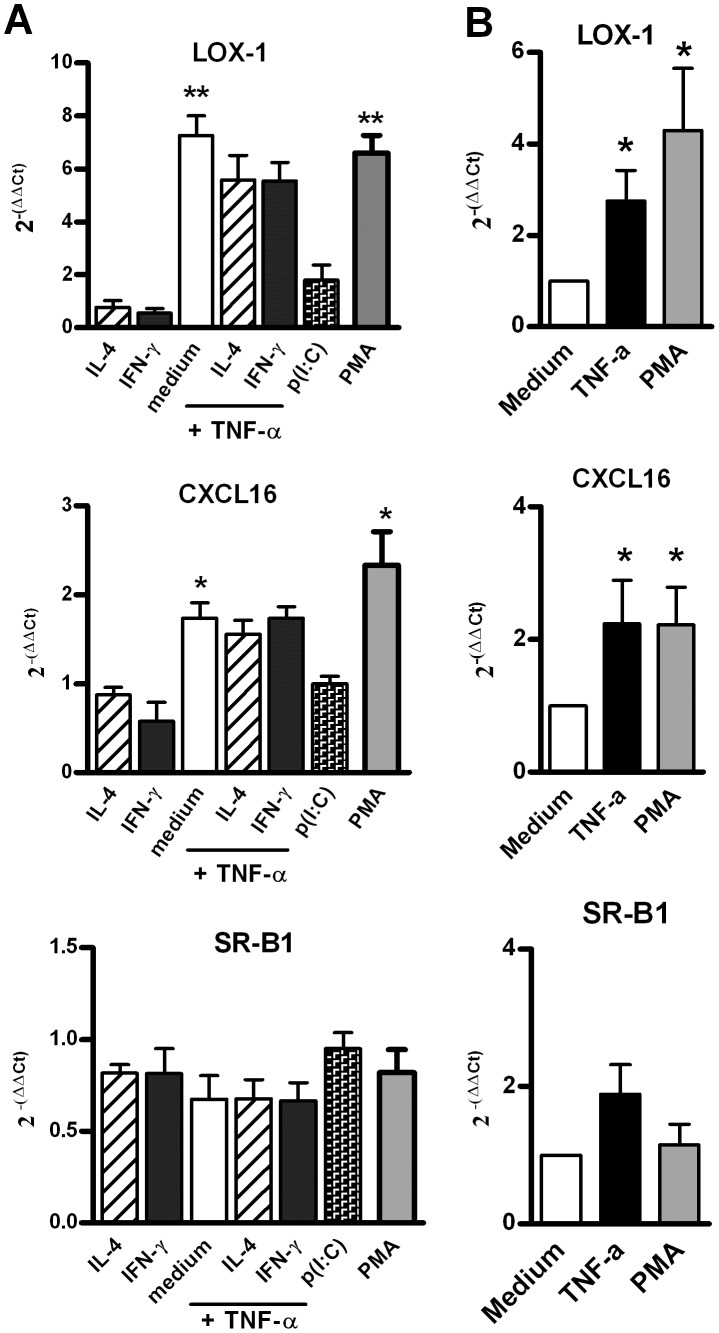
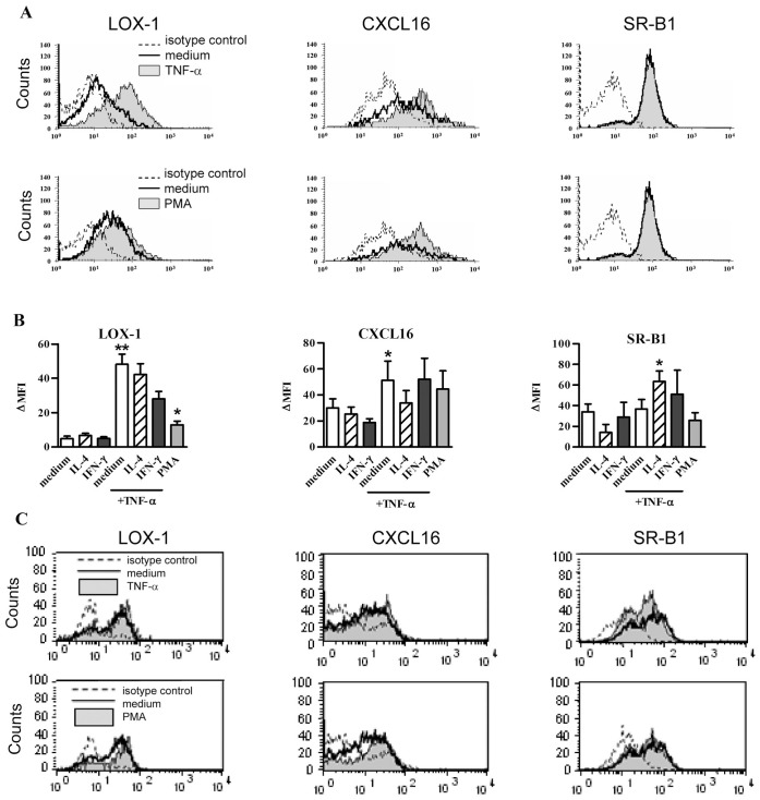
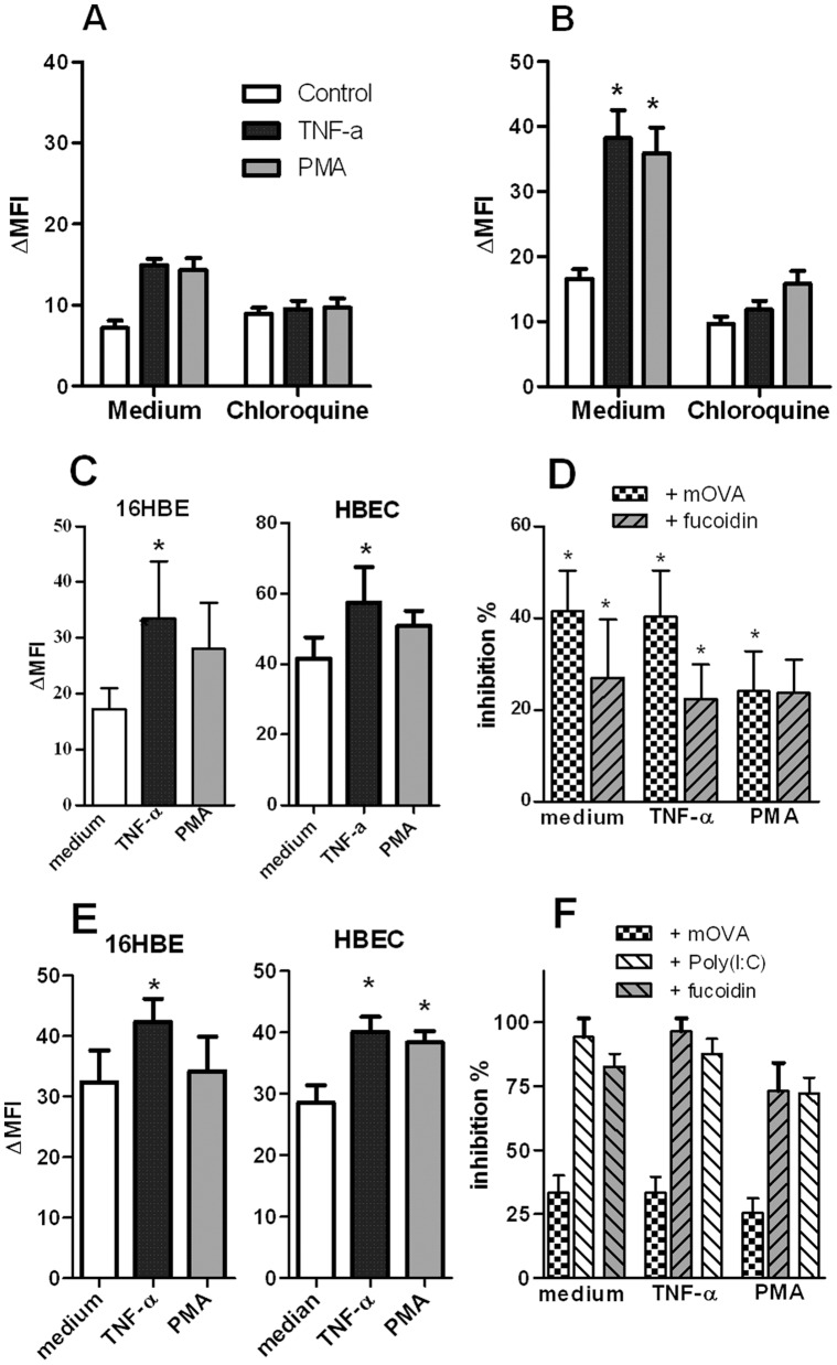
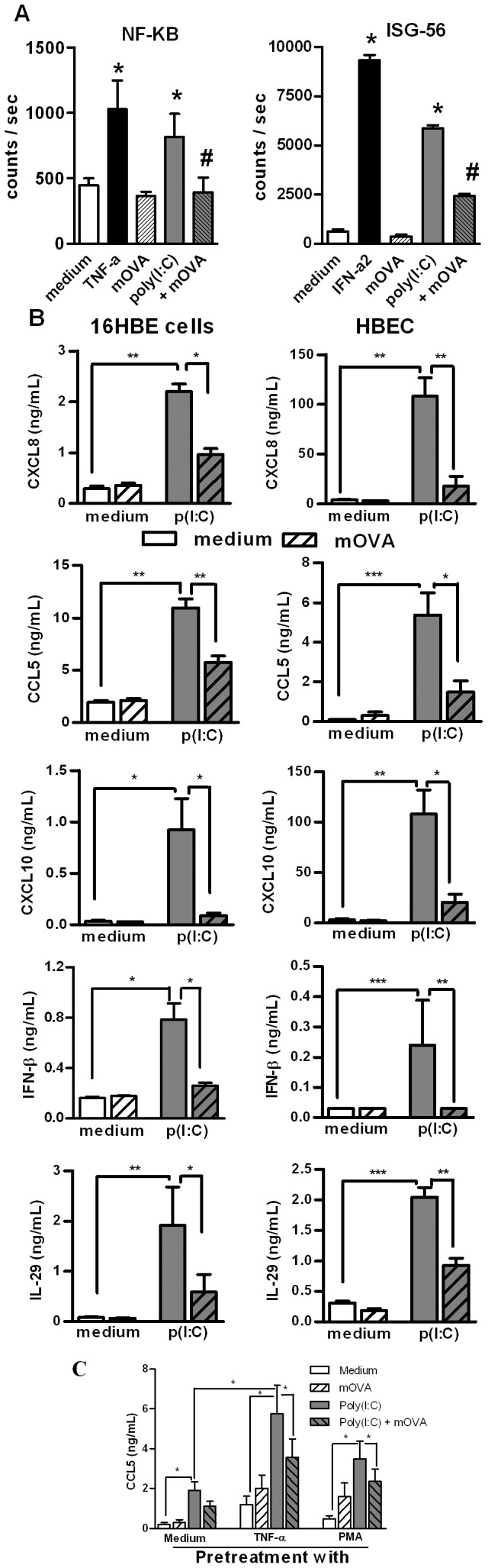
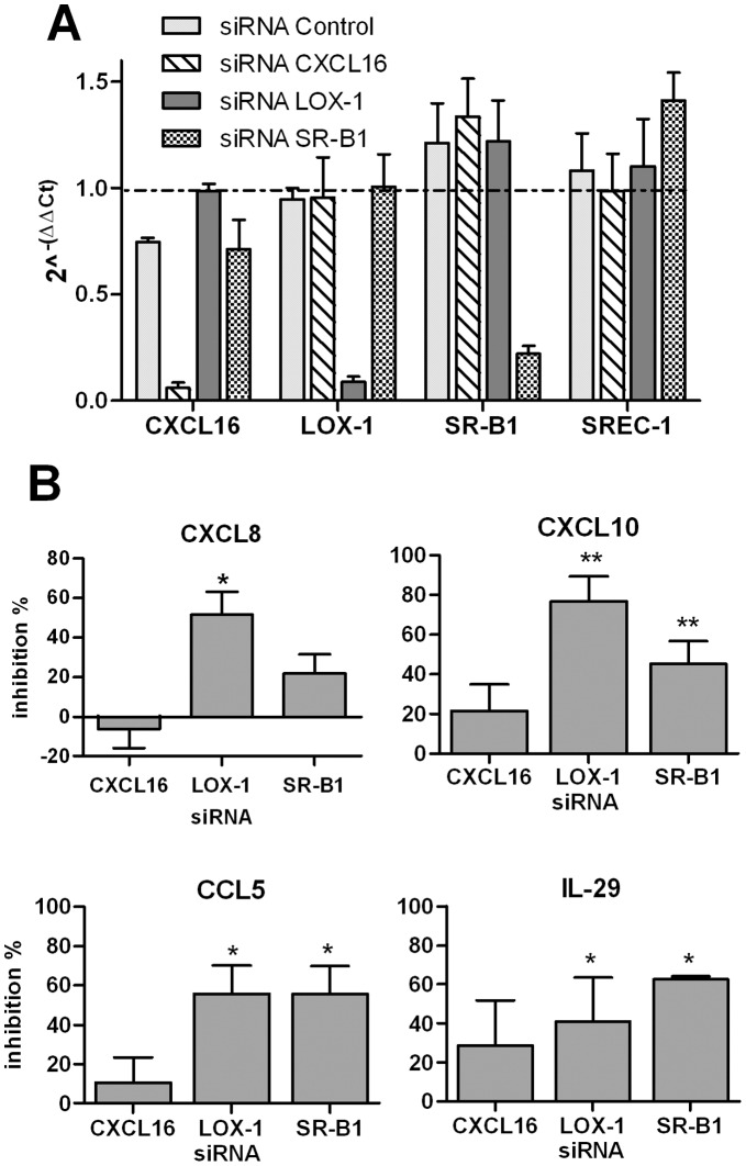
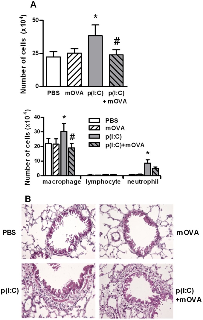
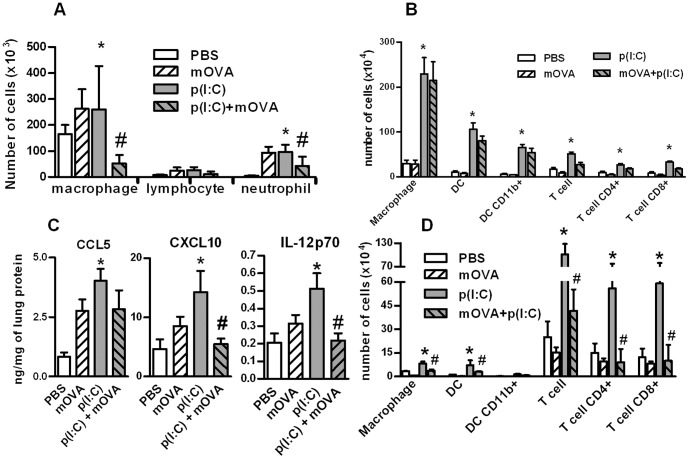
Scavenger receptors and Toll-like receptors (TLRs) cooperate in response to danger signals to adjust the host immune response. The TLR3 agonist double stranded (ds)RNA is an efficient activator of innate signalling in bronchial epithelial cells. In this study, we aimed at defining the role played by scavenger receptors expressed by bronchial epithelial cells in the control of the innate response to dsRNA both in vitro and in vivo. Expression of several scavenger receptor involved in pathogen recognition was first evaluated in human bronchial epithelial cells in steady-state and inflammatory conditions. Their implication in the uptake of dsRNA and the subsequent cell activation was evaluated in vitro by competition with ligand of scavenger receptors including maleylated ovalbumin and by RNA silencing. The capacity of maleylated ovalbumin to modulate lung inflammation induced by dsRNA was also investigated in mice. Exposure to tumor necrosis factor-α increased expression of the scavenger receptors LOX-1 and CXCL16 and the capacity to internalize maleylated ovalbumin, whereas activation by TLR ligands did not. In contrast, the expression of SR-B1 was not modulated in these conditions. Interestingly, supplementation with maleylated ovalbumin limited dsRNA uptake and inhibited subsequent activation of bronchial epithelial cells. RNA silencing of LOX-1 and SR-B1 strongly blocked the dsRNA-induced cytokine production. Finally, administration of maleylated ovalbumin in mice inhibited the dsRNA-induced infiltration and activation of inflammatory cells in bronchoalveolar spaces and lung draining lymph nodes. Together, our data characterize the function of SR-B1 and LOX-1 in bronchial epithelial cells and their implication in dsRNA-induced responses, a finding that might be relevant during respiratory viral infections.


Conflict of interest statement

Competing Interests: The authors have declared that no competing interests exist.

Figures

Figure 1
Figure 1. mRNA expression of SR in human BEC.
(A) mRNA expression of LOX-1, CXCL16 and SR-B1 by stimulated 16 HBE cells. These cells were stimulated for 6 h with IL-4, IFN-γ, TNF-α, poly(I:C) and PMA. mRNA expression was analyzed by quantitative RT-PCR. Results were normalized using β-actin as endogenous control and are shown as fold changes (2−ΔΔCt) relative to unstimulated cells used as calibrator. Data reported the mean ± SEM from 4 independent experiments. (B) mRNA expression of LOX-1, CXCL16 and SR-B1 by stimulated primary cultures of BEC. Cells were stimulated for 6 h with TNF-α, and PMA. Data were expressed as mean ± SEM of 2 -ΔΔCt from 4 independent experiments. *: p<0.05, **: p<0.01 versus control cells.
Figure 2
Figure 2. LOX-1, CXCL16 and SR-B1 surface expression in BEC.
(A) Analysis of SR expression by flow cytometry in 16 HBE cells. These cells were stimulated for 24 h with TNF-α (upper panel) or PMA (lower panel) (filled line, upper and lower histogram, respectively) as compared with cells in medium (bold line). Isotype control is represented with dotted line. Histograms of one representative experiment out of 7 are presented. (B) Modulation by cytokines and PMA of LOX-1, CXCL16 and SR-B1 expression in 16 HBE cells. The mean ± SEM of ΔMFI for 7 independent experiments are reported. *: p<0.05, **: p<0.01 versus control cells. (C) Modulation of LOX-1, CXCL16 and SR-B1 expression in HBEC stimulated for 24 h with TNF-α (upper panel) or PMA (lower panel). Histograms of flow cytometry are reported for HBEC. The filled line showed the stimulated cells as compared with cells in medium (bold line) and to the isotype control (dotted line). This is a representative experiment out of 3.
Figure 3
Figure 3. Endocytosis of SR ligands and dsRNA by BEC.
(A–B) Uptake of AlexaFluor®488-labelled Ac-LDL by 16 HBE cells. 16 HBE cells were stimulated with TNF-α (20 ng/mL) or PMA (10 ng/mL), in the absence or presence of chloroquine (100 µM), and then, were exposed for 30 minutes (A) or 1 hour (B) with labelled-Ac-LDL (10 µg/mL). The binding of the ligand was assessed by flow cytometry (n = 3). (C) mOVA binding is measured in 16 HBE cells and HBEC activated for 24 h with TNF-α and PMA. Cells were incubated for 1 h with FITC-conjugated mOVA. MFI was determined by flow cytometry. (D) Inhibition of mOVA uptake by unlabelled SR ligands. An excess of unlabelled mOVA and fucoidin was added 10 min before addition of FITC-labelled-mOVA. Data were expressed as the mean ± SEM from 5 to 13 independent experiments. (E) Modulation by TNF-α and PMA of the biotinylated poly(I:C) endocytosis in 16 HBE cells (left panel) and HBEC (right panel). Data are expressed as the mean ± SEM of ΔMFI (n = 4 to 5 experiments). (F) Inhibition of poly(I:C) binding by mOVA, fucoidin and unlabelled poly(I:C) on 16 HBE cells activated or not with TNF-α and PMA. Mean ± SEM of the percentages of inhibition were reported (n = 3). *: p<0.05, **: p<0.01 versus control cells.
Figure 4
Figure 4. Modulation by SR ligand of poly(I:C)-induced activation in BEC.
(A) Inhibition by mOVA of dsRNA-induced NF-κB-dependent and ISG-56-dependent luciferase production in BEAS-2B cells transfected with the appropriate plasmid. The values obtained with medium and mOVA alone, with positive control (TNF-α or IFN-α2) were also reported. Results were expressed as mean ± SEM of counts per second (n = 3). *: p<0.05 versus cells in medium alone; ≠: p<0.05 versus cells with poly(I:C). (B) Addition of mOVA modulated dsRNA-induced cytokine secretion in 16 HBE cells and HBEC. Secretion of CXCL8, CCL5, CXCL10, IFN-β and IL-29 was measured in supernatants of BEC in medium alone or stimulated with p(I:C) (mean ± SEM from 6 to 7 independent experiments). * p<0.05, ** : p<0.01; ***: p<0.001. (C) Pretreatment of 16 HBE cells with TNF-α and PMA increased the production of CCL5 in response to poly(I:C). Addition of mOVA inhibited the dsRNA-induced production in TNF-α and PMA-pretreated cells. * p<0.05, n = 4.
Figure 5
Figure 5. Impact of SR gene silencing on poly(I:C)-induced cytokine secretion by BEAS-2B cells.
BEAS-2B cells were treated with siRNA targeting CXCL16, LOX-1 and SR-B1 or the control siRNA (25 nM) and then activated with poly(I:C). (A) Inhibitory activity of RNA silencing on CXCL16, LOX-1, SR-B1 and SREC-1 mRNA expression was controlled by QRT-PCR. The reference corresponds to untreated cells (value  = 1). (B) RNA silencing of these SR inhibits the secretion of CXCL8, CXCL10, CCL5 and IL-29 induced by p(I:C). Data were expressed as the % of inhibition as compared with cells treated with the control siRNA (mean ± SEM) (n = 5). * p<0.05, n = 4.
Figure 6
Figure 6. Administration of mOVA affects the acute effect of poly(I:C) on cell recruitment in BAL.
Mice received one intranasal injection of PBS, mOVA (100 µg), poly(I:C) (20 µg) or mOVA + poly(I:C). BAL fluids and lungs were collected 16 h later. (A) Total and differential cell counts in the BAL were assessed by MGG coloration. Data are expressed as the mean ± SEM from 3 independent experiments including four mice per group. *: p<0.05 versus cells in medium alone; ≠: p<0.05 versus cells with poly(I:C). (B) Representative hematoxylin and eosin staining of lung sections in the four groups of mice. Magnification: ×300.
Figure 7
Figure 7. Repeated administration of mOVA affects the late response to poly(IC) in the lung.
Mice received three intranasal injections of PBS, mOVA (100 µg), poly(I:C) (20 µg), mOVA + poly(I:C). BAL, lungs and lymph nodes were collected 24 h after the last injection. (A) Absolute numbers of cells in the BAL were reported. (B) Numbers of macrophages, dendritic cell (DC) (total number and CD11b+) and T lymphocytes (total number, CD4+ and CD8+ cells) in the lung are shown. (C) Levels of CCL5, CXCL10 and IL-12p70 were measured by ELISA in protein extracts from the lungs. Results were expressed as the ratio of the total concentration of protein. (D) Numbers of macrophages, dendritic cell (DC) (total number and CD11b+) and T lymphocytes (total number, CD4+ and CD8+ cells) in mediastinal lymph nodes were evaluated by flow cytometry. Data are expressed as the mean ± SEM from 3 independent experiments including 4 mice per group. *: p<0.05 versus cells in medium alone; #: p<0.05 versus cells with poly(I:C).

Similar articles

Cited by

References

    1. Park HS, Suh JH, Kim SS, Kwon OJ (2000) Grain dust induces IL-8 production from bronchial epithelial cells: effect on neutrophil recruitment. Ann Allergy Asthma Immunol 84: 623–627. - PubMed
    1. Pichavant M, Delneste Y, Jeannin P, Fourneau C, Brichet A, et al. (2003) Outer membrane protein A from Klebsiella pneumoniae activates bronchial epithelial cells: implication in neutrophil recruitment. J Immunol 171: 6697–6705. - PubMed
    1. Holgate ST, Lackie P, Wilson S, Roche W, Davies D (2000) Bronchial epithelium as a key regulator of airway allergen sensitization and remodeling in asthma. Am J Respir Crit Care Med 162: S113–117. - PubMed
    1. Takizawa H (1998) Airway epithelial cells as regulators of airway inflammation (Review). Int J Mol Med 1: 367–378. - PubMed
    1. Hippenstiel S, Opitz B, Schmeck B, Suttorp N (2006) Lung epithelium as a sentinel and effector system in pneumonia–molecular mechanisms of pathogen recognition and signal transduction. Respir Res 7: 97. - PMC - PubMed

Publication types

MeSH terms